 New Products
 Earth-Friendly Products
 Biotium Choice Antibodies
 Special Offers


Content #1
Content #1
Content #1
Advances in gene therapy increasingly depend on understanding how viral vectors behave within complex, multilayered human tissues. While retinal organoids serve as a powerful model for studying AAV efficacy, their dense, light-scattering architecture has historically limited the ability to visualize and quantify transduction at single-cell resolution. Conventional nuclear stains suffer from rapid photobleaching, cytotoxicity, and shallow imaging depth which hinder repeated live imaging and prevent accurate 3D cell segmentation throughout the organoid. Conventional membrane dyes also pose challenges for staining organoids due to poor penetration, uneven labeling, and rapid internalization by endocytosis.
In a 2025 Small Methods publication, Rogler et. al. developed a longitudinal imaging and deep-learning pipeline to map single-cell AAV transduction dynamics in intact human retinal organoids. This approach required robust and photostable live-cell stains compatible with deep (>100 µm) confocal imaging and repeated imaging over many days. To meet this need, the authors selected Biotium’s far-red NucSpot® Live 650 Nuclear Stain, which provides bright, uniform labeling with minimal phototoxicity and exceptional light penetration compared to blue- or green-excitable DNA dyes. CellBrite® Steady 550, a unique stain for long-term labeling of membranes in live cells, was also used for manual quantification of transduced cells to gauge the performance of their deep-learning method.
Using NucSpot® Live 650, the team captured high-contrast 3D nuclear signals across entire organoids and enabled the use of Cellpose, a deep-learning segmentation algorithm. Paired with GFP-expressing AAV reporters, this allowed precise quantification of transduced cells, as well as quantification of how transduction patterns evolve over time and spatial depth.
The end result revealed heterogeneous AAV penetration profiles, cell-type-specific susceptibility, and spatial gradients of transduction that would have been obscured using conventional methods. Biotium’s NucSpot® Live 650 Nuclear Stain and CellBrite® Steady 550 Membrane Stain enabled high-fidelity, longitudinal imaging in thick living tissues, making quantitative AAV mapping in 3D retinal models possible.
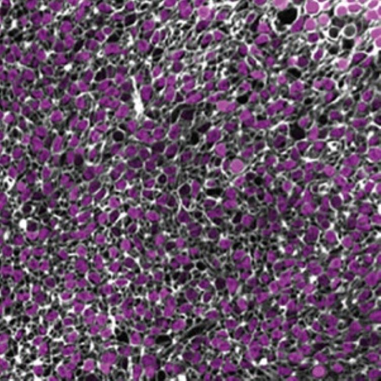
Confocal image of the center plane of the 3D stack of a 264 days old human retinal organoid without virus, stained with NucSpot Live 650 (magenta) and CellBrite Steady 550 (white). Credit: Rogler et al., Small Methods (2025). Reproduced under CC BY 4.0.
Biotium offers an extensive portfolio of bright and specific nuclear and membrane stains, with color options in the near-infrared for deep imaging. View our full selection of cell stains compatible with organoids and other 3D cultures.
Full Citation:
Rogler, T. S., Salbaum, K. A., Brinkop, A. T., Sonntag, S. M., James, R., Shelton, E. R., Thielen, A., Rose, R., Babutzka, S., Klopstock, T., Michalakis, S., & Serwane, F. (2025). 3D quantification of viral transduction efficiency in living human retinal organoids. Small Methods, 2025 Jun 12, e2401050. https://doi.org/10.1002/smtd.202401050

Content #1
Content #1
Content #1
Content #2
Content #3