New Products
 Earth-Friendly Products
 Biotium Choice Antibodies
 Special Offers
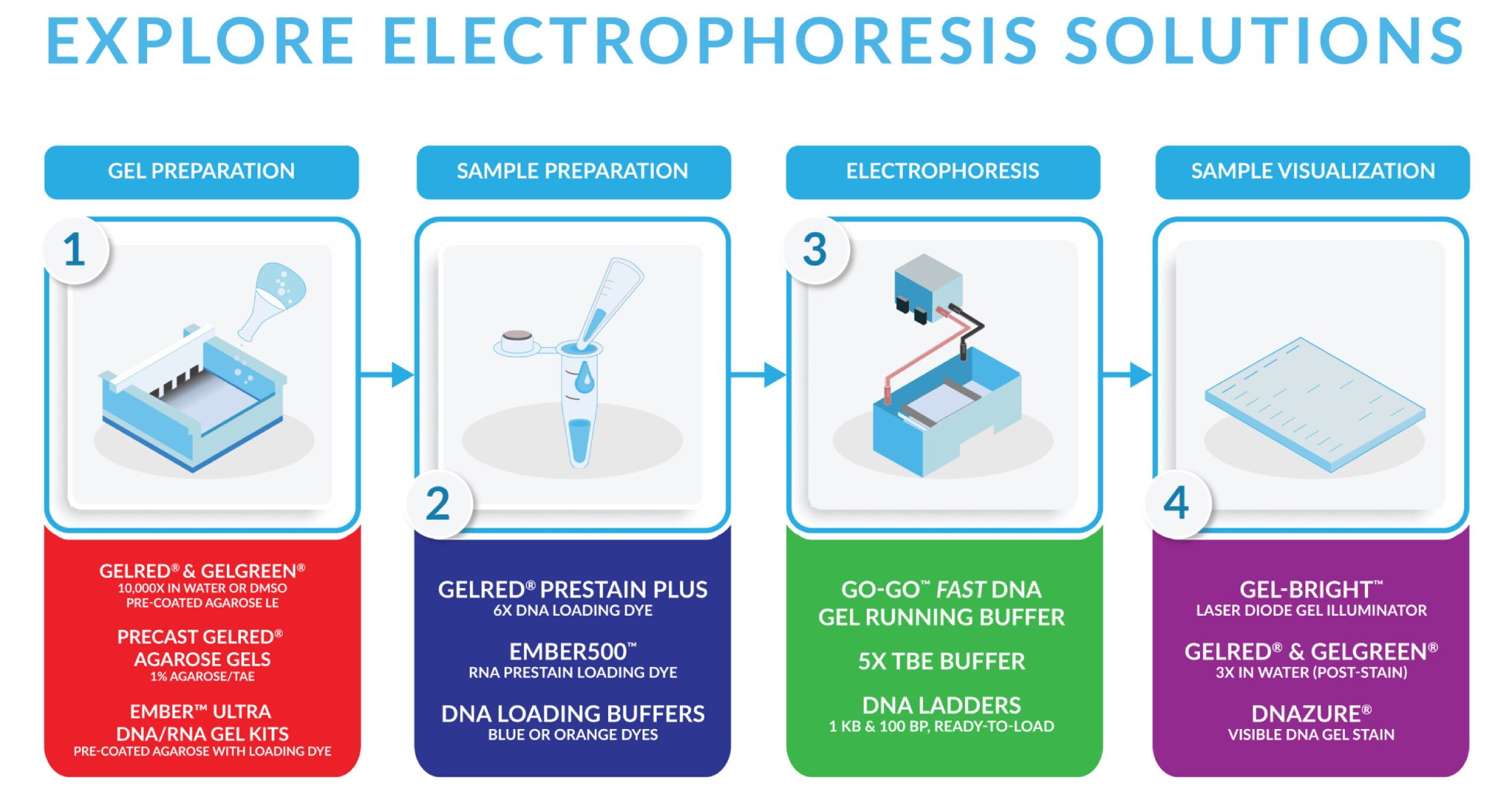
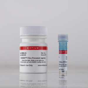
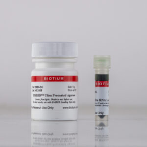
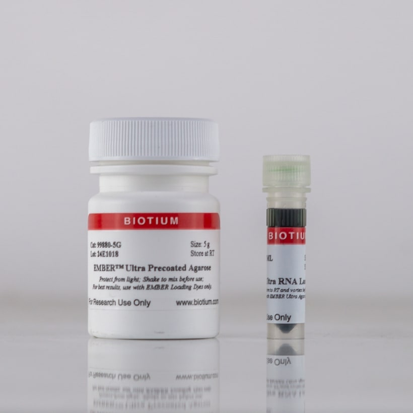
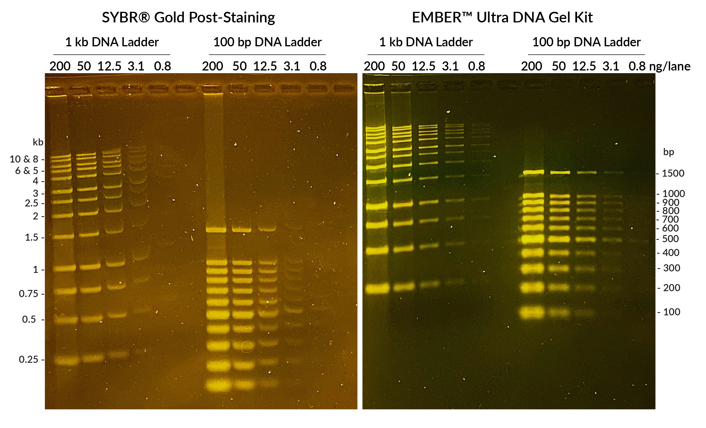
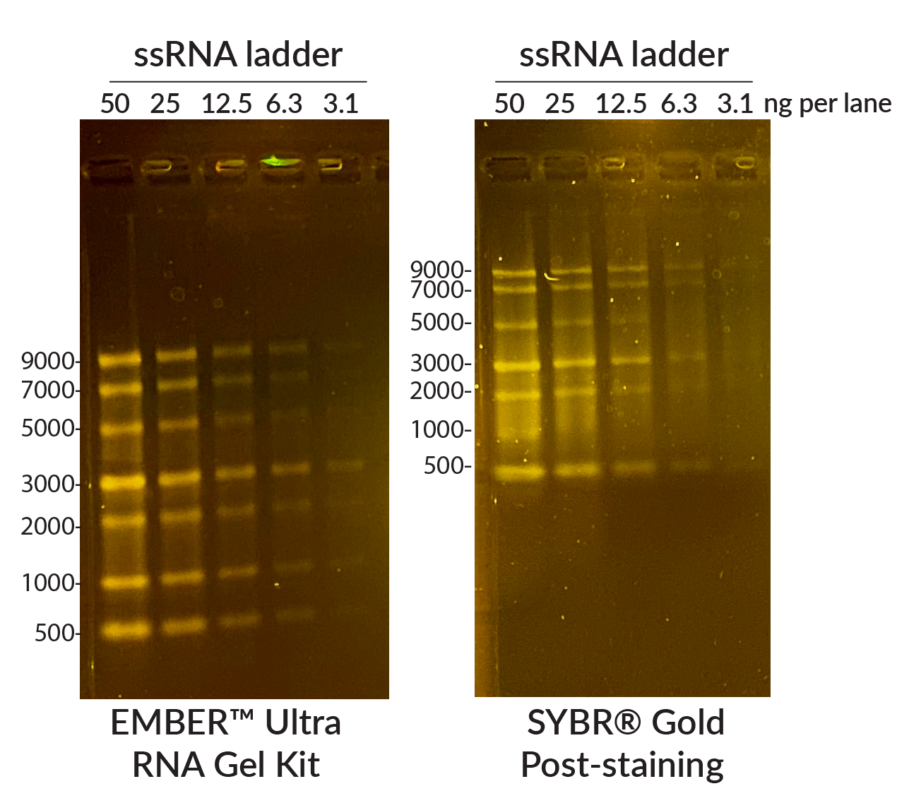
EMBER™ Ultra DNA & RNA Gel Kits
Most sensitive nucleic acid gel stains in a convenient pre-coated agarose format.
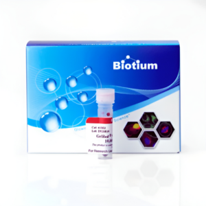
GelRed®
Safer and more sensitive than Ethidium Bromide.
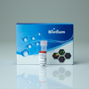
GelGreen®
PAGE GelRed®
DNAzure® 2.0 Visible Blue DNA Gel Stain
Glo-Plate™ White
Go-Go™ Fast DNA Gel Running Buffer
Classic Gel Stains
Gel-Bright™ Gel Illuminator
DNA Ladders, Buffers, & Other Accessories
EMBER™ Ultra DNA & RNA Gel Kits
The Most Sensitive DNA & RNA Gel Stains Available
The EMBER™ Ultra DNA Gel Kit and EMBER™ Ultra RNA Gel Kit provide the highest sensitivity and resolution for DNA or RNA. They are available in a convenient pre-coated agarose format and therefore do not need a post-electrophoresis staining step. Simply cast your gel using the provided EMBER™ Ultra Precoated Agarose and prepare your samples with the include EMBER™ Ultra Loading Dye. Bands can be imaged immediately after electrophoresis.
EMBER™ Ultra Gel Kit Features:
- Easy-to-use pre-coated agarose format for DNA or RNA gel electrophoresis
- Superior sensitivity and resolution over other nucleic acid gel stains
- No need for post-electrophoresis staining
- EMBER™ Ultra RNA Gel Kit includes formamide in the loading dye
- Optimal for blue LED gel imagers
- Also compatible with UV imagers
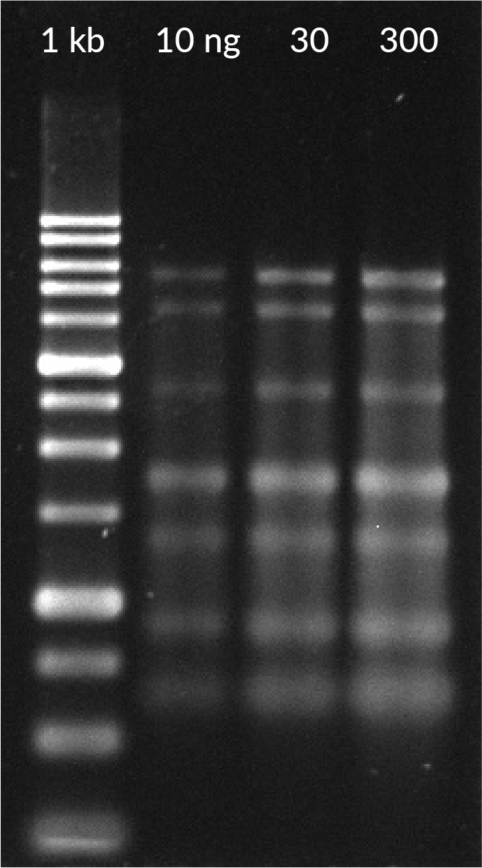
EMBER™ Ultra DNA Gel Kit:
Detect ≤1 ng DNA
Experience unmatched sensitivity and convenience with the EMBER™ Ultra DNA Gel System—featuring pre-coated agarose and loading dye for immediate imaging post-electrophoresis. Detect as little as ≤1 ng DNA with no staining step required.
Learn more about the EMBER™ Ultra DNA Gel Kit.
EMBER™ Ultra RNA Gel Kit:
Detect ≤5 ng RNA
Achieve exceptional RNA detection with the EMBER™ Ultra RNA Gel System—no post-staining required. Pre-coated agarose and denaturing loading dye deliver ≤5 ng sensitivity for fast, reliable results and clear RNA integrity assessment.
Learn more about the EMBER™ Ultra RNA Gel Kit.
View Product Page

GelRed® & GelGreen® Nucleic Acid Gel Stains
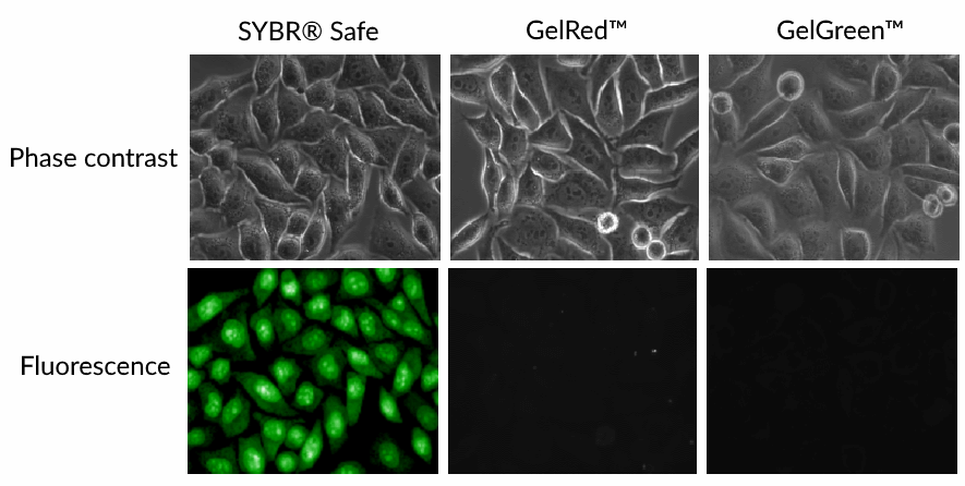
How Safe is Your Gel Stain?
A number of ethidium bromide (EtBr) alternatives are marketed as being safe. In fact, many so-called “safe” gel stains contain dyes that are well known to bind DNA in living cells, with cytotoxic effects. GelRed® and GelGreen® are highly sensitive gel stains designed to be nontoxic and nonmutagenic by virtue of being cell membrane impermeable, so they cannot enter living cells. Download our white paper to learn more.
The Gold Standard of DNA Gel Stains
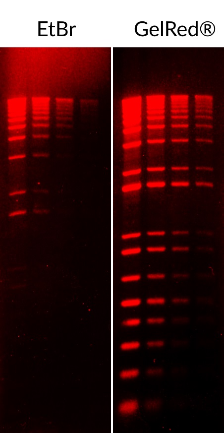
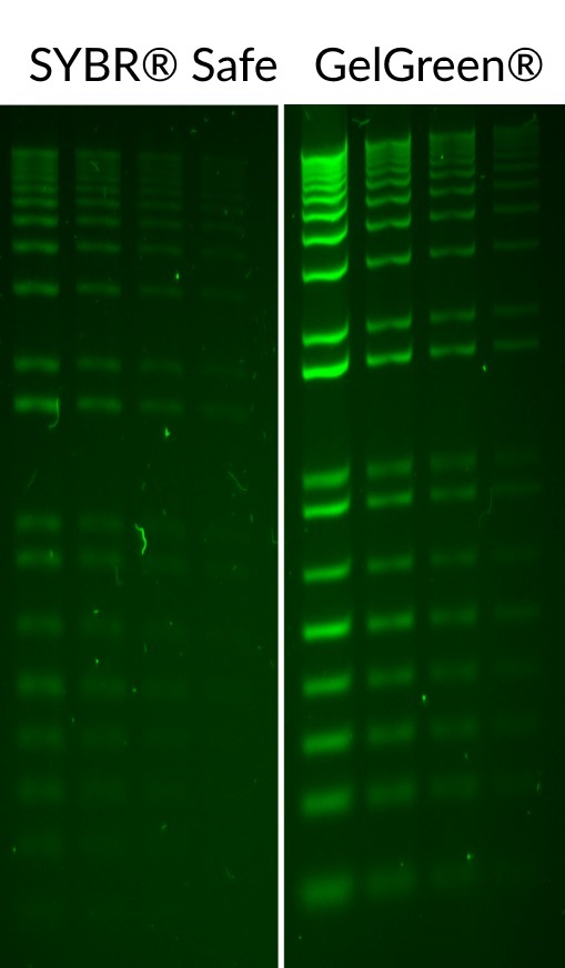
GelRed® and GelGreen® are safer alternatives to EtBr, SYBR® Safe, and others
While EtBr has been widely used for its low cost, but its mutagenicity and associated safety and disposal issues can make it costly overall. Biotium’s GelRed® and GelGreen® dyes were developed to offer superior sensitivity and unmatched safety—proven non-toxic and safer by independent labs. They're also available pre-coated on ultra-pure LE agarose for added convenience, eliminating the need to handle concentrated dye.
GelRed® Features:
- Visualized with UV transilluminators using ethidium bromide detection settings
- Most sensitive fluorescent red DNA gel stain
- Safer and more environmentally friendly than EtBr, as well as other so-called “safe” gel stains
- Can be used as a post-stain, or precast in agarose
- Available as a 10,000X stock, ready-to-use 3X solution, and in 6X loading buffer format
GelGreen® Features:
- Visualized with blue light boxes or UV light box using SYBR® green detection settings
- More sensitive than SYBR® Safe
- Safer for users and the environment than SYBR® Safe and other so-called “safe” gel stains
- Can be used as a post-stain, or precast in agarose
- Available in a concentrated 10,000X stock
GelRed® & GelGreen® DNA Agarose Gel Stain Formats
| Product Name | Catalog Number | Size |
|---|---|---|
| GelRed® 10,000X in water | 41003-T | 0.1 mL (Trial Size) |
| 41003 | 0.5 mL | |
| 41003-1 | 10 mL | |
| GelGreen® 10,000X in water | 41005 | 0.5 mL |
| 41005-1 | 10 mL | |
| GelRed® 3X in water | 41001 | 4 L |
| 6X GelRed® Prestain Loading Buffer, Orange Tracking Dye | 41010 | 1 mL |
| GelRed® Prestain Plus 6X DNA Loading Dye | 41011 | 1 mL |
| GelRed® Agarose LE | 41029-5G | 5 g |
| 41029-50G | 50 g | |
| GelGreen® Agarose LE | 41030-5G | 5 g |
| 41030-50G | 50 g |
PAGE GelRed® for Acrylamide Gels
PAGE GelRed® is a version of our popular GelRed® dye that was specially formulated for staining DNA in polyacrylamide gels. Like the classic GelRed®, PAGE GelRed® has also been found to be safe by independent laboratories.
Download the PAGE GelRed® Safety Report.
- Visualized with UV transilluminators using ethidium bromide detection settings
- Formulated for efficient penetration and staining of polyacrylamide gels
- Proven safe and environmentally friendly
- Compatible with downstream cloning applications
| Product Name | Catalog Number | Size |
|---|---|---|
| PAGE GelRed® 10,000X in Water | 41008-T | 0.1 mL |
| PAGE GelRed® 10,000X in Water | 41008-500uL | 500 uL |
| PAGE GelRed® 1X in Water | 41014 | 4 L |
Choose the Right Stain for Your Application
| Product / Method | Procedure | Advantages | Disadvantages | Recommended for |
|---|---|---|---|---|
| DNA staining with EMBER™ Ultra DNA Gel Kit | Agarose is supplied pre-coated with EMBER™ Ultra Dye, just dissolve, heat, and pour. | • Safer and more convenient, no need to handle concentrated dye • Superior sensitivity, detect as little as ≤1 ng DNA • No need for post-electrophoresis staining • Optimal for blue LED gel imagers | • Not suitable for PAGE, DGGE, EMSA, or PFGE gels • Dye may cause band migration issues when loading larger amounts of DNA (more than ~200 ng/band), or for some restriction digests | • Routine agarose gels |
| RNA staining with EMBER™ Ultra RNA Gel Kit | Agarose is supplied pre-coated with EMBER™ Ultra Dye, just dissolve, heat, and pour. | • Safer and more convenient stain for RNA, no need to handle concentrated dye • Superior sensitivity, detect as little as ≤5 ng RNA • No need for post-electrophoresis staining • Included loading dye contains formamide for denaturing • Optimal for blue LED gel imagers | • Will stain DNA as well as RNA • Dye may cause band migration issues when loading larger amounts of RNA (more than ~200 ng/band) | • Routine RNA gel electrophoresis • Evaluate total RNA integrity and DNA contamination |
| DNA prestaining with GelRed® Prestain Plus 6X DNA Loading Dye | GelRed® loading buffer is added directly to the DNA sample before loading | • Fast & simple: one-step sample loading & DNA staining • Less concentrated dye for safer handling • Can re-run a gel to use empty lanes | • Not recommended for PAGE, DGGE, EMSA, or PFGE gels • Dye may cause band migration issues when loading larger amounts of DNA (more than ~100 ng/band), or for some restriction digests | • Routine agarose gels • Recommended loading 50-200 ng ladder or 2-5 uL PCR product ( ~100 ng/band or less) |
| Precast staining with GelRed® 10,000X in water or GelGreen® 10,000X in water | GelRed® or GelGreen® is mixed with molten agarose before gel casting | Familiar protocol, rapid results | ||
| Precast staining with GelRed® Agarose LE or GelGreen® Agarose LE | Agarose is supplied pre-coated with GelRed® or GelGreen®, just dissolve, heat, and pour | Safer & more convenient, no need to handle concentrated dye | ||
| Post-electrophoresis staining with GelRed® 10,000X in water or GelGreen® 10,000X in water - or - GelRed® 3X in water | No fluorescent dye is added to the gel, it is stained in 3X GelRed® or 3X GelGreen® solution after electrophoresis | • Most accurate sizing/sharpest bands • Staining solution can be re-used • Enhance sensitivity by adding NaCl | Extra staining step (up to 30 minutes) after electrophoresis (some customers report good results after only 5 minutes if dye is not reused) | • Highly accurate band sizing • Gels with more than ~100 ng DNA per band • Analyzing restriction digests |
| Post-electrophoresis staining with DNAzure® 2.0 Visible Blue DNA Gel Stain Kit | No fluorescent dye is added to the gel, it is stained in DNazure® 2.0 solution and then exposed to a bright light source to generate visible blue DNA bands. We recommend the Glo-Plate™ White Photoactivation Device as a light source for developing DNAzure® 2.0-stained gels | • Allows visualization of DNA bands by the naked eye, no need for a UV light source • Detect as little as ~1 ng DNA • Stained bands are stable in gel for weeks • Also emits near-IR fluorescence (~700 nm) for detection on near-IR imaging systems | Extra staining step (up to 30 minutes) followed by a light exposure step (up to 30 minutes) to generate visible blue DNA bands | • Routine DNA agarose gels • Visualizing gels without the UV light or expensive imaging systems • Recommended loading 50-200 ng DNA per lane |
| Post-electrophoresis staining of PAGE gels using PAGE GelRed® 10,000X or 1X in water | No fluorescent dye is added to the gel, it is stained in 1X PAGE GelRed® solution after electrophoresis | • Formulated for efficient penetration and staining of polyacrylamide gels • Like the classic GelRed®, it is safe and environmentally friendly | Extra staining step of approx. 30 minutes after electrophoresis | Staining of nucleic acids in PAGE gels |
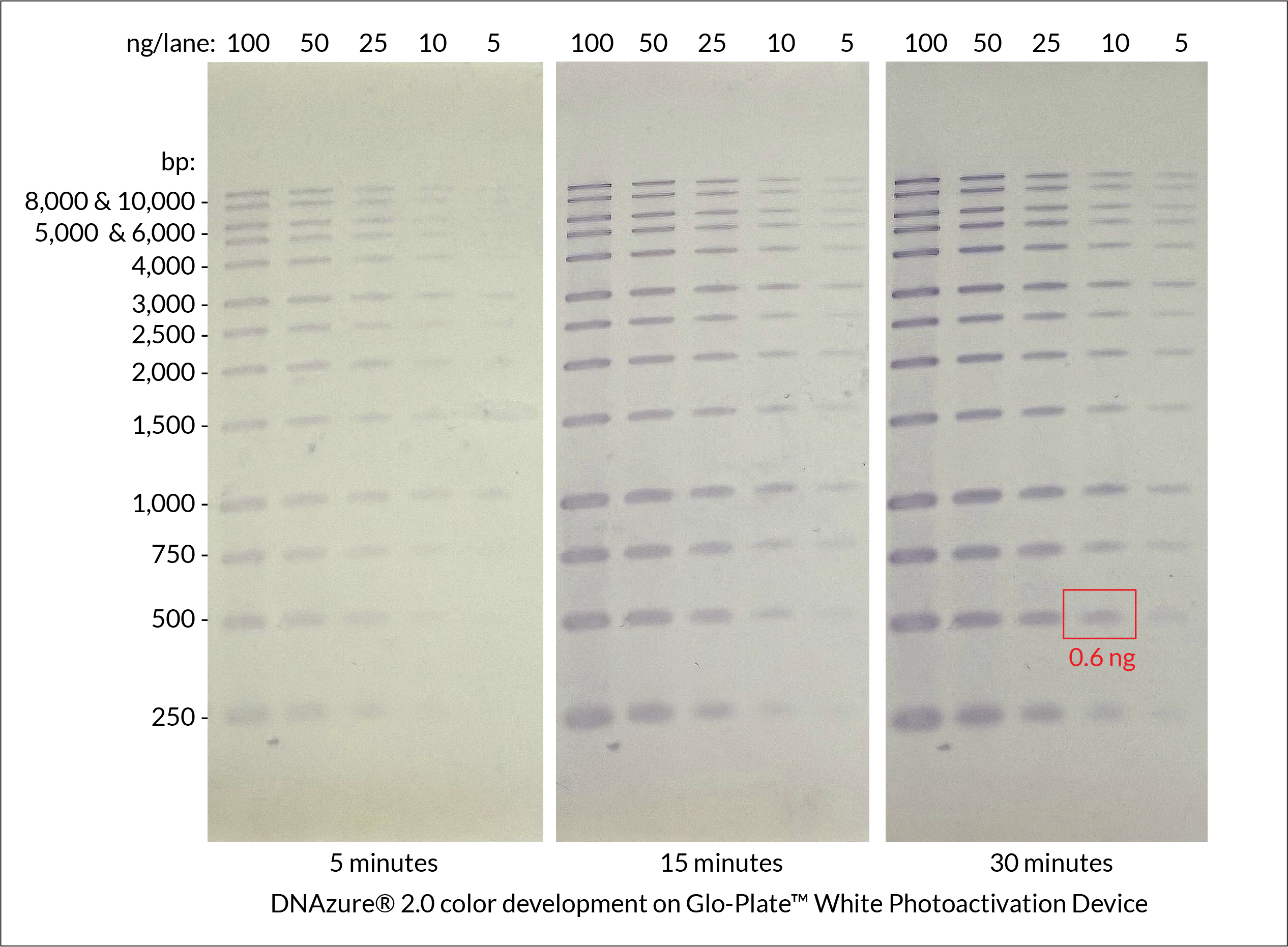
DNAzure® 2.0 Visible DNA Gel Stain
Visualize Your Results with the Naked Eye
Now in New Formulation with Improved Sensitivity
DNAzure® 2.0 Visible Blue DNA Gel Stain Kit is a new and improved formulation of our original DNAzure® Blue Nucleic Acid Gel Stain, delivering enhanced sensitivity for visible staining of double-stranded DNA in both agarose and polyacrylamide gels. DNAzure® 2.0 utilizes a DNA-binding dye that turns deep blue upon exposure to bright light, with sensitivity comparable to most fluorescent stains and the ability to detect less than 1 ng of DNA per band. After light exposure, bands are visible to the naked eye with no need for a UV transilluminator or LED light box. Stained gels remain stable for weeks after color development, making it easy to revisit results on your timeline.
DNAzure® 2.0 Features:
- Visible deep blue bands following light exposure
- Ultrasensitive detection, down to ~1 ng DNA
- Simplified DNA band excision without UV light
- Stained bands are stable in gel for weeks
- Detectable by near-IR imaging systems at ~700 nm
- Stronger signal and higher sensitivity than original DNAzure® Stain
View Product Page
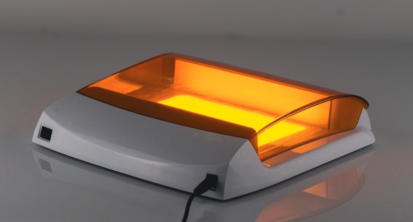
Glo-Plate™ White Photoactivation Device
Reliably Develop DNAzure® Stained Gels
Specifically created to empower educational labs and academic environments, the Glo-Plate™ White offers a cost-effective and user-friendly alternative to expensive DNA gel imaging systems or hazardous UV light. The device delivers even, high-intensity white LED illumination across its panel, optimized for activating the blue dye precipitation in DNAzure® 2.0-stained DNA gels.
Glo-Plate™ White Features
- Bright, uniform illumination: Designed for developing gels stained with DNAzure® 2.0 Visible Blue DNA Gel Stain Kit
- Eliminates UV light hazards: DNAzure® 2.0 allows direct visualization of blue DNA bands without UV light
- Small footprint: Compact, lightweight design ideal for shared lab benches
- Two-year warranty included
View Product Page
Order now and save $340 on the Glo-Plate™ White Photoactivation Device at checkout.
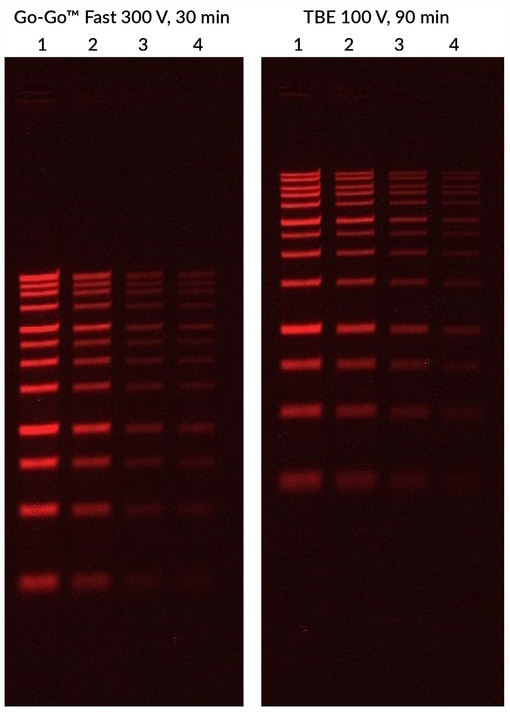
Go-Go™ Fast DNA Gel Running Buffer
Wish Your Agarose Gel Was Done Already?
Speed up routine DNA gel analysis with Biotium’s Go-Go™ Fast DNA Gel Running Buffer. This novel low ionic strength running buffer allows you to run DNA agarose gels at higher voltage to get results up to 3X faster than with TAE or TBE buffer.
- Save time: Run your gel 3X faster than with TAE or TBE
- Clear results: Provides crisp band resolution
- Versatile: Excellent results with GelRed®, GelGreen®, and other popular gel stains
- Supplied as a 50X concentrated stock solution
Go-Go™ Fast DNA Gel Running Buffer gives excellent results with GelRed® and GelGreen® DNA gel stains, as well as with GelRed® Prestain Plus 6X DNA Loading Dye. The buffer can be used with other commonly used gel stains like SYBR® Safe and ethidium bromide.
View Product Page
Classic Gel Stains
| Product Name | Catalog Number | Size |
|---|---|---|
| Thiazole Green (SYBR® Green I), 10,000X in DMSO | 40086-0.5mL | 500 uL |
| 40086-1mL | 1 mL | |
| Oxazole Gold (SYBR® Gold), 10,000X in DMSO | 40094 | 500 uL |
| Ethidium Bromide, 10 mg/mL in H2O | 40042 | 10 mL |
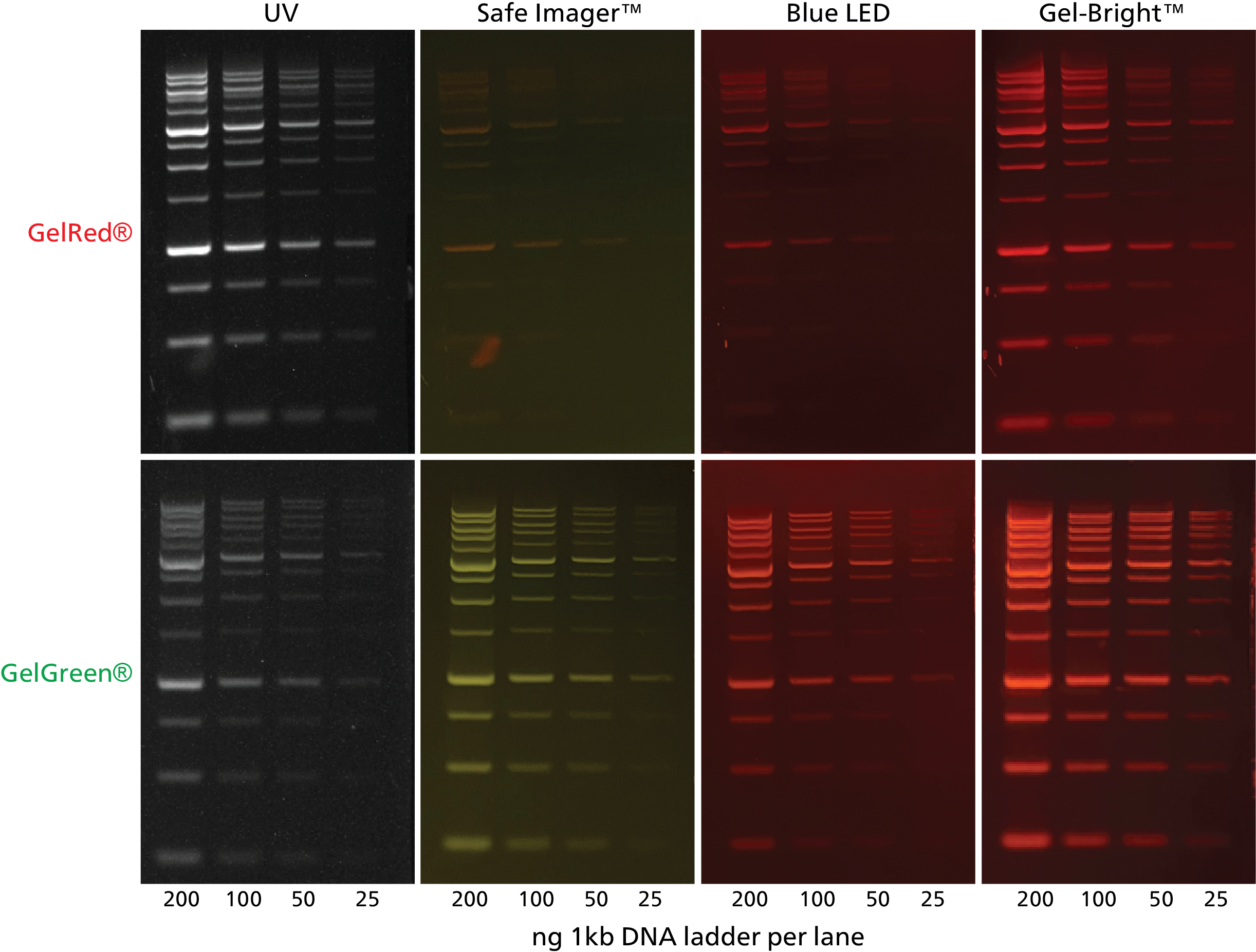
Gel-Bright™ Laser Diode Gel Illuminator
Novel Laser Diode Illumination for Superior Performance
Gel illuminators have traditionally relied on UV illumination for visualization of fluorescently labeled nucleic acids and proteins. However, UV-based imaging can damage skin and eyes, as well as your DNA samples. LED-based illuminators were developed as a safer alternative to UV, but often have high background and dimmer signal due to excessive ambient light and poor excitation efficiency.
In partnership with Biotium, OMEC Medical has developed a new type of gel illuminator that uses laser diodes (LDs) optimized for brighter and clearer imaging of gels.
The new laser diode-based Gel-Bright™ offers better sensitivity over UV-based transilluminators when imaging green dyes such as GelGreen® or SYBR® Green, as well as significantly better performance over blue LED gel illuminators for red dyes such as GelRed®, ethidium bromide (EtBr), and One-Step Lumitein™ Protein Gel Stain. Read the full press release.
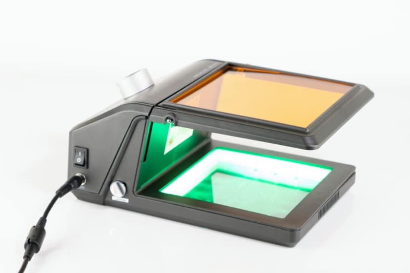
The Laser Diode Advantage:
- Novel LD illumination: Superior performance for imaging fluorescent gels
- Brighter signal: More sensitive than UV for green dyes
- Versatile: Works well with both green and red dyes, unlike blue LED illuminators
- Safer: Eliminates UV light hazards for user and DNA samples
Other Features:
- Optimized lighting angle for even illumination
- Adjustable light intensity
- Multi-hinged adjustable amber filter for optimal signal
- Easy access to the gel for gel slice excision
- Compact and portable design
Sensitive Gel Imaging Without UV Hazards
View Product Page
DNA Ladders, Buffers, & Other Accessories
| Product Name | Catalog Number | Size |
|---|---|---|
| 1 kb DNA Ladder, Ready-to-Load | 31084 | 150 applications (1.5 mL) |
| 100 bp DNA Ladder, Ready-to-Load | 31085 | 150 applications (1.5 mL) |
| 1 kb DNA Ladder in TE Buffer | 31080 | 500 uL |
| 100 bp DNA Ladder in TE Buffer | 31081 | 500 uL |
| 6X DNA Loading Buffer (Blue) | 99962-1 | 4 x 1.5 mL |
| 6X DNA Loading Buffer (Orange) | 99859-1 | 4 x 1.5 mL |
| 1X TAE (1L) Buffer Powder Packets | 22031 | 50 packets |
| 1X TBE (1L) Buffer Powder Packets | 22032 | 50 packets |
| 1X PBS (2L) Buffer Powder Packets | 22033 | 50 packets |
| TBE Buffer, 5X | 41006 | 4 L |
| Water, Ultrapure Molecular Biology Grade | 41024-4L | 4 L |
| Activated Charcoal Decontamination Bags | 22007 | 25 bags |
| DNA Gel Extraction Kit | 31030-50 | 50 assays |
| 31030-250 | 250 assays | |
| Agarose LE, Ultra-Pure Molecular Biology Grade | 41028-25G | 25 g |
| 41028-100G | 100 g | |
| 41028-500G | 500 g |