New Products
 Earth-Friendly Products
 Biotium Choice Antibodies
 Special Offers
Introduction
Flow cytometry is a prolific and powerful analytical tool used for biological research and the clinic. At its core, flow cytometry enables researchers to obtain highly specific information on individual cells within a sample. Recent advancements in this field have led to the development of spectral flow cytometry, a rapidly growing technology with significantly enhanced multiplexing capabilities over conventional flow cytometry. Several companies have capitalized on spectral flow with the release of the Sony ID7000™ and the well-known Cytek® Aurora. Where conventional flow cytometer instruments can detect panels with more than a dozen fluorophores, these new spectral flow cytometers are capable of accommodating multiparametric panels with upwards of 30 different fluorophores.
How Does Spectral Flow Cytometry Work?
Spectral flow cytometry shares much of the same hardware associated with conventional flow cytometry. Both systems employ standard fluidic and laser technologies that enable cell-by-cell spectral analysis. For both spectral and conventional flow cytometry methods, the process begins by delivering a sample stream through a flow chamber where cells move in a single file at a constant velocity, a process known as hydrodynamic focusing. This allows uniform and efficient excitation by a set of monochromatic lasers. Emitted photons are then detected and analyzed by a combination of optics and software to identify unique spectral emissions from fluorophore labeled biomolecules.
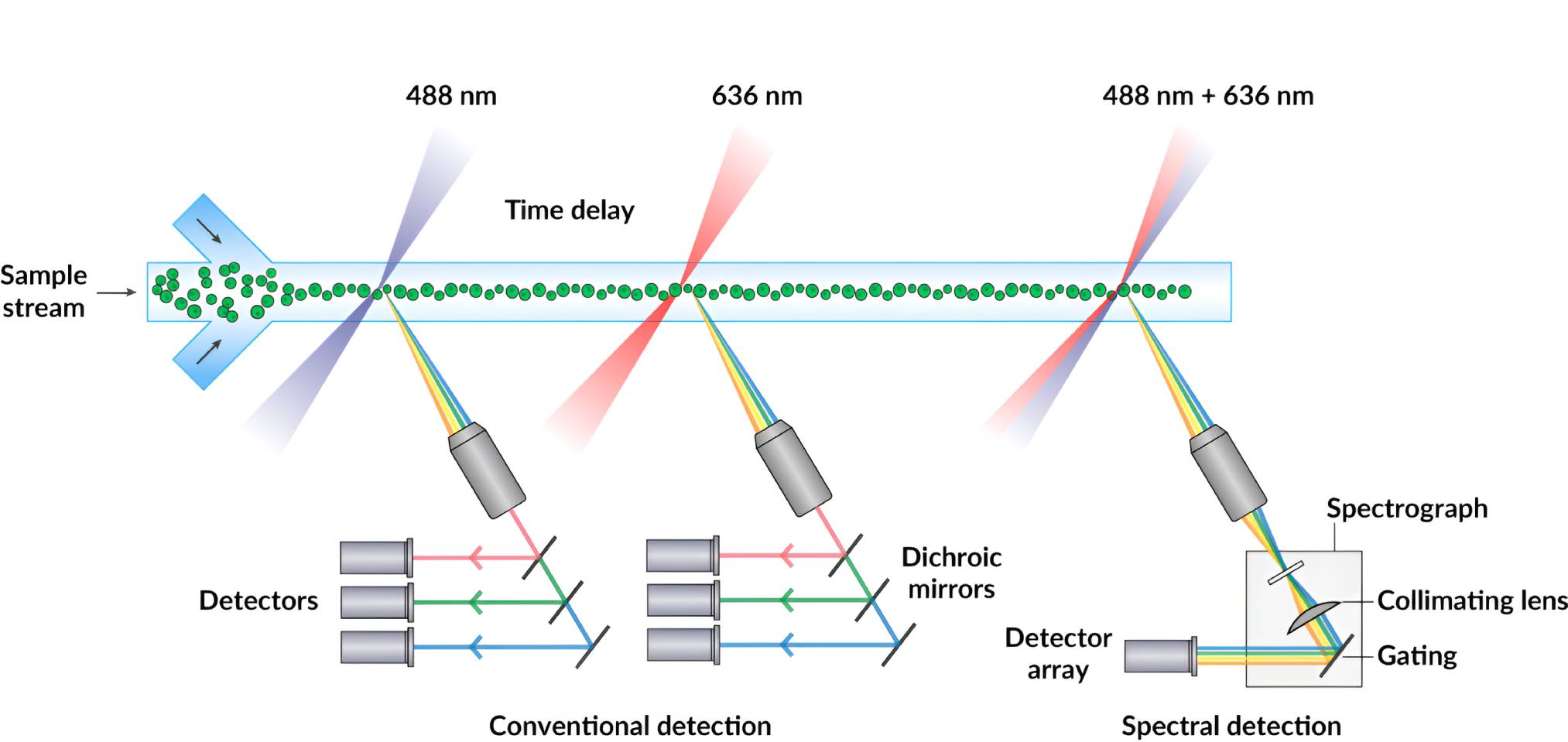
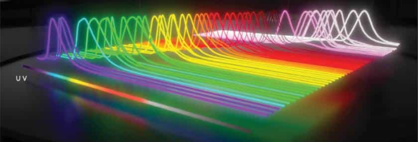
Capturing the Full Spectrum
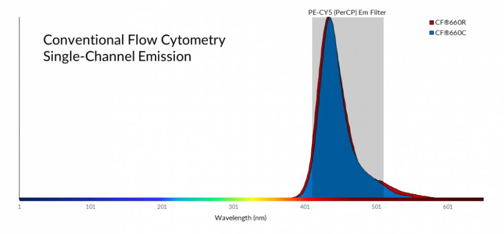
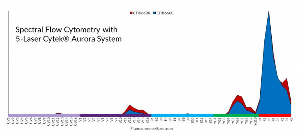
Spectral flow differs most significantly from conventional flow cytometry in the optical configuration and software analysis. In conventional flow cytometry, emitted photons are funneled through dichroic mirrors and band pass filters that partition the light into a narrow bandwidth for detection by a set of photomultiplier tubes. Spectral flow uses dispersive optics that disperse photons according to wavelength across an array of detectors, which broadens a fluorophore’s spectral profile by capturing the entire visible and near-IR spectrum and allows for higher resolution spectral analysis. Consequently, fluorophores with very similar emission spectra that were once impossible to separate by conventional flow cytometry methods, can now be distinguished through spectral flow cytometry.
Compensation vs. Spectral Unmixing
The same flow cytometry principles of panel design and spectral spillover are applicable to spectral flow cytometry. Designing multicolor panels requires careful consideration of fluorophores with distinct spectral profiles that can be separated by the instrument. In conventional flow, when a panel with a dozen or more fluorophores are used, overlap of emission spectra is unavoidable. This leads to noise from emissions of unintended fluorophores, known as spectral spillover. Single-color control samples are run with each experiment to determine the amount of spectral overlap present in each detector. In conventional flow cytometry, a mathematical method known as compensation applies these controls to subtract the overlapping spectra and isolate a fluorophore’s emission profile.
In spectral flow cytometry, more complex mathematical models are required to deal with spectral spillover. This is because the instrument must distinguish between multiple fluorescent profiles across the entire visible spectrum, rather than from a few distinct channels. The process of deconvoluting fluorophore emission spectra across an array of detectors is known as spectral unmixing. This type of compensation also requires experimental reference controls in addition to noise reducing mathematical algorithms such as the least squares method. This is particularly useful for cell culture samples prone to high autofluorescence. If you are interested in learning more about spectral flow cytometry, Nolan et al. have published a helpful review in Current Protocols in Cytometry.
| Conventional Flow Cytometry | Spectral Flow Cytometry | |
|---|---|---|
| Optics | Dichroic mirrors/band pass filters | Dispersive optics: prisms or spectrographs |
| Multiplexing | Limited by number of detectors and filters as well as emission overlap of fluorophores | Improved spectral resolution allows for more fluorophore probes per experiment |
| Spectral Resolution | Acquires only a narrow emission bandwidth from a single-laser excitation | Acquires the entire fluorophore spectral profile for each laser |
| Fluorophore Separation | Compensation | Spectral unmixing |
Take Full Advantage of Spectral Flow with CF® Dyes
Validated & Published on the Cytek® Aurora
In a study published by Cytek in collaboration with Biotium, researchers acknowledge the limited number of spectrally unique fluorophores presents a challenge when designing panels that take full advantage of spectral flow cytometry technology. To overcome this challenge the study focuses on developing new flow cytometry reagents using CF® Dyes to accommodate the enhanced multiplexing capabilities of spectral flow cytometry. The authors conclude the addition of spectrally unique CF® Dyes enable support of high dimensional flow cytometry analysis of 30-40 fluorescent colors, a record in flow cytometry.
“The CF® Dyes enable the expansion of flow cytometry reagent panels to support high dimensional flow cytometry analysis of the resulting emissions of 30-40 fluorescent colors, a record in flow cytometry."
Jiang et al.
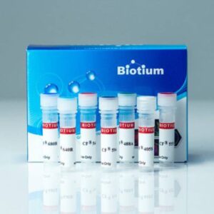
High-Performance CF® Dye Antibodies & Bioconjugates
We have a growing collection of over 2000 monoclonal CF® Dye primary and secondary antibody conjugates, as well as other bioconjugates. Our Biotium Choice line of primary antibodies are developed and optimized specifically for flow cytometry and are available with a selection of widely published clones against common targets.
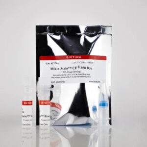
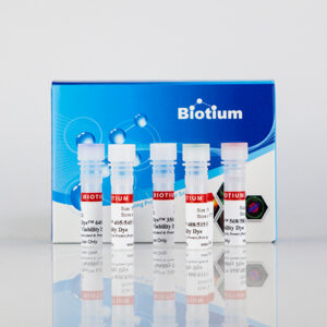
Also see our Mix-n-Stain™ CF® Dye Antibody Labeling Kits for rapid and efficient conjugation to one of our CF® Dyes in as little as 15 minutes. For cell viability, please view our bright and extremely stable Live-or-Dye™ Fixable Viability Stains available in 16 different colors. Live-or-Dye™ stains 510/550, 665/685, 375/600, and 615/740 were designed specifically for spectral flow.
View our flow cytometry page to see a full listing of probes for flow cytometry and related products.
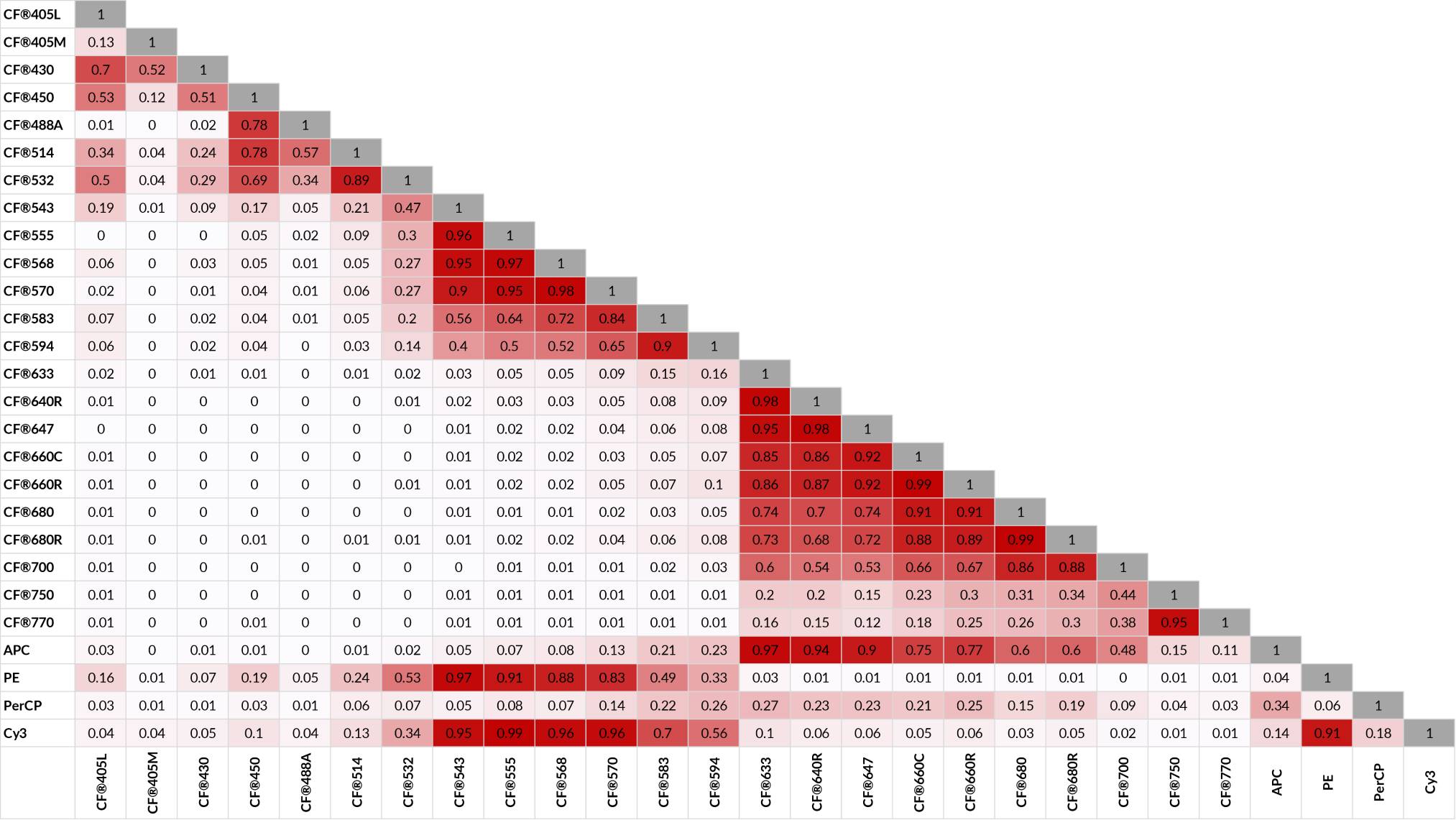
CF® Dye Similarity Index
Download our CF® Dyes for Flow Cytometry Poster

Poster
CF® Dyes for Flow Cytometry Poster