New Products
 Earth-Friendly Products
 Biotium Choice Antibodies
 Special Offers
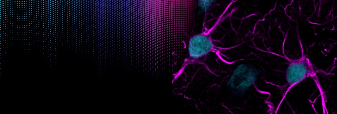
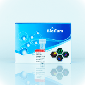
TrueBlack® Lipofuscin Autofluorescence Quenchers
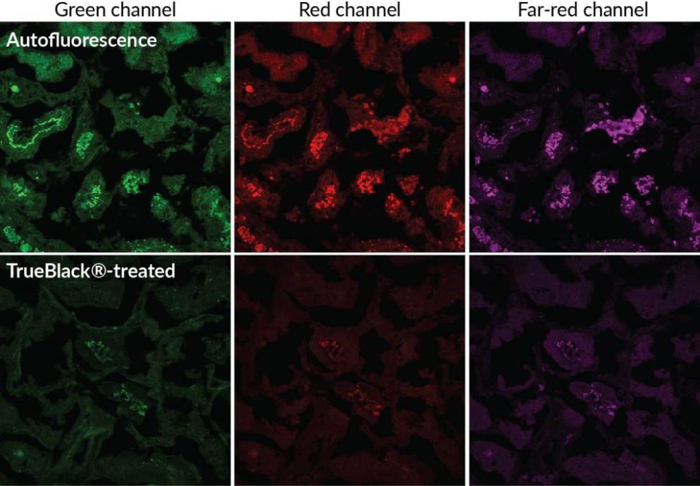
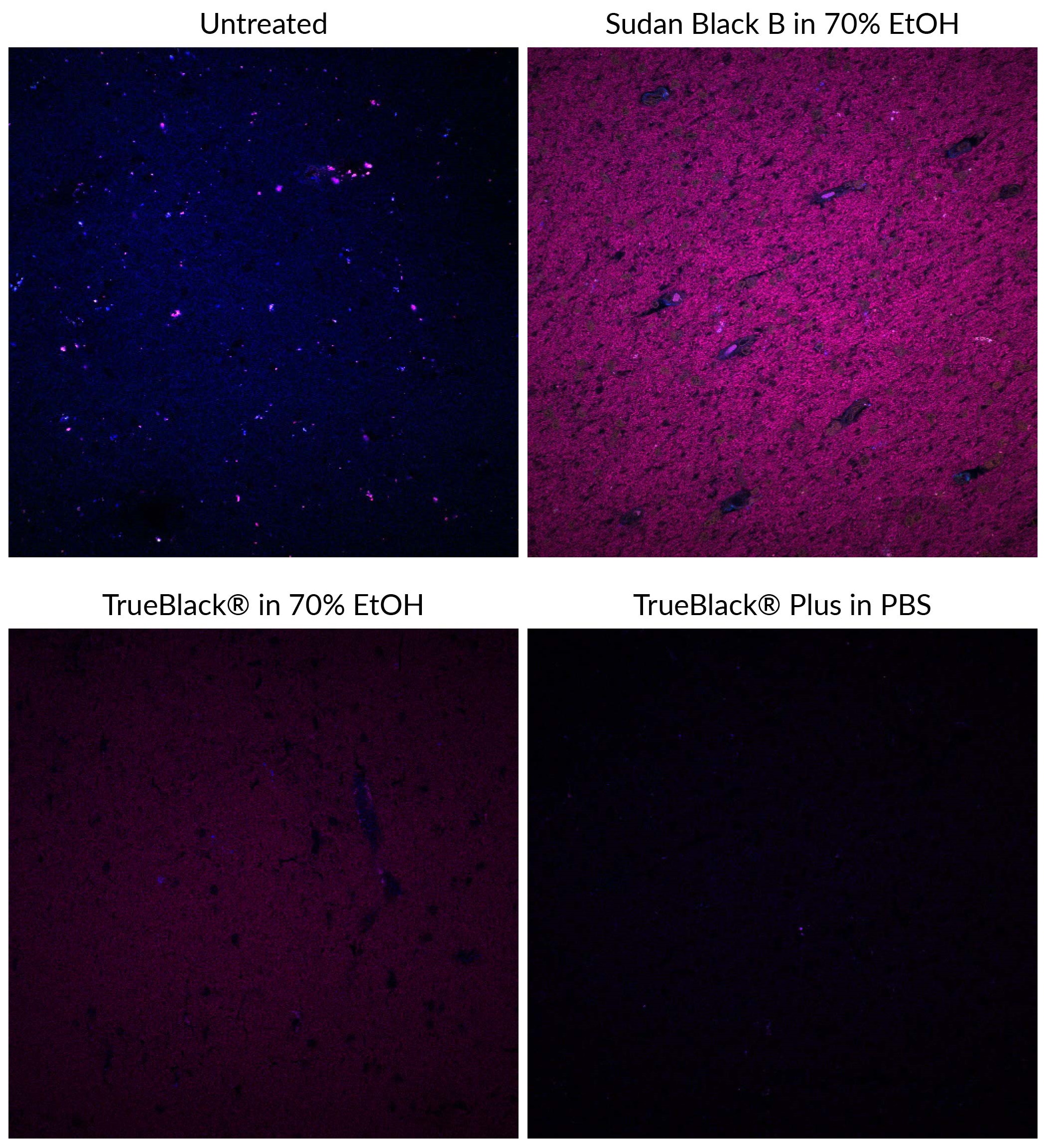
Lipofuscin can make fluorescence imaging of human tissues virtually impossible
Lipofuscin consists of highly autofluorescent granules of oxidized proteins and lipids built up in lysosomes of aging cells in various tissues. The granules fluoresce brightly in all channels used for fluorescence microscopy. Consequently, immunofluorescence in many human tissues or aged animal tissues can be virtually impossible unless lipofuscin fluorescence is masked.
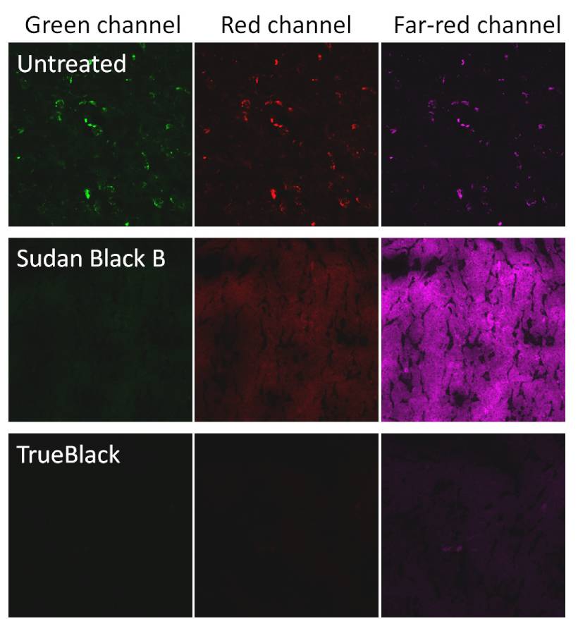
TrueBlack® eliminates lipofuscin autofluorescence, clearing the way for immunofluorescence
Traditionally, Sudan Black B has been used to quench lipofuscin autofluorescence. However, Sudan Black B introduces non-specific red and far-red fluorescence, limiting the use of dyes in those wavelengths. TrueBlack® Lipofuscin Autofluorescence Quencher is a superior alternative to Sudan Black B to quench autofluorescence with much lower background.
- Eliminates lipofuscin autofluorescence
- Reduces autofluorescence from non-lipofuscin sources
- Doesn’t cause high background, unlike Sudan Black B
- Can be used before or after immunofluorescence staining
- Clears the way for fluorescence imaging of human and aged animal tissues
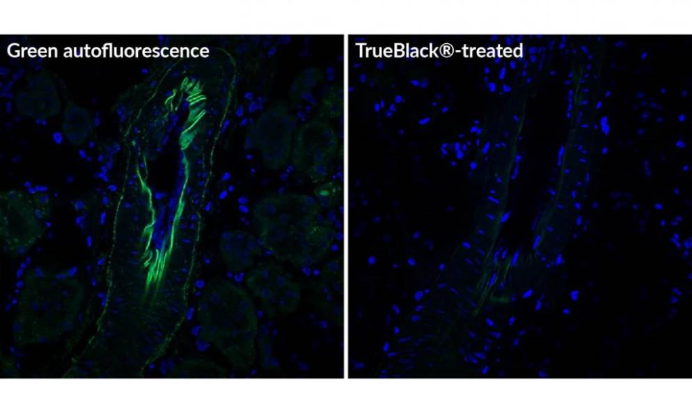
TrueBlack® treatment can be performed before or after immunostaining. It is rapid, simple, and has minimal effect on signal from fluorescent antibodies or nuclear dyes, thus preserving specific staining. Quenching is stable and compatible with commonly used wet-set and hardset fluorescence mounting media, so slides can be stored after staining. See TrueBlack® FAQs for more information.
TrueBlack® eliminates lipofuscin autofluorescence in tissues, and also can reduce autofluorescence from other sources, such as collagen, elastin, and red blood cells. It is not as effective at quenching these sources of autofluorescence as it is for lipofuscin, but it can improve background. It also has been used to quench fluorescence on polycarbonate filters used as cell supports (Futia et al. 2016). Download a list of TrueBlack® references.
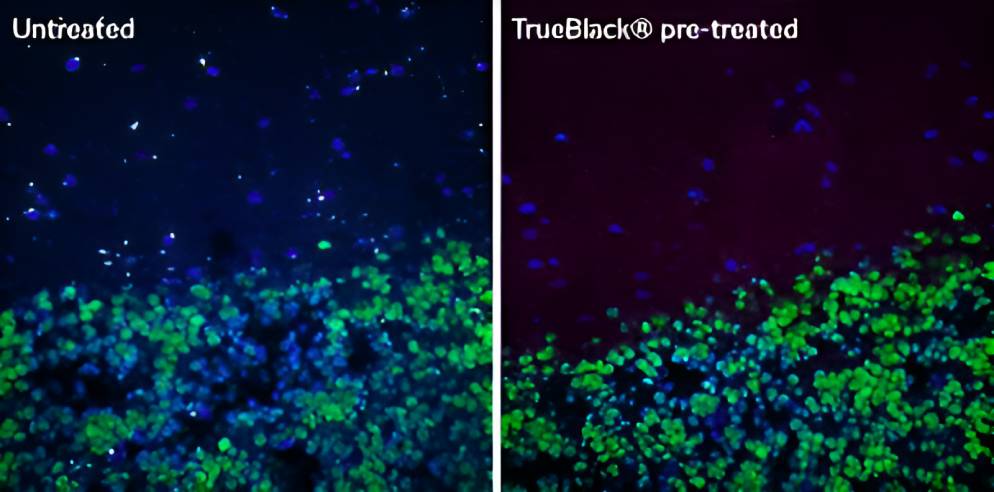
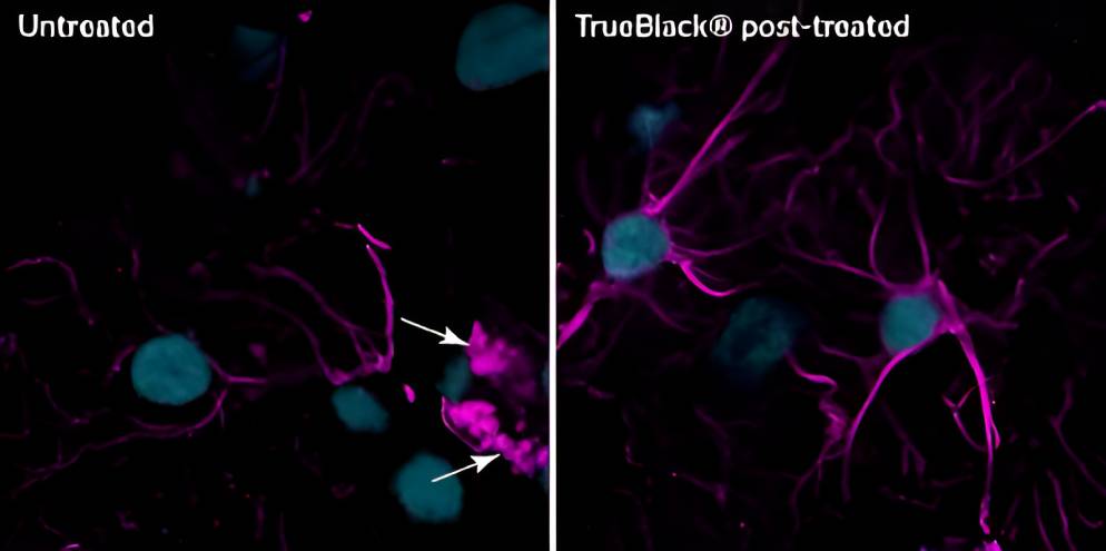
TrueBlack® can be used before or after antibody staining
TrueBlack® reduces autofluorescence from multiple sources
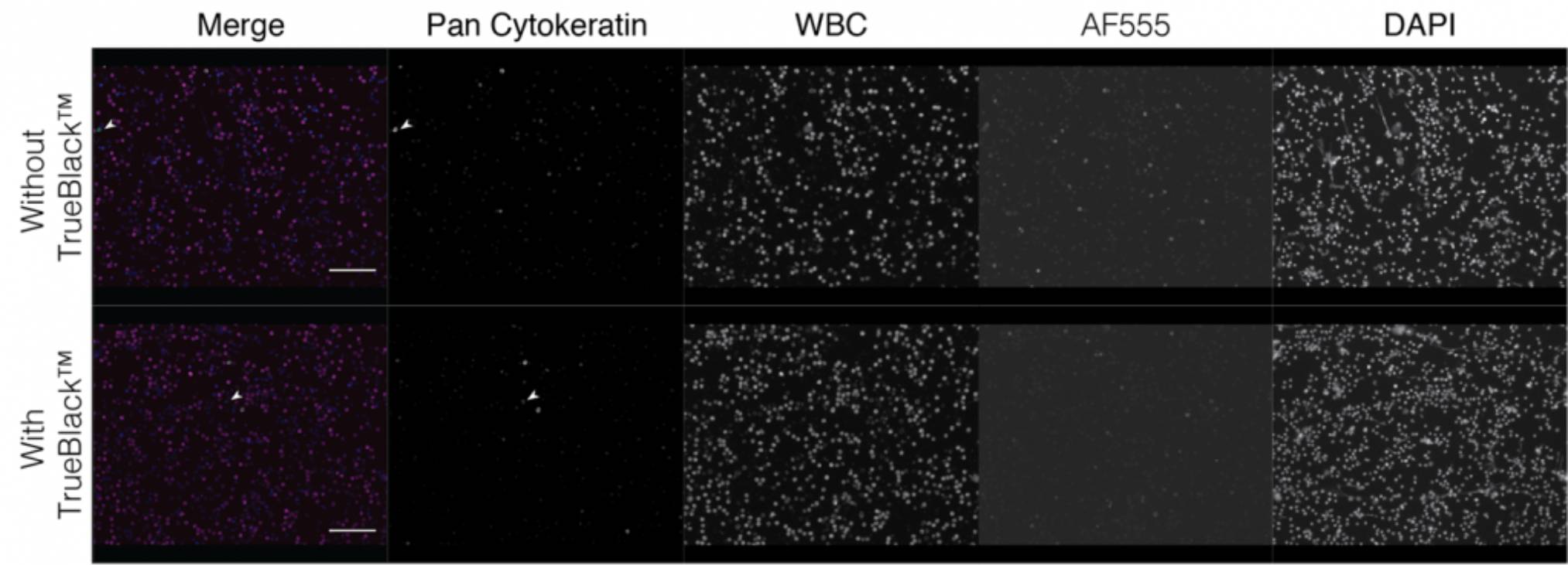
Highlighted Citation
Reports have indicated that autofluorescence due to lipofuscin present in macrophages can also contribute to background signal…Addition of a TrueBlack® blocking step reduced autofluorescence particularly in the AF555 channel.
Axelrod et al.
Check Out More Publications Validating TrueBlack®

Literature Digest
TrueBlack® Lipofuscin Autofluorescence Quencher in Diverse Neuroscience Applications
Even Lower Far-Red Background with TrueBlack® Plus
TrueBlack® Plus is a next-generation lipofuscin quencher developed by Biotium chemists. This new quencher was designed to allow lipofuscin quenching in aqueous buffer with even lower background than the original TrueBlack®. Quenching in PBS allows longer incubation times for thick samples without shrinkage, and is compatible with hydrophobic stains.
TrueBlack® Plus Features
- Quenches lipofuscin with even lower far-red background than our original TrueBlack®
- The only lipofuscin quencher that can be used in aqueous buffer instead of 70% EtOH
- Reduces autofluorescence from non-lipofuscin sources
- Fast and simple treatment before or after immunostaining
- Stable quenching, compatible with commonly used fluorescence mounting media
- Clears the way for multi-color imaging in human tissue
Original TrueBlack® Vs. TrueBlack® Plus
| Product | Catalog no. | Supplied as | Pros | Cons |
|---|---|---|---|---|
| TrueBlack® Lipofuscin Autofluorescence Quencher | 23007 | 20X in DMF | • Complete quenching of lipofuscin autofluorescence • Ultra-low background in blue and green channels • Quenching takes only 30 seconds | • Introduces some red/far-red background • Quenching must be done in 70% EtOH • Some quenching of fluorescent dyes |
| 23011 | 30X in DMSO | |||
| TrueBlack® Plus Lipofuscin Autofluorescence Quencher | 23014 | 40X in DMSO | • Greatly reduces lipofuscin autofluorescence • Has lower red/far-red background than the original TrueBlack® • The only lipofuscin quencher that can be used in PBS and other aqueous buffers | • Titration recommended for optimal quenching • May not be as effective as the original TrueBlack® for high-lipofuscin samples • Some quenching of fluorescent dyes |
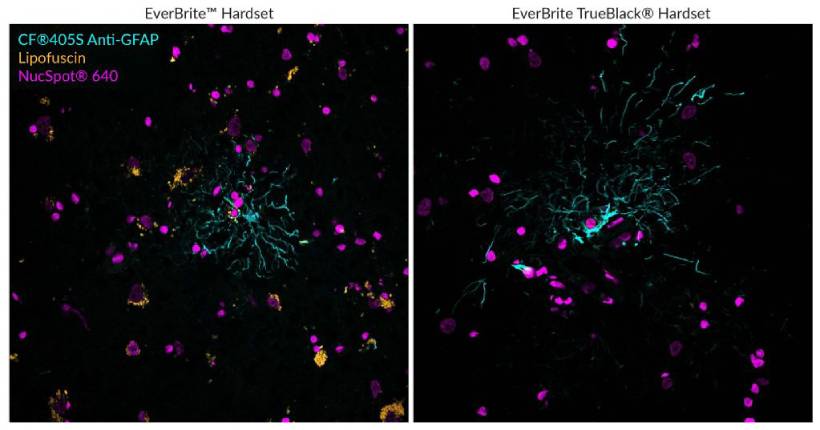
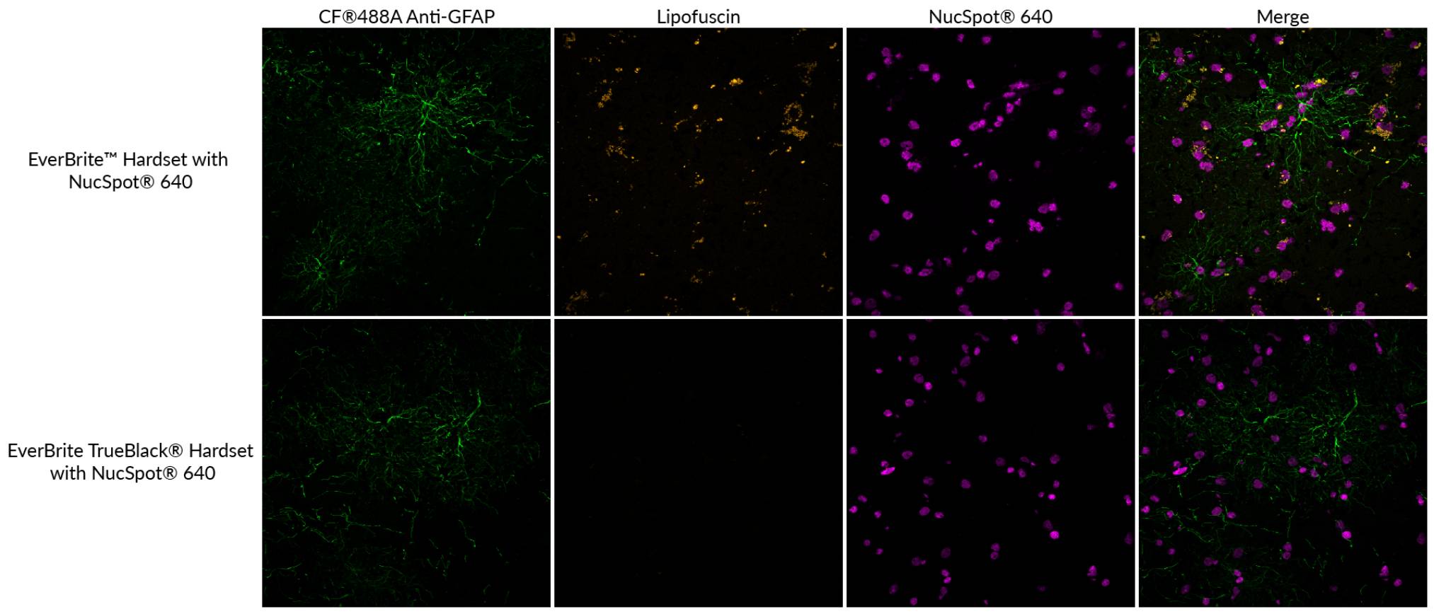
EverBrite TrueBlack® Hardset Mounting Medium
All-in-one mounting medium
Lipofuscin are autofluorescent granules that are a common source of background that make fluorescence imaging virtually impossible in human and aged animal tissues, such as brain and retina. EverBrite TrueBlack® Hardset Mounting Medium is the only mounting medium optimally formulated for quenching lipofuscin fluorescence while offering the same protection against photobleaching as our original EverBrite™ Hardset. The mounting medium is available without nuclear stain, with DAPI, or with NucSpot® 640 nuclear counterstains.
Features
- The only mounting medium with autofluorescence quenching
- Quenches as it hardens, with low background fluorescence
- Optimally formulated for protecting CF® dyes and other dyes from photobleaching
- Refractive index well-matched to coverglass (1.46 after curing)
- Choice of DAPI, NucSpot® 640, or without nuclear counterstain
View Product Page

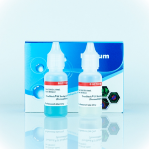
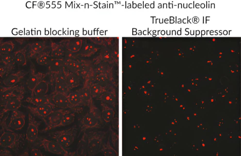
TrueBlack® IF Background Suppressor System
The TrueBlack® Background Suppressor System is a buffer system designed for optimal blocking of non-specific staining for immunofluorescence (IF). The buffers are designed to block background from both non-specific antibody binding as well as direct interaction of fluorescent dyes on antibodies with cells or tissue sections.
- Suppresses background from non-specific antibody binding and charged fluorescent dyes
- More efficient than Image-iT® FX, block and permeabilize in just 10 minutes
- Complete system for blocking, permeabilizing, and antibody dilution
- Non-mammalian blocking agents, for broad secondary antibody compatibility
- For IF on cells or tissue sections
Fluorescent dyes can cause
non-specific antibody binding
Non-specific signal in IF can arise from antibody cross-reactivity, non-specific adsorption, and tissue autofluorescence. Also, fluorescent dyes often carry multiple negative charges to improve dye solubility and brightness, which can result in non-specific antibody binding, particularly for low abundance targets. BSA or gelatin can reduce non-specific protein binding, but they don’t block background from charged dyes.
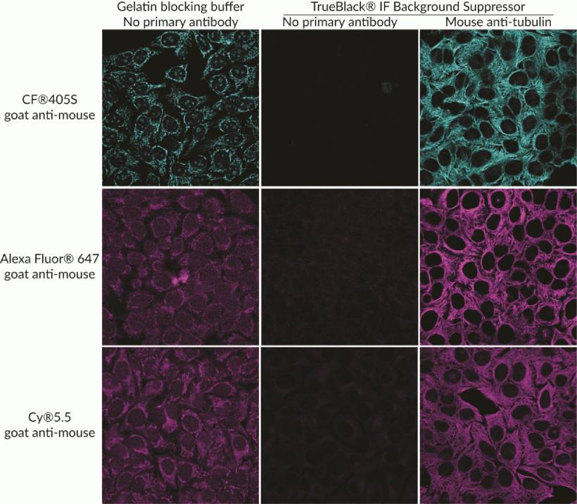
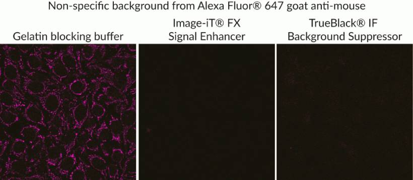
TrueBlack® blocks multiple sources of background
TrueBlack® Background Suppressor includes reagents for blocking both non-specific protein binding as well as background from charged dyes. Examples of charged dyes that show improved signal to noise with the Background Suppressor are CF®405S, CF®405M, CF®555, Alexa Fluor® 647, and Cy®5.5. One-step blocking and permeabilization takes only 10 minutes, and the buffers contain no mammalian proteins, for broad antibody compatibility.
For IF blocking of cells or tissue sections
View Product Page
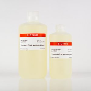
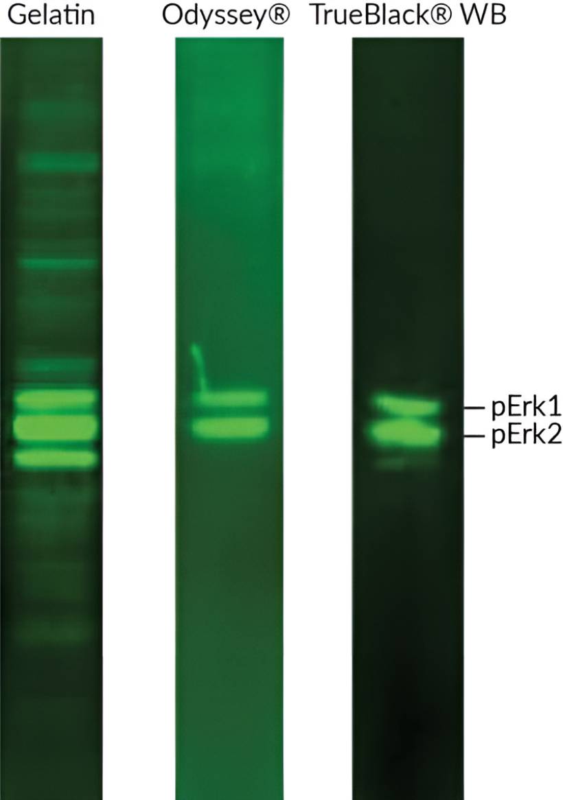
TrueBlack® WB Blocking Buffer Kit
The TrueBlack® WB Blocking Buffer Kit is a ready-to-use buffer system for fluorescence-based western blotting (WB). The buffers yield optimal specificity and sensitivity by blocking non-specific interactions of dye-labeled antibodies with proteins and the blotting membrane.
Features
- Blocks as well or better than Odyssey® Blocking Buffer, at a lower price
- Reduces non-specific bands and background
- Suppresses background from charged dyes better than BSA, gelatin, or casein
- Compatible with PVDF and nitrocellulose
- Contains no mammalian proteins, for broad antibody compatibility
- For visible and near-IR fluorescent westerns
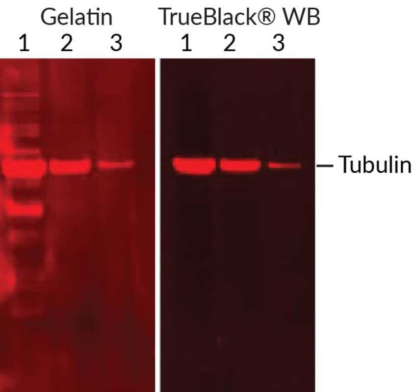
Superior WB Blocking for Charged Fluorescent Dyes
Non-specific signal in WB can arise from antibody cross-reactivity, non-specific adsorption, and membrane autofluorescence. Also, fluorescent dyes often carry multiple negative charges to improve dye solubility and brightness, which can result in non-specific binding. The TrueBlack® WB blocks background from multiple sources, including charged dye conjugates, and is especially advantageous for phosphoprotein detection.
Switch from Odyssey® Blocking Buffer and Save
TrueBlack® WB Blocking Buffer performs as well or better for fluorescent WB compared to LI-COR’s Odyssey® Blocking Buffer (Figure 1), and is priced lower on a per membrane basis.
Compare TrueBlack® WB & Odyssey® Blocking Buffer
| Product | TrueBlack® WB Blocking Buffer Kit | Odyssey® Blocking Buffer |
|---|---|---|
| Trial Size | For 10 membranes | 125 mL for 4 membranes |
| Full Size | For 50 membranes | 500 mL for 16 membranes |