 New Products
 Earth-Friendly Products
 Biotium Choice Antibodies
 Special Offers


 Powered by Bioz


Content #1
Content #1
Content #1
No-wash, fluorescent DNA nuclear stains with low toxicity for long-term live cell imaging or fixed cell staining. Choose green or far-red fluorescence.
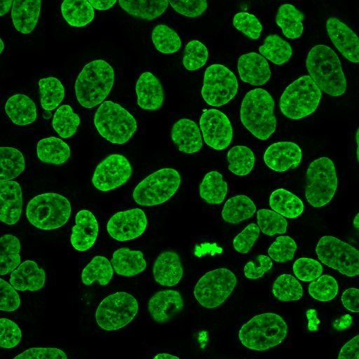
NucSpot® Live Cell Nuclear Stains are cell membrane-permeant DNA dyes that specifically stain nuclei in live or fixed cells. They have excellent specificity for DNA without the need for a wash step, with low toxicity for live cell imaging.
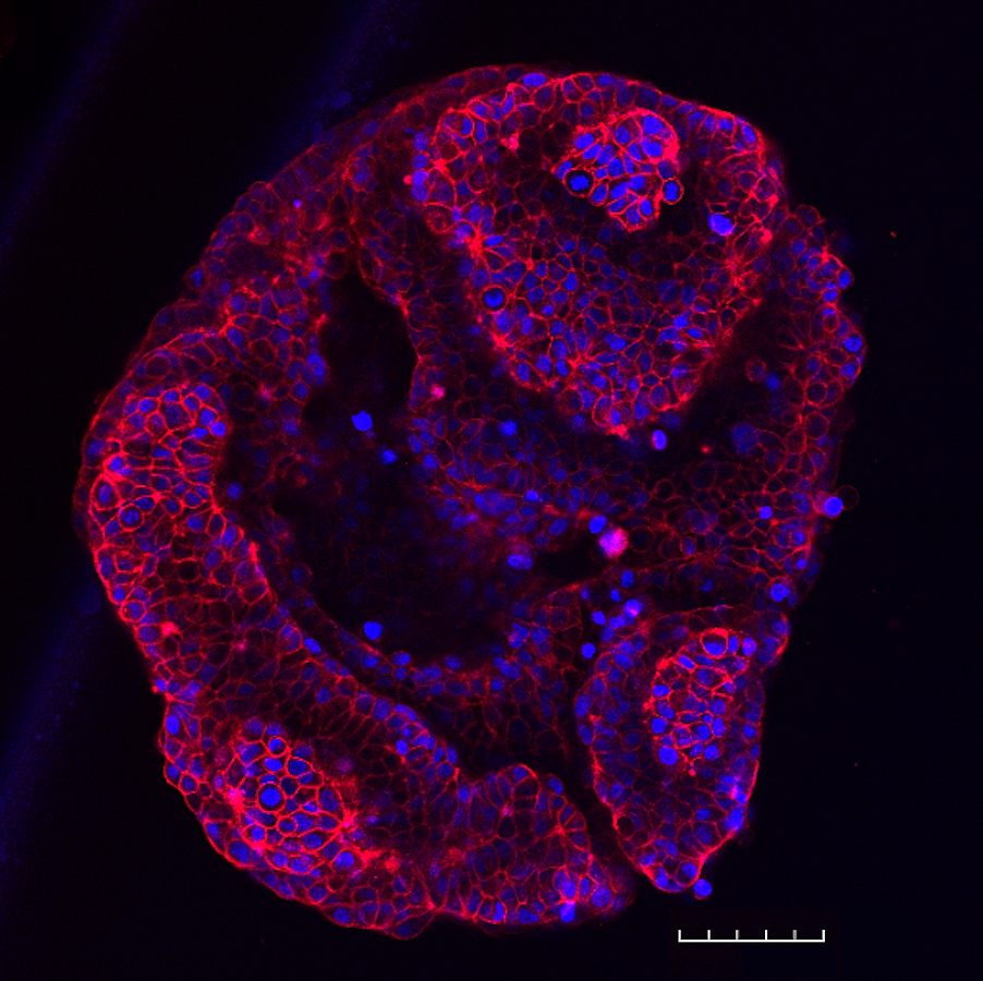
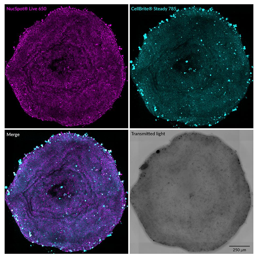
NucSpot® Live 488 stains nuclei with green fluorescence for the FITC channel, while NucSpot® Live 650 has far-red fluorescence for the Cy®5 channel. NucSpot® Live 650 dye also is compatible with super-resolution imaging by SIM or STED, and has been validated in STORM (Ref. 4). The dyes are supplied as 1000X stock solutions in DMSO, and are supplied with a vial of the efflux pump inhibitor verapamil for optional use, which may increase probe retention and live cell staining in some cell types. Note: NucSpot® Live 488 and NucSpot® Live 650 have dim blue fluorescence in the DAPI channel, and may not be suitable for multicolor imaging with blue probes. NucSpot® Live 650 has also been validated for live imaging of 3D organoid cultures (see figure below). NucSpot® Live Cell Nuclear Stains can be used to stain live gram-positive bacteria, but do not stain live gram-negative bacteria or yeast. See our Cellular Stains Table for more information on how our dyes stain various organisms.
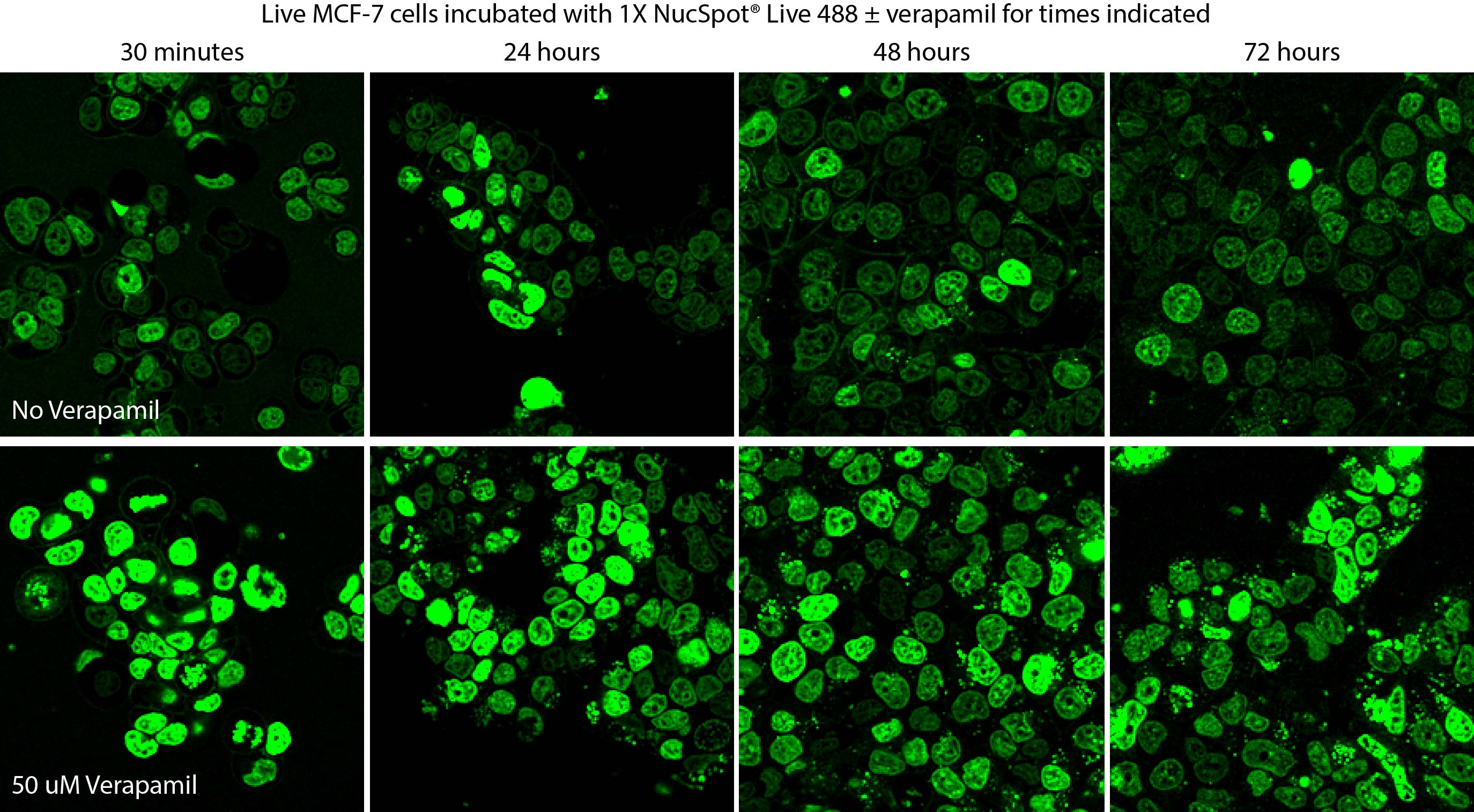
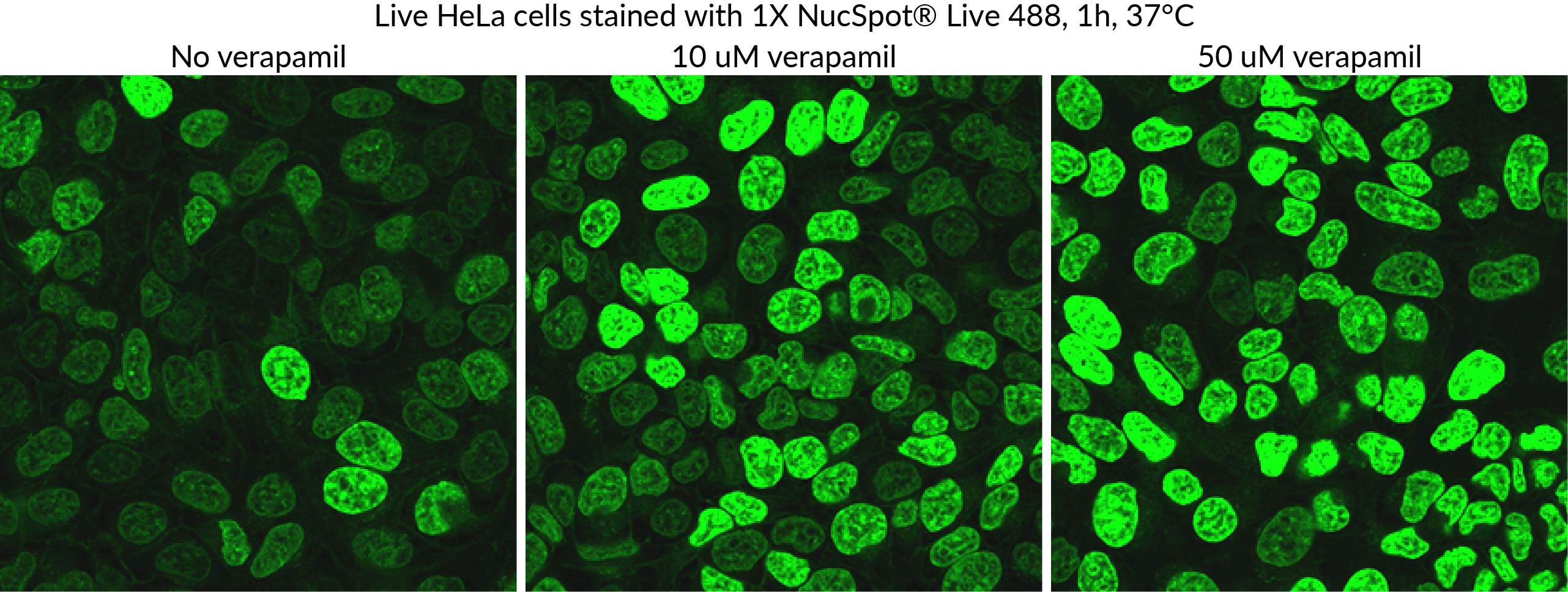
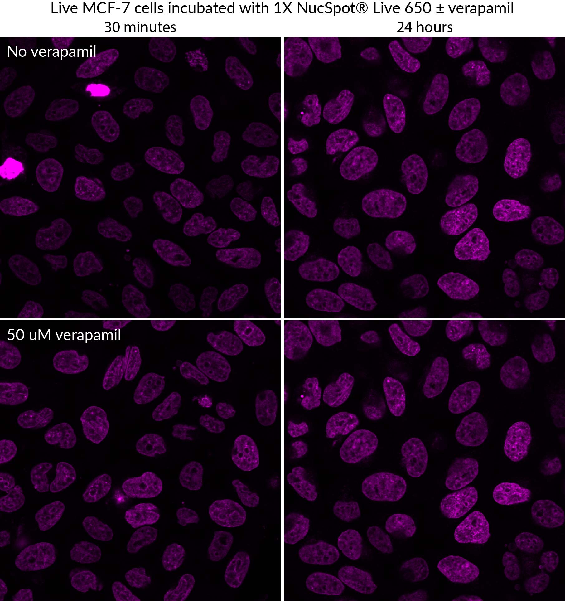
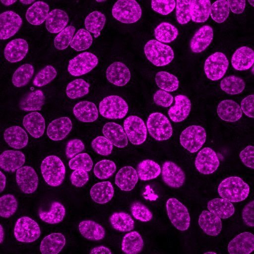
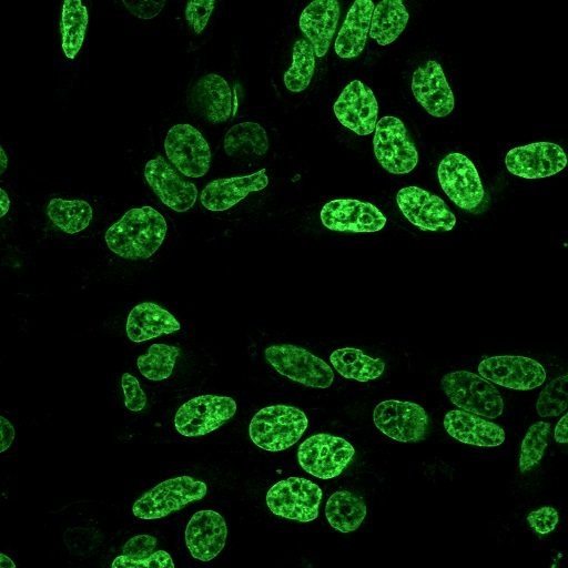
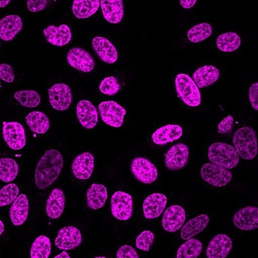
Figure 1. Time course of live MCF-7 cell staining with NucSpot® Live 488, with or without verapamil. Dye and verapamil were added at time 0 and incubated with cells continuously for three days. Verapamil improves dye retention and staining. NucSpot® Live 488 with verapamil showed no obvious toxicity in MCF-7 cells over the course of 72 hours. With longer incubation times, some extranuclear staining can be observed.
Figure 2. 3D tissue dynamics of branching mouse mammary gland organoids. Organoids were incubated with NucSpot® Live 650 and treated with growth factors to trigger post-natal remodeling. The organoids were imaged for 48 hours on the Leica Viventis SCAPE light sheet microscope. Credit: Ayushi Bhanushali, and Sergio P. Acebrón.
Biotium also offers NucSpot® Nuclear Stains for fixed cells in a wide selection of colors from green to near-IR. Also see our wide selection of other Membrane and Organelle Stains, or download the Cellular Stains Brochure.
Cy Dye is a registered trademark of Cytiva.
The mechanism of binding for RedDot™ 1 and RedDot™ 2 to DNA has not been characterized. However, based on the dye structure, it may bind by a similar mechanism as DRAQ®5, which has been reported in the literature to be a concentration-dependent intercalator and minor groove binder.
DRAQ is a registered trademark of Biostatus, Ltd.
Mitochondrial dyes, including MitoView™ Mitochondrial Dyes, are positively charged and lipophilic. They passively diffuse across cellular membranes and are presumed to accumulate in the mitochondrial matrix due to the proton gradient in the mitochondria (for a detailed review, see Cytometry Part A79A: 405-425, 2011).
However, some dyes are still retained in mitochondria after depolarization. Our dye chemists hypothesize that this is because some of the dyes are more lipophilic than others. Once they accumulate in the mitochondria because of their charge, they are less likely to diffuse back into the cytoplasm due to their hydrophobicity, even after the proton gradient that attracted them is dissipated by mitochondrial depolarization. Probably they associate with the mitochondrial membranes instead.
The so-called potential-independent dyes like MitoView™ Green, MitoTracker® Green, and Nonyl Acridine Orange are much more hydrophobic than potential-responsive dyes like MitoView™ 633, Rhodamine 123, and JC-1. The former dyes are retained after mitochondrial depolarization, and can be used to measure mitochondrial mass independent of potential. However, it would be more accurate to call these dyes relatively potential-insensitive, rather than potential-independent, because mitochondrial potential still plays a role in their localization. These dyes have been reported to show some loss of signal upon depolarization (Cytometry 39(3):203-10, 2000).
There is another class of mitochondrial dyes that accumulate in mitochondria based on charge, but also have a reactive group that can covalently link the dye to protein targets within the mitochondria, allowing them to be well-retained after fixation and permeabilization. Our MitoView™ Fix 640 is this type of dye.
Some dyes, like MitoView™ Green can stain mitochondria in cells that are already fixed. The mechanism by which this occurs is not well-understood. After fixation, there should be no proton gradient in the mitochondria to attract the dyes at all. Our chemists suspect that there may be some residual membrane potential in fixed mitochondria that is not due to the proton gradient (which would disappear following fixation), but instead arises from uneven distribution of proteins that have different isoelectric points (net charge). There are reports that the net charge of resident proteins in organelles differs based on the pH of the cellular compartment (Proc Natl Acad Sci USA 115(46):11778-11783, 2018). Charge differences may be sufficient to attract cationic lipophilic dyes to mitochondria in the absence of a proton gradient, due to a combination of weak electrostatic and hydrophobic interactions with mitochondrial proteins and membranes.
However, currently there is no direct evidence to suggest this is the mechanism for MitoView™ Green staining of fixed cells. There may be other targets that the dye is binding. For example, Nonyl Acridine Orange is reported to bind cardiolipin, a lipid that is enriched in mitochondrial membranes. It's possible that MitoView™ Green binds to particular molecules in mitochondria with some degree of specificity. However, staining of fixed cells with mitochondrial dyes generally is not as specific as staining of live cells. That's why we recommend using mitochondrial marker antibodies instead of dyes to stain fixed cells when possible.
Mounting medium can alter the staining of lipophilic dyes like LipidSpot™, due to interaction of the dyes with glycerol or other components that help form the interface between the coverslip and slide. The antifade compounds in mounting medium are generally compatible with the dyes. In our tests, LipidSpot™ staining was well preserved in EverBrite™ Mounting Medium (catalog. nos. 23001/23002) for up to 24 hours after mounting, but lipid droplet size and staining intensity were somewhat altered after samples were stored in mounting medium for several days. Therefore, if mounting medium is required to image samples, we'd recommend imaging as soon as possible after mounting.
LipidSpot™ is not compatible with FluoroShield mounting medium (staining is lost immediately after mounting). We have not tested other types of mounting medium.
Bioscience kits
The guaranteed shelf life from date of receipt for bioscience kits is listed on the product information sheet. Some kits have an expiration date printed on the kit box label, this is the guaranteed shelf life date calculated from the day that the product shipped from our facility. Kits often are functional for significantly longer than the guaranteed shelf life. If you have an older kit in storage that you wish to use, we recommend performing a small scale positive control experiment to confirm that the kit still works for your application before processing a large number of samples or precious samples.
Antibodies and other conjugates
The guaranteed shelf life from date of receipt for antibodies and conjugates is listed on the datasheet sheet which can be downloaded on the product page. Antibodies and other conjugates often are functional for significantly longer than the guaranteed shelf life. If you have an older conjugate in storage that you wish to use, we recommend performing a small scale positive control experiment to confirm that the product still works for your application before processing a large number of samples or precious samples.
For lyophilized antibodies, we recommend reconstituting the antibody with glycerol and antimicrobial preservative like sodium azide for the longest shelf life (note that sodium azide is not compatible with HRP-conjugates).
Chemicals, dyes, and gel stains
Biotium guarantees the stability of chemicals, dyes, and gel stains for at least a year from the date you receive the product. However, the majority of these products are highly stable for many years, as long as they are stored as recommended. Storage conditions can be found on the product information sheet or product safety and data sheet, material safety data sheet, and on the product label. Fluorescent compounds should be protected from light for long term storage.
If you have a Biotium compound that has been in storage for longer than one year that you wish to use, we recommend performing a small scale positive control experiment to confirm that the compound still works for your application before processing a large number of samples or precious samples.
Expiration date based on date of manufacture (DOM)
If your institution requires you to document expiration date based on date of manufacture for reagents, please contact [email protected] for assistance.
Chemical products with special stability considerations:
Esters
Ester compounds include the following:
Ester dyes are stable in solid form as long as they are protected from light and moisture. Esters are not stable in aqueous solution. Concentrated stock solutions should be prepared in anhydrous DMSO (see Biotium catalog no. 90082). Stock solutions in anhydrous DMSO can be stored desiccated at -20°C for one month or longer. Esters should be diluted in aqueous solution immediately before use. Succinimidyl esters (SE) should be dissolved in a solution that is free of amine-containing compounds like Tris, glycine, or protein, which will react with the SE functional group. AM esters and diacetate compounds should be dissolved in a solution that is free of serum, because serum could contain esterases that would hydrolyze the compound.
A note on CF® Dye succinimidyl ester stability
Succinimidyl esters (SE) are generally susceptible to hydrolysis, which can result in lower labeling efficiency. Many commercially available fluorescent dyes used for life science research are heavily sulfonated dyes which makes them particularly hygroscopic, worsening the hydrolysis problem. In addition, for several commercially available SE reactive dyes, the SE group is derived from an aromatic carboxylic acid, while the SE group in all of Biotium’s CF® Dyes is prepared from an aliphatic carboxylic acid. This structural difference reduces the susceptibility of CF® Dye SE reactive groups to hydrolysis, resulting in relatively stable reactive dyes with consistently higher labeling efficiency compared to other SE derivatives of other fluorescent dyes.
Maleimides, MTS and thiosulfate dyes
Like the succinimidyl ester dyes, these dyes are also susceptible to hydrolysis, although generally to a much lower degree. Thus, for long term storage, anhydrous DMSO is recommended for making stock solutions.
Other reactive dyes
Amines, aminooxy (also known as oxylamine), hydrazide, azide, alkyne, BCN, and tyramide reactive dyes, as well as dye free acids, are generally stable in aqueous solution when stored at -20°C for 6-12 months or longer, as long as no compounds are present that may react with the dye’s functional group. See the product information sheets for specific reactive dyes more information.
Coelenterazines and D-luciferin
Coelenterazines are stable in solid form when stored as recommended; they are not stable in aqueous solution. Concentrated coelenterazine stock solutions (typically 1-100 mg/mL) should be prepared in ethanol or methanol; do not use DMSO or DMF to dissolve coelenterazines, because these solvents will oxidize the compounds. Ethanol or methanol stocks of coelenterazine can be stored at -20°C or below for six months or longer; alcohol stocks may evaporate during storage, so use tightly sealing screw cap vials and wrap the vials with Parafilm for long term storage. Propylene glycol also can be used as a solvent to minimize evaporation. If the solvent evaporates, the coelenterazine will still be present in the vial, so note the volume in the vial prior to storage so that you can adjust the solvent volume to correct for evaporation if needed. Prepare working solutions in aqueous buffers immediately before use. Coelenterazines are stable for up to five hours in aqueous solution.
Aquaphile™ coelenterazines are water soluble formulations of coelenterazines. They are stable in solid form when stored as recommended. Aquaphile™ coelenterazines should be dissolved in aqueous solution immediately before use. They are stable for up to five hours in aqueous solution.
Note that coelenterazines are predominantly yellow solids, but may contain dark red or brown flecks. This does not affect product stability or performance. If your coelenterazine is uniformly brown, then it is oxidized and needs to be replaced.
D-luciferin is stable in solid form and as a concentrated stock solution when stored as recommended; it is not stable at dilute working concentrations in aqueous solution. Prepare concentrated D-luciferin stock solutions (typically 1-100 mg/mL) in water, and store in aliquots at -20°C or below for six months or longer. Prepare working solutions immediately before use.
For dyes or reagents that are supplied lyophilized (as solids), it is hard to compare quantities based on appearance of the dye in the tube, because during the lyophilization process the dye can dry down in different ways, either spread out all over the tube, clumped together, or coating the sides or bottom of the tube. Centrifugation of the tube may not help in collecting the dye solid to the bottom of the tube as this generally works for solutions. However, lyophilized solids are packaged based on highly accurate absorbance measurement of the reagent solution prior to drying, so the vial will contain the correct amount of dye.
Biotium ships all antibodies (primary, secondary and conjugates) at room temperature. We guarantee their quality and performance under these conditions based upon our stability testing. Antibodies were subjected to accelerated stability testing by storing them at various temperatures (4°C, room temperature, or 37°C) for 1 week to mimic simulated shipping conditions and tested in immunostaining experiments. All antibodies showed the expected brightness and specificity, even after storage at sub-optimal temperatures for a week or longer. You can also download our Product Storage Statement here.
In line with our goal to be more environmentally friendly by reducing the use of excess packaging, and lowering shipping costs for our customers, products that have passed our stability testing are shipped at room temperature.
Once you have received the antibody vial, please follow the long-term storage instructions on the product information (PI) sheet.
