New Products
 Earth-Friendly Products
 Biotium Choice Antibodies
 Special Offers

Membrane Potential Dyes
Slow-Response Membrane Potential Dyes
Translational (slow-response) voltage-sensitive dyes change membrane distribution in response to voltage shifts. DiBAC4(3) shows a slower response but increased fluorescence with depolarization, offering larger signal changes than faster dyes like ANEPPS. DiOC2(3), used in bacteria, shifts from green to red fluorescence as membrane potential rises, enabling ratiometric measurements. DiOC5(3) and DiOC6(3) are widely used carbocyanine dyes for membrane potential. TMRE and TMRM are rhodamine-based dyes suitable for quantitative assessment of membrane and mitochondrial membrane potential.
Fast-Response Membrane Potential Dyes
Fast-response membrane voltage-sensitive dyes are styryl dyes that undergo changes in fluorescence intensity in response to changes in membrane potential. These dyes undergo spectral shift with changes in membrane potential, allowing ratiometric measurements, and have been used to measure electrical activity in neural and cardiac cells.
Di-4-ANNEPS is used in stem cell-derived cardiomyocytes. Di-8-ANNEPS is more hydrophobic, photostable, and less phototoxic, making it better for long-term studies. Di-2-ANEPEQ is water-soluble and typically microinjected. Di-8-ANEPPQ and Di-12-ANEPPQ are more hydrophobic, used in retrograde neuronal labeling. RH237, RH414, RH421, and RH795 are fast potentiometric probes often used for functional imaging of neurons.
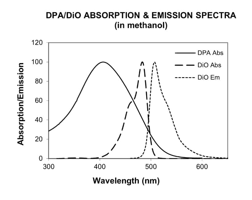
DiO/DPA Membrane Potential Kit
The membrane localization of dipicrylamine (DPA) is a function of the polarity and magnitude of membrane potential. The DiO/DPA system detects cytoplasmic membrane potential changes using the principle of fluorescence resonance energy transfer (FRET). The green fluorescent membrane dye DiO is a “stationary” FRET donor while DPA acts as a mobile FRET acceptor, resulting in a membrane potential-dependent quenching of fluorescence by FRET.
Slow-Responding Membrane Potential Dyes
Fast-Responding Membrane Potential Dyes
| Product | Ex/Em | Catalog number |
|---|---|---|
| Di-4-ANEPPS | 496/705 nm1 | 61010 |
| Di-8-ANEPPS | 498/713 nm1 | 61012 |
| Di-2-ANEPEQ (JPW 1114) | See Note 2 | 61013 |
| Di-8-ANEPPQ | 61014 | |
| Di-12-ANEPPQ | 61015 | |
| RH237 | 528/782 nm | 61018 |
| RH414 | 532/706 nm | 61016 |
| RH421 | 515/704 nm | 61017 |
| RH795 | 530/712 nm | 61019 |
| DiO/DPA Membrane Potential Kit | 484/501 nm | 30037 |
2Spectrally similar to the ANEPPS dyes.
Nerve Terminal Dyes
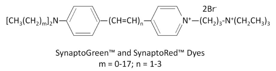
SynaptoRed™ & SynaptoGreen™
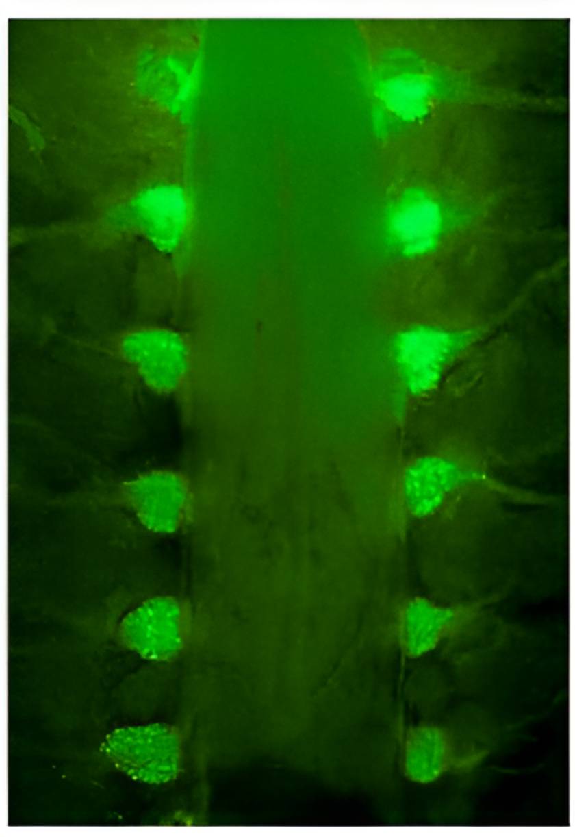
SynaptoGreen™ and SynaptoRed™ (formerly FM® dyes) are fluorescent styryl dyes used to trace endocytic vesicles and monitor synaptic activity. They label synaptic vesicles in neurons and other cells in an activity-dependent manner. These dyes have hydrophilic, cationic head groups and lipophilic tails, becoming highly fluorescent in membranes but not in solution. Upon nerve stimulation, they are internalized into endocytic vesicles and released during exocytosis, causing a fluorescence decrease. The fluorescence on/off rates vary by dye. AM and HM variants are fixable, allowing for post-staining fixation and immunostaining. See the table below for dye properties.

Background Reducers & Nerve Terminal Staining Kits
Nerve terminal dyes can cause background fluorescence from residual membrane staining. To reduce this, Biotium offers three clearing agents. ADVASEP-7 forms a washable complex with SynaptoGreen™ C4. SCAS reduces background immediately without washing. Sulforhodamine 101 quenches SynaptoGreen™ background via FRET. These are available individually or in kits with dyes and clearing agents.
Properties of Nerve Terminal Dyes
| Nerve Terminal Dye | m* | n* | Fixable? | Size | Catalog number | Features |
|---|---|---|---|---|---|---|
| SynaptGreen™ Dyes (Ex/Em ~480/660 nm in membranes) | ||||||
| SynaptoGreen™ C1 | 0 | 1 | No | 5 mg, 5 x 1 mg | 70042, 70043 | • Green nerve terminal probe • Shortest tail for slowest on-rate & fastest off-rate |
| SynaptoGreen™ C2 (equivalent to FM®2-10) | 1 | 1 | No | 70044, 70045 | • Equivalent to FM®2-10 | |
| SynaptoGreen™ C3 | 2 | 1 | No | 70023, 70026 | • Green nerve terminal probe | |
| SynaptoGreen™ C4 (equivalent to FM®1-43) | 3 | 1 | No | 70020, 70022 | • Equivalent to FM®1-43 | |
| SynaptoGreen™ C5 (equivalent to FM®1-84) | 4 | 1 | No | 70046, 70047 | • Equivalent to FM®1-84 | |
| SynaptoGreen™ C18 (equivalent to FM®3-25) | 17 | 1 | No | 70048, 70049 | • Equivalent to FM®3-25 | |
| AM1-43 | 3 | 1 | Yes | 1 mg | 70024 | • Fixable version of SynaptoGreen C4 • Equivalent to FM®1-43FX |
| AM1-44 | 4 | 1 | Yes | 70038 | • Improved fixability over AM1-43 | |
| AM2-10 | 1 | 1 | Yes | 70036 | • Fixable analog of SynaptoGreen™ C2 | |
| AM3-25 | 17 | 1 | Yes | 70051 | • Fixable far-red nerve terminal probe | |
| HM1-43 | 3 | 1 | Yes | 70053 | • Fixable red nerve terminal probe | |
| SynaptoRed Dyes™ (Ex/Em ~510/750 nm in membranes) | ||||||
| SynaptoRed™ C1 | 0 | 3 | No | 5 mg, 5 x 1 mg | 70040, 70041 | • One carbon shorter than SynaptoRed™ C2 |
| SynaptoRed™ C2 (equivalent to FM®4-64) | 1 | 3 | No | 70021, 70027 | • Equivalent to FM®4-64 | |
| SynaptoRed™ C2M** (equivalent to FM®5-95) | 1 | 3 | No | 70019, 70028 | • More water soluble than SynaptoRed™ C2 • Equivalent to FM®5-95 |
|
| AM4-64 | 1 | 3 | Yes | 1 mg | 70025 | • Fixable version of SynaptoRed™ C2 |
| AM4-65 | 3 | 3 | Yes | 70039 | • Fixable version of SynaptoRed™ C2 | |
| AM4-66 | 4 | 3 | Yes | 70050 | • Fixable and spectrally identical to SynaptoRed™ C2 | |
**The positively-charged end of SynaptoRed C2M is a trimethylammonium group.
FM is a registered trademark of Thermo Fisher Scientific.
Nerve Terminal Staining Kits
| Nerve Terminal Staining Kit | Nerve Terminal Dye | Background Reducer | Catalog number |
|---|---|---|---|
| Nerve Terminal Staining Kit I | SynaptoGreen™ C4 (5 x 1 mg ) | ADVASEP-7 (250 mg) | 70030 |
| Nerve Terminal Staining Kit II (A) | AM1-43 (1 mg) | ADVASEP-7 (100 mg) | 70031 |
| Nerve Terminal Staining Kit II (B) | AM1-43 (1 mg) | SCAS (100 mg) | 70031-1 |
| Nerve Terminal Staining Kit III | SynaptoGreen™ C4 (5 x 1 mg) | Sulforhodamine 101 (100 mg) | 70032 |
| Nerve Terminal Staining Kit V | SynaptoRed™ C2 (5 x 1 mg) | ADVASEP-7 (250 mg) | 70034 |