 New Products
 Earth-Friendly Products
 Biotium Choice Antibodies
 Special Offers
Nucleus
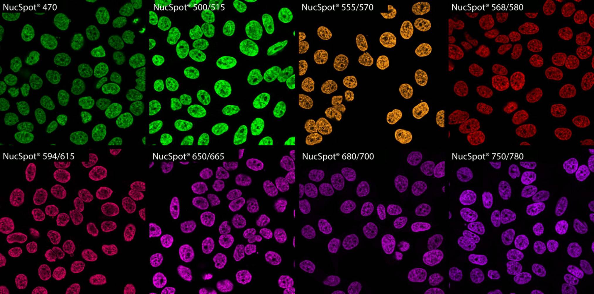
NucSpot® Nuclear Stains
NucSpot® Nuclear Stains are cell membrane-impermeant fluorescent DNA stains available in a variety of colors from green to near-infrared (near-IR). NucSpot® Stains have minimal fluorescence until they bind to DNA and can be used for no-wash nuclear staining.
Unlike other nucleic acid dyes such as propidium iodide (PI), TOTO®, TO-PRO®, and similar dyes that stain both the nucleus and cytoplasm, NucSpot® Nuclear Stains selectively stain the nucleus in fixed and permeabilized cells without the need for RNase treatment.
NucSpot® Nuclear Stains also can be used for selective fluorescent staining of dead cells in unfixed cell cultures for analysis by flow cytometry or fluorescence imaging.
Several of the dyes can be continuously incubated with cells for multi-day imaging. NucSpot® 470 and NucSpot® Far-Red can be used for DNA content analysis of cell cycle by flow cytometry in fixed and permeabilized cells without requiring RNase treatment, unlike propidium iodide.
NucSpot® Live 488 and NucSpot® Live 650 Nuclear Stains
NucSpot® Live Nuclear Stains specifically stain nuclei in live or fixed cells with no need for washing. NucSpot® Live 488 has green fluorescence (Ex/Em 500/515 nm), while NucSpot® Live 650 has far-red fluorescence (650/675 nm) for detection in the Cy®5 channel.
Unlike Draq5™, NucSpot® Live 650 has low cytotoxicity and can be used for longer term cellular staining and imaging. NucSpot® Live 650 dye is also compatible with super-resolution imaging by SIM and STED. NucSpot® Live Nuclear Stains are also validated for live imaging or organoids and other 3D cultures.
RedDot™1 and RedDot™2 Far-Red Nuclear Stains
RedDot™1 and RedDot™2 are far-red nuclear counterstains for the Cy®5 channel. RedDot™1 is an alternative to DRAQ5™ that rapidly and specifically stains nuclei in live cells. It can be used for cell cycle analysis by flow cytometry. It also can be used for cell normalization for In Cell Western™. RedDot™1 shows cytotoxicity within a few hours of staining, so for long-term live cell imaging, we recommend NucSpot® Live 650.
RedDot™2 is membrane impermeant and can be used as a selective dead cell stain, or as a nuclear counterstain for fixed cells. RedDot™2 shows better nuclear specificity in fixed cells than DRAQ7™, which requires a blocking step for nuclear-specific counterstaining. RedDot™2 has also been validated for tissue clearing protocols such as CUBIC.
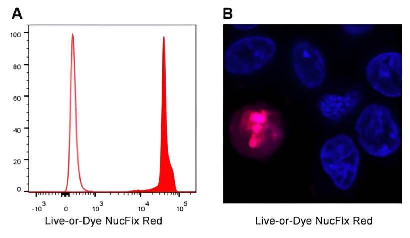
Live-or-Dye NucFix™ Red
Live-or-Dye NucFix™ Red is a unique, cell membrane-impermeant dye that specifically stains the nuclei of dead cells. The dye can enter dead cells that have compromised membrane integrity and covalently label the cell nucleus, allowing for clear differentiation of live and dead cells by either microscopy or flow cytometry.
Unlike other commonly used nuclear stains such as propidium iodide or DRAQ7™, NucFix™ covalently attaches to DNA, allowing the cells to be fixed and permeabilized without loss of fluorescence or dye transfer between cells.
Classic Blue Nuclear Fluorescent Stains
DAPI and Hoechst are widely used blue fluorescent nuclear counterstains. They are minor groove-binding DNA dyes that are minimally fluorescent in solution, but have strong fluorescence enhancement upon binding DNA.
- DAPI is less membrane-permeant than Hoechst, and is typically used as a fixed cell stain at concentrations around 1 ug/mL.
- Antifade mounting medium with DAPI, like Biotium’s EverBrite™ Mounting Media, can be used for one-step mounting and counterstaining.
- Staining live cells with DAPI requires higher concentration (~10 ug/mL). We offer DAPI dilactate, a more water soluble DAPI salt, which can be used at higher concentrations.
- Hoechst dyes are membrane-permeant and can be used for live or fixed cellular staining and cell cycle analysis.
- Hoechst 33342 and Hoechst 33528 both are quenched by BrdU labeled DNA, and have been used in cell division studies. The two dyes are spectrally similar.
- Hoechst 33342 is slightly more cell permeable than Hoechst 33258, but both dyes are commonly used as nuclear stains for live or fixed cells at 1 ug/mL.
While Hoechst and DAPI show less cytotoxicity than intercalating DNA dyes, they bind DNA in living cells and are potentially hazardous. Biotium offers Hoechst 33342, Hoechst 33258, and DAPI as 10 mg/mL solutions in water, for greater convenience and safety compared to weighing out the powdered dyes.
DAPI and Hoechst undergo photoconversion by UV excitation to form green fluorescent dyes, which can lead to artifacts in multi-color imaging. See our Tech Tip Avoiding Artifacts from UV Photoconversion of DAPI and Hoechst for more information.
Asymmetric Cyanine Dyes and Other Dead Cell Nuclear Stains
Biotium offers a selection of cyanine dyes for nucleic acids. Cyanine dyes have high affinity for DNA and RNA. Several of the membrane-impermeant dyes are useful dead cell stains. Oxazole Yellow (equivalent to YO-PRO®-1) is unique in that it selectively stains early apoptotic cells.
Membrane-impermeant cyanine dyes also can be used as nuclear counterstains, but unlike RedDot™ or NucSpot® dyes, they stain both RNA and DNA, and so require RNase treatment for selective nuclear staining.
We also offer a variety of other cell-membrane impermeant nuclear stains for dead cell staining. See the table below for more information. For more application specific information, please see our nuclear stains comparison table.
Nuclear Stains | Catalog | Color (Ex/Em*) | Cell | For live / | Fix after | Features |
|---|---|---|---|---|---|---|
| EverBrite™ Mounting Medium w/DAPI EverBrite™ Hardset w/DAPI Drop-n-Stain EverBrite™ w/DAPI | 23002 23004 23009 | Blue (358/461 nm) | N/A | Mounting fixed samples | N/A | • Hardset and wet-set mounting media • Broad dye compatibility • Available with DAPI for one-step mounting and staining • Drop-n-Stain EverBrite™ comes in convenient dropper bottle |
| DAPI DAPI, 10 mg/mL in H2O DAPI, dilactate 10 mg in H2O | 40011 40043 40009 | Blue (358/461 nm) | Membrane permeant | Live cells Fixed cells | Yes | • Classic nuclear counterstain for fixed cells • Can be used at higher concentrations to stain live cells • Dilactate salt has improved water solubility |
| Hoechst 33258, 10 mg/mL in H2O Hoechst 33258, pentahydrate Hoechst 33342, 10 mg/mL in H2O Hoechst 33342, trihydrochloride trihydrate | 40044 40045 40046 40047 | Blue (358/461 nm) | Membrane permeant | Live cells Fixed cells | Yes | • Classic nuclear counterstain for live cells • Can also be used on fixed cells |
| Oxazole Gold (SYBR® Gold) | 40094 | Green (496/539) | Membrane permeant | Live cells | No 2 | • Aka SYBR® Gold, an ultrasensitive DNA and RNA gel stain • Can be used for live cell staining of nuclei and mitochondrial DNA |
| NucSpot® 470 Nuclear Stain | 40083 | Green (460/546 nm) | Membrane impermeant | Dead cells Fixed cells | No 1 | • Nuclear-specific green counterstain for fixed cells • Selectively stains dead cells in live cultures • Excellent match for blue LED excitation sources |
| Thiazole Green (SYBR® Green I) | 40086 | Green (498/522 nm) | Membrane permeant | Live cells Fixed cells | No 2 | • Aka SYBR® Green I, a well known DNA gel stain and qPCR dye • Can be used as a green nuclear stain for all cells in live cultures • Loses nuclear specificity after fixation • Can be excited by 488 nm laser line |
| NucSpot® Live 488 Nuclear Stain | 40081 | Green (500/515 nm) | Membrane permeant | Live cells Fixed cells | Yes | • Low-toxicity nuclear stain • Fix before or after labeling • Live cell staining may require VRP (included) |
| Live-or-Dye NucFix™ Red | 32010 | Red (520/610 nm) | Membrane impermeant | Dead cells | Yes | • Reactive nuclear stain for dead cells • Specifically stains dead cell nuclei • Fix/permeabilize without dye transfer between cells |
| NucSpot® 555/570 Nuclear Stain | 41033 | Red (559/566 nm) | Membrane impermeant | Dead cells Fixed cells | No 1 | • Nuclear-specific red counterstain for fixed cells • Specifically stains dead cells in live cultures |
| NucSpot® 568/580 Nuclear Stain | 41036 | Red (572/583 nm) | Membrane impermeant | Dead cells Fixed cells | No 1 | • Nuclear-specific red counterstain for fixed cells • Specifically stains dead cells in live cultures • Suitable for multi-day live cell imaging |
| NucSpot® 594/615 Nuclear Stain | 41037 | Red (603/613 nm) | Membrane impermeant | Dead cells Fixed cells | No 1 | • Nuclear-specific red counterstain for fixed cells • Specifically stains dead cells in live cultures • Suitable for multi-day live cell imaging |
| NucSpot® 650/665 Nuclear Stain | 41034 | Far-red (653/671 nm) | Membrane impermeant | Dead cells Fixed cells | No 1 | • Nuclear-specific far-red counterstain for fixed cells • Specifically stains dead cells in live cultures |
| NucSpot® Live 650 Nuclear Stain | 40082 | Far-red (650/675 nm) | Membrane permeant | Live cells Fixed cells | Yes | • Low-toxicity nuclear stain for the Cy®5 channel • Fix before or after labeling • Live cell staining may require VRP (included) • Compatible with SIM, STED, or STORM |
| RedDot™1 Far-Red Nuclear Stain | 40060 | Far-red (662/694 nm) | Membrane permeant | Live cells | No 2 | • For short-term live cell staining (≤4 hours) • Analyze DNA content/cell cycle by flow cytometry • Useful for cell number normalization for In Cell Western® • Can be excited at wavelengths between 488 and 647 nm • Detect in Cy®5 or APC channel |
| RedDot™2 Far-Red Nuclear Stain | 40061 | Far-red (665/695 nm) | Membrane impermeant | Dead cells Fixed cells | No 1 | • Far-red nuclear stain for dead or fixed cells • Selectively stains dead cells • Specific nuclear counterstain for fixed cells • Can be excited at wavelengths between 488 and 647 nm • Detect in Cy®5 or APC channel |
| NucSpot® 680/700 Nuclear Stain | 41035 | Near-IR (683/707 nm) | Membrane impermeant | Dead cells Fixed cells | No 1 | • Nuclear-specific near-IR counterstain for fixed cells • Specifically stains dead cells in live cultures |
| NucSpot® 750/780 Nuclear Stain | 41038 | Near-IR (757/780 nm) | Membrane impermeant | Dead cells Fixed cells | No 1 | • Nuclear-specific near-IR counterstain for fixed cells • Specifically stains dead cells in live cultures • Suitable for multi-day live cell imaging |
1 Dye can transfer from dead to live cells after fixation.
2 Loses nuclear specificity after fixation.
In Cell Western is a registered trademark of LI-COR® Bioscience. Cy Dye is a registered trademark of Cytiva.
Dead Cell Nucleic Acid Stains | Catalog No. | Color (Ex/Em*) | Features |
|---|---|---|---|
| Oxazole Blue, 1 mM in DMSO | 40091 | Blue (434/457 nm) | • Blue cell-impermeant dye • Selectively stains early apoptotic cells • Equivalent to PO-PRO™-1 Iodide |
| Oxazole Blue Homodimer, 1 mM in DMSO | 40093 | Blue (433/457 nm) | • Blue cell-impermeant dye • Equivalent to POPO™-1 Iodide |
| NucSpot® 470, 1000X in DMSO | 40083 | Green (460/546 nm) | • Green cell-impermeant dye • Nuclear-specific counterstain in fixed cells • Useful for cell cycle analysis in fixed cells • Excellent match for blue LED excitation sources |
| Oxazole Yellow, 1 mM in DMSO | 40089 | Green (491/506 nm) | • Green cell-impermeant dye • Selectively stains early apoptotic cells • Equivalent to YO-PRO®-1 Iodide |
| Oxazole Yellow Homodimer, 1 mM in DMSO | 40090 | Green (491/508 nm) | • Green cell-impermeant dye • Equivalent to YOYO®-1 Iodide |
| TO Iodide, 1 mM in DMSO | 40088 | Green (515/531 nm) | • Green cell-impermeant dye • Equivalent to TO-PRO®-1 Iodide |
| Thiazole Orange Homodimer, 1 mM in DMSO | 40079 | Green (514/531 nm) | • High affinity dimeric cyanine dye • Dead cell stain and electrophoresis dye • Equivalent to TOTO®-1 Iodide |
| NucSpot® 555/570 Nuclear Stain | 41033 | Red (559/566 nm) | • Red cell-impermeant dye for the Cy®3 or PE channels • Nuclear-specific counterstain in fixed cells |
| NucSpot® 568/580 Nuclear Stain | 41036 | Red (572/583 nm) | • Red cell-impermeant dye for the Cy®3 or PE channels • Nuclear-specific counterstain in fixed cells • Suitable for multi-day live cell imaging |
| NucSpot® 594/615 Nuclear Stain | 41037 | Red (603/613 nm) | • Red cell-impermeant dye for the Texas Red® or PE-Texas Red® channels • Nuclear-specific counterstain in fixed cells • Suitable for multi-day live cell imaging |
| Propidium Iodide | 40016, 40017, 40048 | Red (530/622 nm) | • Widely used dead cell stain • Can be excited by 488 nm laser line for detection in the PE channel by flow cytometry • Useful for cell cycle analysis in fixed cells (with RNase treatment) |
| Ethidium Homodimer I | 40010, 40014 | Red (527/624 nm) | • High-affinity membrane-impermeant nucleic acid stain • >30-fold fluorescence enhancement upon binding to DNA/RNA • High-purity grade not available from other manufacturers |
| Ethidium Homodimer III | 40050, 40051 | Red (532/625 nm) | • Developed at Biotium as an alternative to Ethidium Homodimer I • 45% brighter than EthDI when bound to DNA |
| Oxazole Red, 1 mM in DMSO | 40105 | Far-red (613/629 nm) | • Far-red cell-impermeant dye for the PE-Cy®5, or APC channel • Useful dead cell stain • Equivalent to YO-PRO®-3 |
| Oxazole Red Homodimer, 1 mM in DMSO | 40106 | Far-red (612/631 nm) | • Far-red cell-impermeant dye for the PE-Cy®5, or APC channel • Useful dead cell stain • Equivalent to YOYO®-3 |
| 7-AAD | 40037, 40084 | Far-red (546/647 nm) | • Far-red dye for flow cytometry detection in the PE-Cy®5 channel • Can be excited by the 488 nm or 532 nm laser line • Useful for cell cycle analysis in fixed cells |
| NucSpot® Far-Red, 1000X in DMSO | 40085 | Far-red (597/667 nm) | • Designed as improved replacement for 7-AAD • For flow cytometry in the PE-Cy®5 or APC channel • Useful for cell cycle analysis in fixed cells • Less bleed into the PE-Texas Red® channel |
| RedDot™2 Far-Red Nuclear Stain | 40061 | Far-red (665/695 nm) | • Far-red cell-impermeant dye for the Cy®5 channel • Nuclear-specific counterstain in fixed cells • Replaces Draq7™ |
| Thiazole Red, 1 mM in DMSO | 40087 | Far-red (642/657 nm) | • Far-red cell-impermeant dye for the Cy®5 channel • Dead cell stain and electrophoresis dye • Equivalent to TO-PRO®-3 Iodide |
| Thiazole Red Homodimer, 1 mM in DMSO | 40080 | Far-red (642/661 nm) | • High affinity dimeric cyanine dye for the Cy®5 channel • Useful dead cell stain • Equivalent to TOTO®-3 Iodide |
| NucSpot® 650/665 Nuclear Stain | 41034 | Far-red (653/671 nm) | • Far-red cell-impermeant dye for the Cy®5 or APC channels • Nuclear-specific counterstain in fixed cells |
| NucSpot® 680/700 Nuclear Stain | 41035 | Near-IR (683/707 nm) | • Near-IR cell-impermeant dye for the Cy®5.5 channel • Nuclear-specific counterstain in fixed cells |
| NucSpot® 750/780 Nuclear Stain | 41038 | Near-IR (757/780 nm) | • Near-IR cell-impermeant dye for the Cy®7 or APC-Cy®7 channels • Nuclear-specific counterstain in fixed cells • Suitable for multi-day live cell imaging |
SYBR, PO-PRO, POPO, Texas Red, TOTO, TO-PRO, YO-PRO, and YOYO are trademarks or registered trademarks of Thermo Fisher Scientific. Cy Dye is a registered trademark of Cytiva. Draq7 is a trademark of Biostatus Ltd.
Dyes for Cell Cycle Analysis | Catalog No. | Live or Fixed Cells? | RNase treatment required? | Color (Ex/Em*) | Features |
|---|---|---|---|---|---|
| NucSpot® 470 Nuclear Stain, 1000X in DMSO | 40083 | Fixed | No | Green (460/546 nm) | • Nuclear-specific green counterstain for fixed cells • Selectively stains dead cells in live cultures • Excellent match for blue LED excitation sources |
| Propidium Iodide, 100 mg | 40016 | Fixed | Yes | Red (530/622 nm) | • Widely used dead cell stain • Can be excited by 488 nm laser line for detection in the PE channel |
| Propidium Iodide, 1 mg/mL in Water | 40017 | ||||
| Propidium Iodide Buffer, 50 ug/mL | 40048 | ||||
| 7-AAD, 1 mg | 40037 | Fixed | No | Far-red (546/647 nm) | • Far-red dye for detection in the PE-Cy®5 channel • Can be excited by the 488 nm or 532 nm laser line |
| 7-AAD, 1 mg/mL solution | 40084 | ||||
| NucSpot® Far-Red, 1000X in DMSO | 40085 | Fixed | No | Far-red (597/667 nm) | • Designed as improved replacement for 7-AAD • For the PE-Cy®5 or APC channel • Less bleed into the PE-Texas Red® channel |
| RedDot™1 Far-Red Nuclear Stain, 200X in Water | 40060 | Live | No | Far-red (662/694 nm) | • For short-term live cell staining (≤4 hours) • Useful for cell number normalization for In Cell Western® • Can be excited at wavelengths between 488 and 647 nm • Detect in Cy®5 or APC channel |
Cy Dye is a registered trademark of Cytiva. In Cell Western is a registered trademarks of LI-COR Inc.

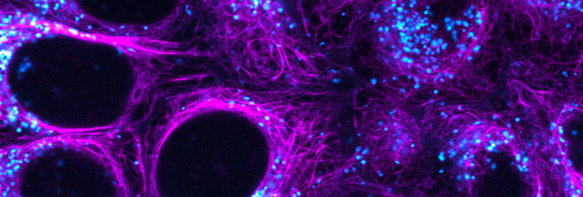
Membrane & Cell Surface
CytoLiner™ Fixed Cell Membrane Stains
While classic lipophilic membrane dyes like DiI can be used to stain formaldehyde-fixed cells, staining can be highly variable due to the poor solubility of the dyes. CytoLiner™ Dyes are a new generation of membrane dyes uniquely engineered to permit selective fluorescent staining of the plasma membrane in fixed cells.
CytoLiner™ staining is compatible with formaldehyde fixation and mild detergent permeabilization before cellular staining. Staining can tolerate blocking agents used during immunofluorescence staining protocols, allowing subsequent staining with antibodies and other probes.
The dyes also are compatible with poly-L-lysine coated cultureware and Transwell® permeable filter supports. CytoLiner™ Dyes are available in a selection of 6 colors from blue to near-IR.
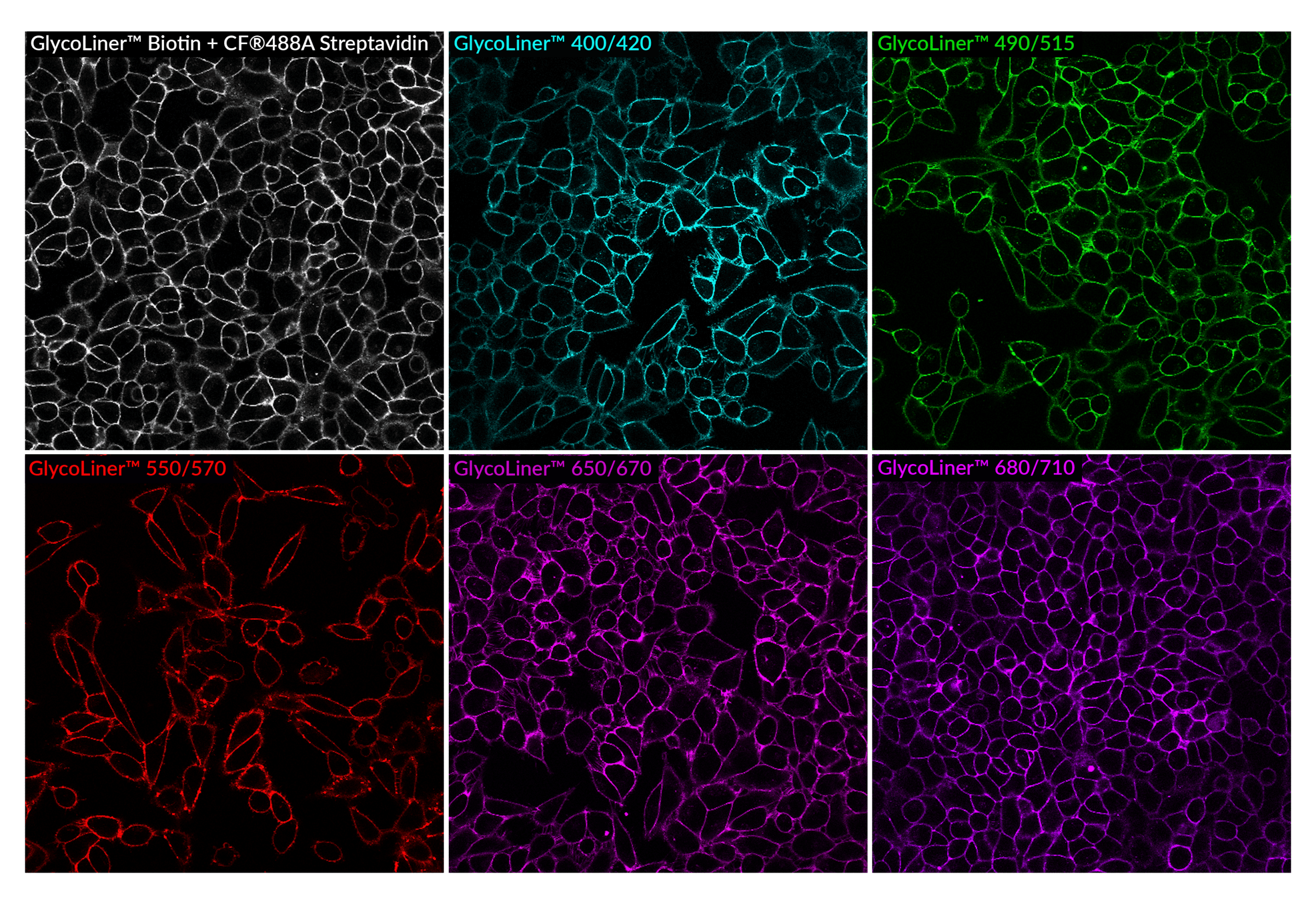
GlycoLiner™ Cell Surface Glycoprotein Labeling Kits
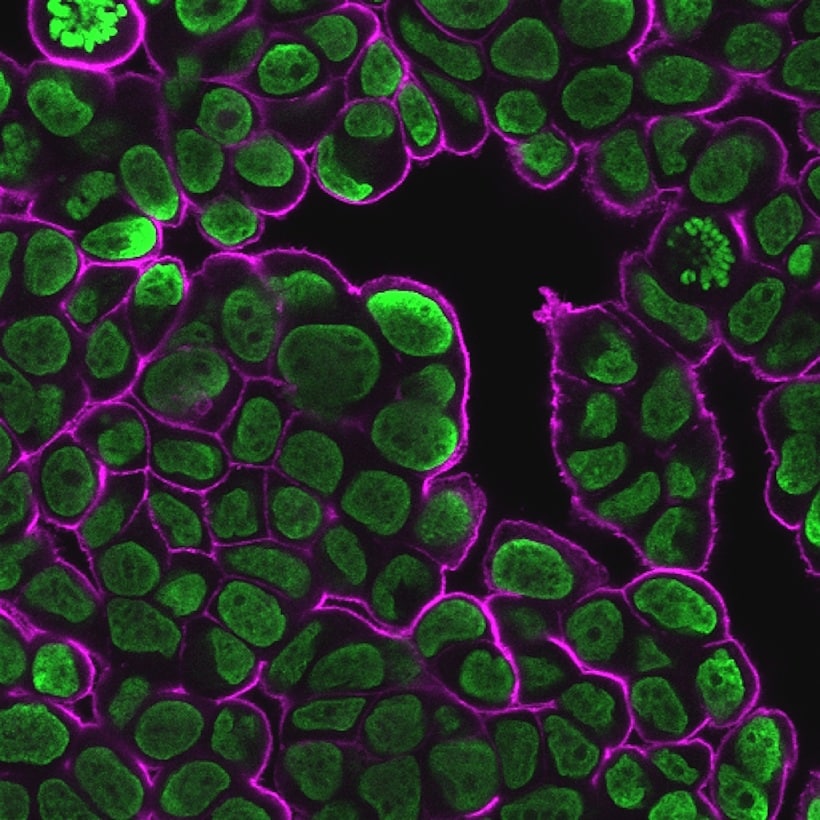
GlycoLiner™ Kits were designed as a superior alternative for covalent labeling of cell surfaces in live cultures. Unlike other covalent cell surface labels which can have significant cytoplasmic background, GlycoLiner™ staining of dead cell cytoplasm is less intense, providing easier imaging of cell surfaces.
GlycoLiner™ technology uses novel self-catalyzing aminooxy groups that are highly reactive at neutral pH without the need to add a catalyst. Unlike glycoprotein labeling using conventional aminooxy labels, which is not only slow but also requires the use of high concentrations of reactive dye and catalyst in acidic buffer. The GlycoLiner™ labeling procedure is complete in about 20 minutes, and can be performed at room temperature or 4°C.
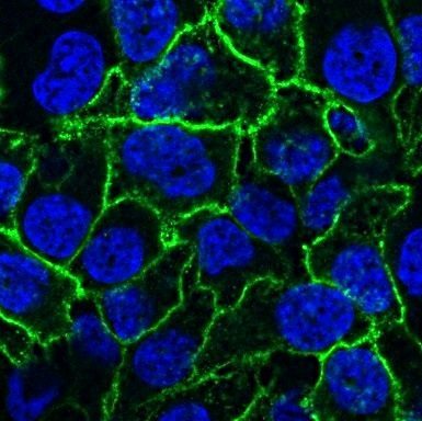
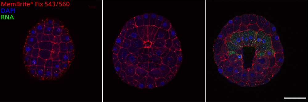
CellBrite® Fix and MemBrite® Fix:
Surface Stains That Tolerate Fix & Perm
CellBrite® Fix and MemBrite® Fix dyes are new classes of dyes developed by Biotium to rapidly stain the outer plasma membranes of live cells. While other lipophilic membrane dyes can be fixed with formaldehyde, they have poor tolerance for detergent permeabilization or methanol fixation. In contrast, CellBrite® Fix and MemBrite® Fix stains are unique in that they can withstand permeabilization and methanol fixation, allowing surface staining to be combined with intracellular immunofluorescence.
CellBrite® Fix dyes are fluorogenic membrane dyes that accumulate in the cell membrane, where they become fluorescent. The dyes have an amine-reactive group for covalent attachment to membrane proteins.
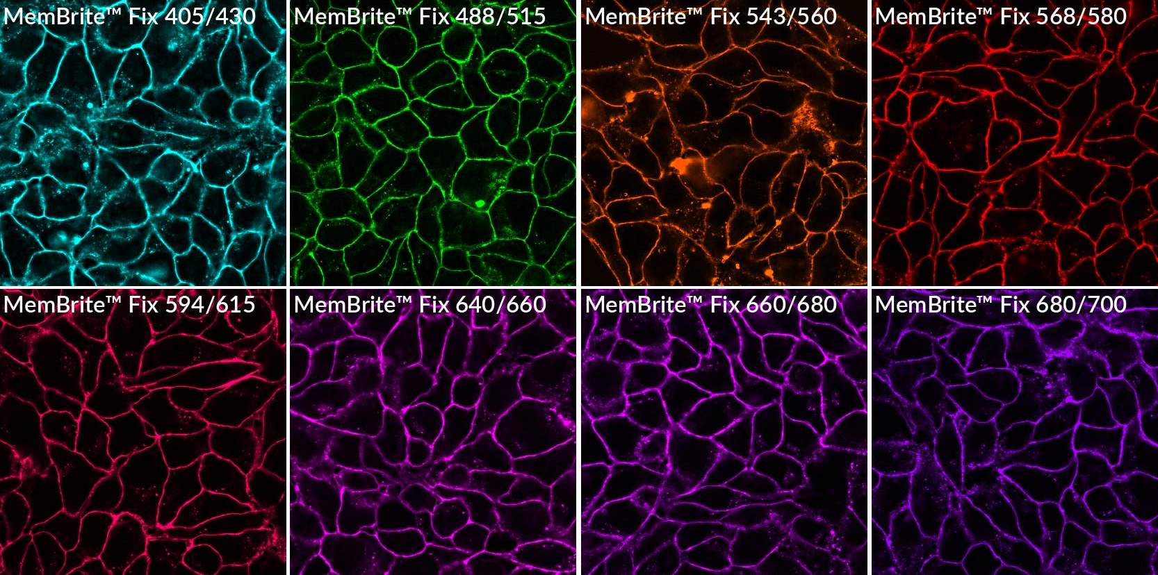
MemBrite® Fix are non-lipophilic dyes that react covalently with cell surface proteins by a different mechanism than CellBrite® Fix, and are available with a wider selection of colors.
MemBrite® Fix labels cells more evenly than traditional membrane stains, and tolerates permeabilization. Unlike lipophilic dyes, MemBrite® Fix labeling is covalent, so it does not redistribute or wash off after permeabilization.
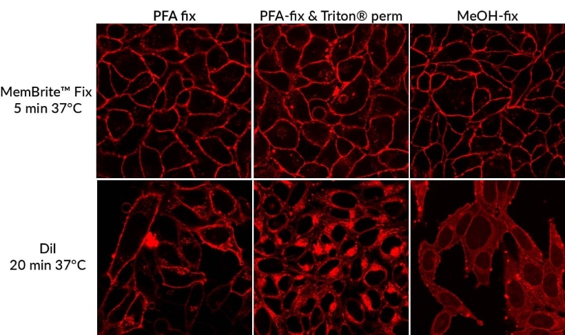
Due to their high water solubility, CellBrite® Fix and MemBrite® Fix dyes yield much more uniform fluorescent staining than lipophilic dyes like DiO and DiI. They are non-cytotoxic and do not readily transfer between cells, and also stain yeast and bacteria. Surface staining is well-retained after permeabilization or methanol fixation, with only a slight increase in intracellular fluorescence.
Unlike lectins, which bind specific targets, CellBrite® Fix and MemBrite® Fix are general surface stains. However, CellBrite® Fix and MemBrite® Fix dyes cannot be used as cell stains post-fixation.
CellBrite® Steady Membrane Staining Kits for Multi-Day Cell Surface Imaging
Our recently released CellBrite® Steady Membrane Staining Kits are unique fluorescent membrane probes designed for multi-day cell surface imaging. Unlike other membrane/cell surface stains that are rapidly lost from the cell surface by endocytosis, CellBrite® Steady Dyes equilibrate between intracellular compartments and the plasma membrane. This allows the cells to retain surface staining as well as intracellular staining for several days.
These kits also include CellBrite® Steady Enhancer as an optional reagent that can be used to mask intracellular fluorescence of CellBrite® Steady Dyes, providing more selective visualization of cell boundaries. Fluorescent stains are available from blue to near-IR, with STORM-compatible options.
CF® Dye Lectin Conjugates
Biotium’s CF® Dye Lectin Conjugates offer bright, photostable labeling for clear visualization of cell-surface glycans and tissue structures. Cell surfaces can be selectively labeled using Concanavalin A (Con A) which binds to mannose/glucose residues and Wheat Germ Agglutinin (WGA) which binds sialic acid and N-acetylglucosamine. Biotium also offers Peanut Agglutinin (PNA) for β-galactose, Tomato Lectin (LEL/TL) for vasculature staining, Ulex Europaeus Agglutinin I (UEA I) for staining endothelial cells, Datura Stramonium Lectin (DSL) for GlcNAc oligomers, Sambucus Nigra Lectin (SNA) for sialic acid, and Phaseolus Vulgaris Leucoagglutinin (PHA-L) for stimulating lymphocyte and T cell proliferation. These conjugates are ideal for live or fixed cell staining, glycan profiling, microbiology, and labeling blood vessels or microglia in tissue sections.
Surface Stains for Yeast and Bacteria
CellBrite® Fix stains yeast and bacteria cell surface. MemBrite® Fix and Con A conjugates can also be used for staining yeast. WGA conjugates can be used as yeast bud scar stains, while in bacteria they are useful fluorescent Gram stains.
See our full selection of Microbiology Products.
Other Membrane and Lipid Probes
We offer a number of other probes for specialized membrane staining applications, such as labeled phospholipids, cholera toxin conjugates for lipid raft labeling, vesicle trafficking probes, nerve terminal dyes, and membrane potential dyes.
Download our Cell Stains Brochure to learn more.
Stain Cell Surfaces in Whole Organisms
Find the Right Stain for Your Application or Workflow

Membrane & Cell Surface Stains | Features |
|---|---|
| GlycoLiner™ Cell Surface Glycoprotein Labeling Kits | • Fast, simple, and gentle labeling of cell surface glycoproteins • Highly selective labeling of live cell boundaries • Covalent labeling compatible with subsequent fix/perm, or cell lysis • Novel self-catalyzing aminooxy for fast labeling at neutral pH • Choice of 5 bright fluorescent dyes or biotin |
| CytoLiner™ Fixed Cell Membrane Stains | • Robust and consistent staining of plasma membrane in formaldehyde-fixed cells • Suitable for antibody co-staining • Compatible with poly-L-lysine coated cultureware • Available in 6 colors from blue to near-IR |
| CellBrite® Fix Membrane Stains | • The only fluorogenic membrane dyes that can be fixed & permeabilized for IF • Bright, uniform live cell surface staining in 15 minutes • Available with green, red, and far-red fluorescence • Stain yeast and bacteria |
| MemBrite® Fix Cell Surface Staining Kits | • Covalent, rapid, and uniform live cell surface protein labeling • Can be fixed & permeabilized for IF staining • Stain yeast and gram-positive bacteria • Choice of 8 colors from blue to near-IR |
| CellBrite® Steady Membrane Staining Kits | • Image live cell surface membranes for 24 hours or longer • Optional Enhancer masks intracellular signal for selective imaging of cell surface • Rapid, even staining in complete cell culture medium • Dye colors from blue to near-IR with STORM-compatible options |
| CellBrite® Cytoplasmic Membrane Dyes | • Ready-to-use solutions of traditional lipophilic membrane stains • Fix before or after labeling • Blue, green, red, and far-red options • Don't tolerate permeabilization or methanol fixation |
| CellBrite® NIR Cytoplasmic Membrane Dyes | • CellBrite® dyes with near-IR emission ranging from 724-820 nm • Compatible with NIR small animal imaging systems • Don't tolerate permeabilization or methanol fixation |
| CF® Dye WGA Conjugates | • Binds to sialic acid and N-acetylglucosamine • Stain cell surface glycoproteins in live or fixed cells • Bacterial Gram stain and yeast bud scar stain • 13 CF® dye colors plus HRP |
| CF® Dye Con A Conjugates | • Binds α-mannopyranosyl and α-glucopyranosyl residues • Stain cell surface glycoproteins in live or fixed cells • Stains yeast cell wall • 10 CF® dye colors from UV to NIR |
| CF® Dye Cholera Toxin Subunit B | • Labels lipid rafts • Withstands fixation and permeabilization • Wide selection of CF® dye colors |
Mitochondria
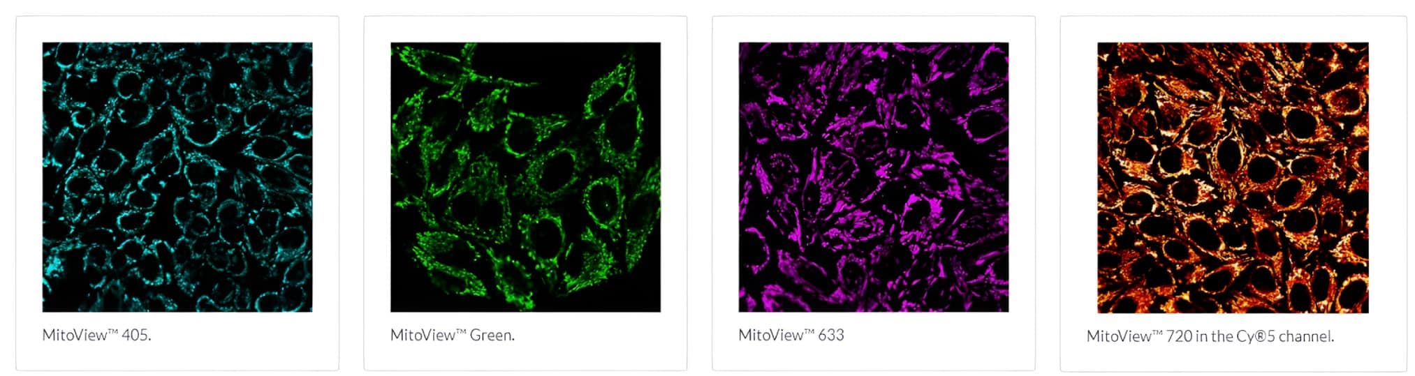
MitoView™ Dyes
In metabolically active cells, mitochondria produce a membrane potential by maintaining a proton gradient across the inner and outer membranes. Loss of mitochondrial membrane potential is a hallmark for apoptosis.
Biotium’s MitoView™ mitochondrial dyes are available in a variety of colors. MitoView™ 633 is a cationic membrane potential-sensitive dye for mitochondria in mammalian cells and yeast. The dye preferentially localizes to the matrix in mitochondria with intact membrane potential in live cells. Staining is lost when mitochondria become depolarized during cell death, allowing monitoring of cell viability.
JC-1 and Other Classic Mitochondrial Dyes
Ratiometric dyes like JC-1 constitute another class of potential-dependent mitochondrial dyes. In healthy cells, JC-1 dye aggregates in mitochondria as a function of membrane potential, resulting in red fluorescence with brightness proportional to the membrane potential.
Conversely, in apoptotic and necrotic cells with diminished mitochondrial membrane potential, JC-1 exists in a green fluorescent monomeric form in the cytosol, allowing cell viability to be assessed by measuring the ratio of red to green fluorescence by flow cytometry or fluorescence microplate reader.
Green fluorescent Rhodamine 123 is a potentiometric dye that accumulates in the mitochondria and is widely used for flow cytometry studies of mitochondrial membrane potential. Red fluorescent TMRM and TMRE are similar potentiometric dyes that are preferred for quantitative membrane potential measurements.
Nonyl acridine orange (NAO) is a membrane potential-independent dye that is reported to bind the phospholipid cardiolipin in the inner mitochondrial membrane. We also offer a number of other classic mitochondrial dyes. See the Cell Stains Brochure for more information.
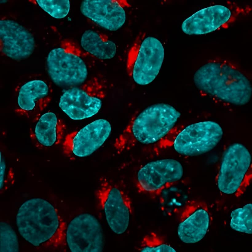
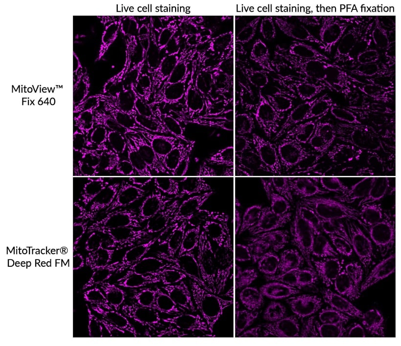
MitoView™ Fix 640
MitoView™ Fix 640 is a far-red-mitochondrial stain suitable for long-term mitochondrial imaging. The cell membrane-permeant dye is non-toxic and offers mitochondrial staining that is well retained for at least 72 hours. In addition, MitoView™ Fix 640 has a reactive group that can covalently attach to proteins inside the mitochondria, allowing for stable staining that withstands fixation and permeabilization for downstream immunofluorescence workflows.
In our testing, MitoView™ Fix 640 offers better specificity after PFA fixation when compared to the similar MitoTracker® Deep Red FM dye. For staining mitochondria in fixed cells or tissue sections, we recommend using one our Mitochondrial Marker Antibodies, available with a wide selection of bright and photostable CF® Dyes and other conjugations.
| Mitochondria Dyes | Abs/Em | Detection channel | Mitochondrial Localization | Potentiometric? | Fixable? | Catalog no. | Size |
|---|---|---|---|---|---|---|---|
| MitoView™ 405 | 398/440 nm | DAPI | Matrix | No1 | No | 70070-T | 50 ug |
| 70070 | 20x50 ug | ||||||
| MitoView™ Green | 490/523 nm | FITC, GFP | Matrix | No | No2 | 70054-T | 50 ug |
| 70054 | 20x50 ug | ||||||
| MitoView™ 633 | 622/648 nm3 | Cy®5, APC3 | Matrix | Yes | No | 70055-T | 50 ug |
| 70055 | 20x50 ug | ||||||
| MitoView™ 650 | 644/670 nm | Cy®5, APC | Matrix | No | No | 70075-50ug | 50 ug |
| 70075 | 20x50 ug | ||||||
| MitoView™ 720 | 720/758 nm4 | Cy®5, Cy®74 | Matrix | No1 | No | 70068-T | 50 ug |
| 70068 | 20x50 ug | ||||||
| MitoView™ Fix 640 | 648/670 nm | Cy®5 | Matrix | No5 | Yes | 70082-50ug | 50 ug |
| 70082 | 10x50 ug | ||||||
| Nonyl Acridine Orange (NAO) | 495/522 nm | FITC/GFP | IMM | No | Yes6 | 70012 | 50 mg |
| Aquaphile™ JC-1 | 510/527 nm (monomer) 585/590 nm (aggregate) | Green/Red7 | Matrix | Ratiometric7 | No | 70076 | 50 uL |
| JC-1 (iodide salt) | 70014 | 5 mg | |||||
| JC-1 (chloride salt) | 70011 | 5 mg | |||||
| Rhodamine 123 | 505/534 nm | FITC/GFP | Matrix | Yes | No | 70010 | 50 mg |
| TMRE | 549/574 nm | Cy®3 | Matrix | Yes | No | 70016 | 25 mg |
| TMRE, 2 mM in DMSO | 70005 | 0.5 mL | |||||
| TMRM | 549/574 nm | Cy®3 | Matrix | Yes | No | 70017 | 25 mg |
| DASPEI | 461/589 nm | Red | Matrix | Yes | No | 70018 | 100 mg |
| DiIC1(5) | 638/658 nm | Cy®5 | Matrix | Yes | No | 70015 | 100 mg |
2. MitoView™ Green is not fixable but can be used to stain cells after formaldehyde fixation.
3. MitoView™ 633 also has red fluorescence in the Cy®3 channel and is not recommended for use with other red dyes.
4. While optimal for Cy®7 settings, MitoView™ 720 is bright enough to be imaged in the Cy®5 channel.
5. MitoView™ Fix 640 reacts covalently with mitochondrial proteins for staining that is retained after fixation and permeabilization.
6. Formaldehyde fixation only. Not compatible with solvent-based fixation or permeabilization.
7. JC-1 forms red fluorescent aggregates in polarized mitochondria, and green fluorescent monomers in cytoplasm.
Matrix: Mitochondrial matrix; IMM: Inner mitochondrial membrane
Cy Dye is a registered trademark of Cytiva.
Lysosomes
LysoView™ Dyes
LysoView™ dyes are fluorescent stains for imaging lysosome localization and morphology in live cells. LysoView™ dyes belong to a family of lysosomotropic dyes that contain weakly basic amines that accumulate in acidic organelles in mammalian cells and yeast.
Red-fluorescent LysoView™ 540 and far-red fluorescent LysoView™ 633 dye fluorescence is pH-sensitive, resulting in specific lysosomal staining without a wash step. We also offer LysoView™ 650, a far-red lysosome dye that is compatible with super-resolution imaging by SIM and STED.
“Light-On” LysoView™ 555
“Light-On” LysoView™ 555 is a UV-activatable lysosome stain. In cells, the dye initially shows low fluorescence, but brief exposure to UV excitation from a mercury arc lamp activates bright red fluorescence localizing to lysosomes.
Lysosomal fluorescence fades over the course of several minutes after UV exposure, but can be re-activated in the same cells multiple times by exposure to UV light. Therefore the dye provides a novel tool for UV-activated, reversible fluorescence imaging of lysosomes.
| Lysosome Stains | Abs/Em (nm) (pH ≤ 5) | Detection channel | Features | Catalog number | Size |
|---|---|---|---|---|---|
| LysoView™ 405, 1000X in DMSO | 318, 400/ 464 | DAPI, Pacific Blue™ | Blue fluorescent lysosome stain | 70066-T | 10 uL |
| 70066 | 50 uL | ||||
| LysoView™ 488, 1000X in DMSO | 496/526 | GFP, FITC | Green fluorescent dye validated in SIM | 70067-T | 10 uL |
| 70067 | 50 uL | ||||
| LysoView™ 540, 1000X in DMSO | 540/634 | TRITC, Cy®3, PE | Orange, pH-sensitive fluorescence** | 70061-T | 10 uL |
| 70061 | 50 uL | ||||
| LysoView™ 550, 1000X in DMSO | 542/567 | TRITC, Cy®3, PE | Bright & photostable orange dye | 70083-T | 10 uL |
| 70083 | 50 uL | ||||
| LysoView™ 594, 1000X in DMSO | 585/634 | Texas Red® | Bright & photostable red dye | 70084-T | 10 uL |
| 70084 | 50 uL | ||||
| LysoView™ 633 (lyophilized solid) | 634/657 | Cy®5, APC | Far-red, pH-sensitive fluorescence | 70058-T | 1 vial* |
| 70058 | 10 vials* | ||||
| LysoView™ 640, 1000X in DMSO | 640/671 | Cy®5, APC | Bright & photostable far-red dye | 70085-T | 10 uL |
| 70085 | 50 uL | ||||
| LysoView™ 650, 1000X in DMSO | 650/675 | Cy®5, APC | Photostable far-red dye compatible with SIM, STED | 70059-T | 10 uL |
| 70059 | 50 uL | ||||
| LysoView™ 680, 1000X in DMSO | 674/711 | Cy®5.5 | Unique near-IR lysosome stain | 70086-T | 10 uL |
| 70086 | 50 uL |
** LysoView™ 540 has limited photostability and may not be suitable for all microcopy applications.
Cy Dye is a registered trademark of Cytiva; Pacific Blue is a trademark and Texas Red is a registered trademark of Thermo Fisher Scientific.
Lipid Droplets
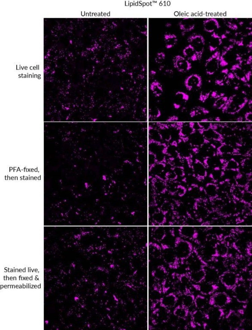
LipidSpot™ Lipid Droplet Stains
Intracellular lipid droplets are cytoplasmic organelles involved in the storage and regulation of triglycerides and cholesterol esters. LipidSpot™ dyes are fluorogenic neutral lipid stains that rapidly stain lipid droplets. The dyes can be used to stain lipid droplets in both live and fixed cells, with no wash step required.
Cells also can be fixed and permeabilized after staining. LipidSpot™ fluorescent stains show minimal background staining of cellular membranes or other organelles, unlike dyes like Nile Red. Available with green or deep-red/far-red fluorescence.
LipidSpot™ Stains Features
- Rapidly and specifically stain lipid droplets
- For live or fixed cellular staining, or fix and permeabilize after staining
- Available with green or deep-red/far-red fluorescence
Cytoskeleton
Visualize Microtubules in Live Cells
ViaFluor® Live Cell Microtubule Stains bind to polymerized tubulin and may be used to visualize microtubules in live cells for up to 24 hours in immortalized cell types. ViaFluor® Microtubule Stains are supplied with a vial of VRP, an efflux pump inhibitor that may improve probe retention and staining in certain cell types.
Biotium offers green fluorescent ViaFluor® 488 and far-red fluorescent ViaFluor® 647 Live Cell Microtubule Stains. ViaFluor® 647 Microtubule Stain is compatible with super-resolution imaging by STED.
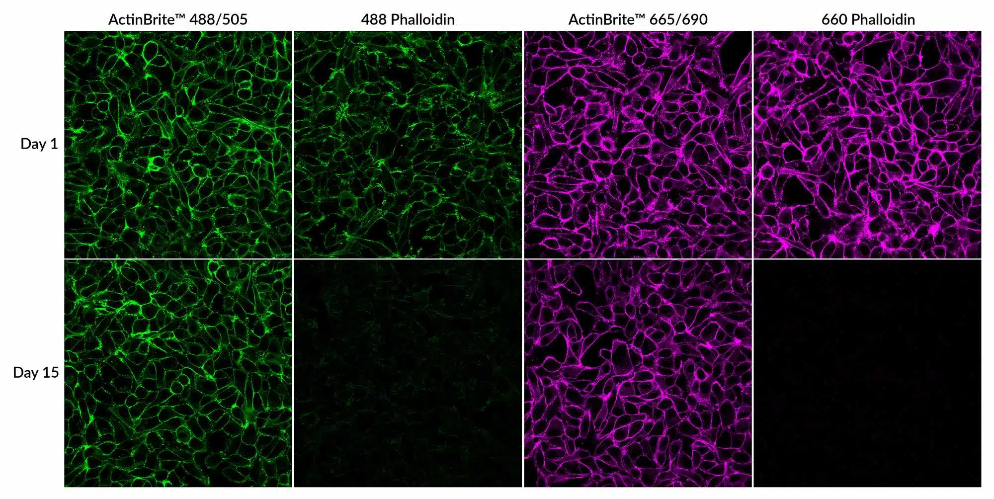
Reliable F-actin Staining in Fixed Cells, No Fading
Fluorescent phalloidins are widely used to stain F-actin in fixed cells and tissues, but traditional dye conjugation can weaken their binding, resulting in dim, short-lived staining. ActinBrite™ High Affinity Phalloidin Conjugates overcome this by maintaining strong F-actin binding, providing bright, stable fluorescent staining that lasts for over a month—making delayed imaging more reliable.
CF® Dye conjugates of Vitamin D-Binding Protein (Vitamin D-BP, also known as GC Globulin) are also available for visualizing monomeric G-actin in fixed and permeabilized cells. They can also be co-stained with phalloidin for comparing G-actin and F-actin distribution
Biotium also offers Phalloidin Conjugates with a wide selection of CF® Dyes as well as traditional dyes and biotin. A number of our CF® Dyes have been validated in super-resolution imaging by STORM, STED, SIM, and other methods.
ActinBrite™ Features
- Novel phalloidin conjugates that preserve strong F-actin binding
- Enables sample storage for 1+ month, depending on conjugate
- Offered in 5 colors from green to near-IR for easy multiplexing
- Direct replacements for any phalloidin conjugate
Cytoskeletal Probes | Features |
|---|---|
| ViaFluor® Live Cell Microtubule Stains | • Live cell microtubule stains • Blue fluorescent ViaFluor® 405, green fluorescent ViaFluor® 488 and far-red fluorescent ViaFluor® 647 Microtubule Stains • ViaFluor® 647 is compatible with super-resolution imaging by STED |
| ActinBrite™ High Affinity Phalloidin Conjugates | • Novel phalloidin conjugates with high F-actin binding affinity • Allows stained samples to be stored for over a month depending on the conjugate • Available in 5 colors from green to near-IR |
| Phalloidin Conjugates | • Actin filament stains for fixed and permeabilized cell • Choice of over 15 bright & photostable CF® dyes • Also available with biotin, FITC, and rhodamine dyes |
| Recombinant Human Vitamin D-Binding Protein Conjugates | • Stains monomeric G-actin in fixed and permeabilized cells • Can be co-stained with phalloidin for comparing G-actin and F-actin distribution • Choice of 5 bright & photostable CF® dyes |
Vesicles & Membrane Trafficking
Fluid phase tracers
Dextrans are water-soluble branched-chain polysaccharides.
Fluorescently labeled dextrans are used as markers for the trafficking of fluid-phase endocytic cargo to lysosomes. They are also used as tracers for epithelial and endothelial permeability and microinjected tracers for neuronal morphology.
CF® Dye Dextrans are available with a variety of dye colors and molecular weights ranging from 3,500 to 250,000.
While formaldehyde fixation preserves the localization of dextrans in endosomes, the labeling generally cannot tolerate detergent permeabilization, likely because the dextran molecules remain in the fluid phase and do not associate closely enough with cellular proteins to become cross-linked by fixative.
CF® Dye Bovine Serum Albumin (BSA) can be used as a tracer for protein uptake and permeability.
Fluorescent hydrazides are formaldehyde-fixable, water-soluble fluid-phase tracers. They are also used as injectable tracers for neuronal morphology. We offer a wide selection of bright and photostable CF® Dye Hydrazides. Learn more about our CF® Dyes.
CF® Dye Transferrin Conjugates
Transferrin is a glycoprotein iron carrier that delivers iron to vertebrate cells via receptor-mediated endocytosis. After binding to its receptor on the cell surface, transferrin is rapidly internalized by invagination of clathrin-coated pits.
Inside endocytic vesicles, the acidic environment favors dissociation of iron from the transferrin-receptor complex. Following the release of iron, the apotransferrin is recycled to the plasma membrane, where it is released from its receptor to scavenge more iron.
We offer human transferrin labeled with a selection of our bright and photostable CF® Dyes for imaging of recycling endosomes by microscopy.
CF® Dye Conjugated Cholera Toxin Subunit B
Cholera toxin subunit B binds to ganglioside GM1 in lipid rafts on the plasma membrane. Cholera toxin subunit B is reported to be internalized by clathrin-dependent and independent pathways depending on cell type.
The conjugates can also be used to label lipid rafts on the cell surface and as a retrograde neuronal tracer. Learn more about our CF® Dyes.
Nerve Terminal Dyes
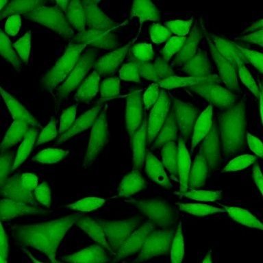
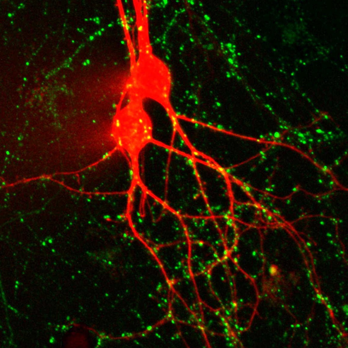
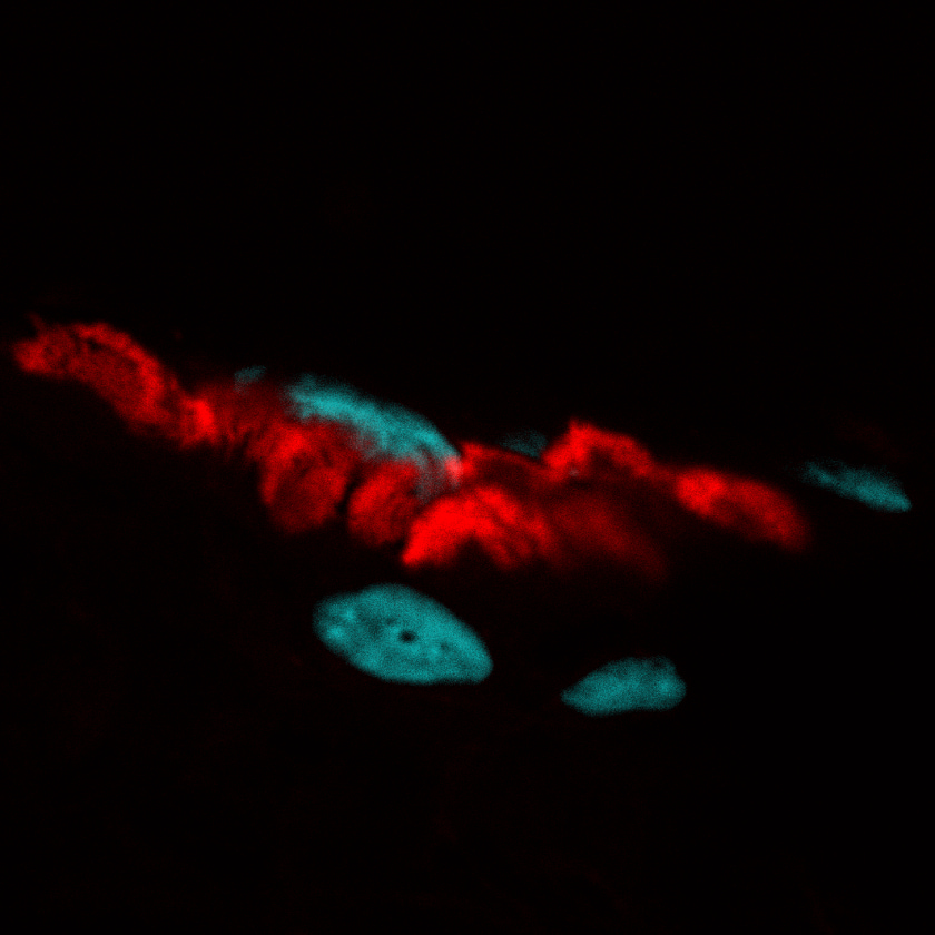
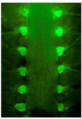
SynaptoGreen™ and SynaptoRed™ nerve terminal probes (originally called FM® dyes) are membrane dyes used to trace endocytic vesicles. While traditionally used to monitor activity-dependent vesicle release at synapses, they can be used to label and track vesicles in non-neuronal cell types as well. See our page on probes and reagents for neuroscience for more information.
Vesicle and Membrane Trafficking Probes | Features |
|---|---|
| CF® Dye Dextrans | • Water-soluble fluid phase markers for tracking bulk endocytosis and cell permeability • Available with molecular weights of 10 kD, 40 kD, 70 kD, 150 kD, or 250 kD • Choose from a selection of bright & photostable CF® dyes |
| Bovine Serum Albumin CF® Dye Conjugates | • Fluorescent BSA for endocytosis and permeability studies • Choice of 4 bright & photostable CF® dyes |
| CF® Dye Hydrazides | • Bright & photostable fixable polar tracers • Wide choice of dye colors |
| CF® Dye Human Transferrin Conjugates | • Recycling endosome tracers with a selection of bright & photostable CF® dyes |
| CF® Dye Cholera Toxin Subunit B | • Lipid raft and endocytosis probe • Withstands fixation and permeabilization • Choice of 6 CF® dye colors |
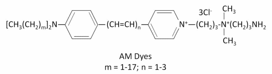
| Nerve Terminal Staining Kits | • SynaptoGreen™ & SynaptoRed™ can be used label vesicles in both neuronal and non-neuronal cells • AM1-43 and other AM dyes are fixable • Available in kits with background reducing agents |
Extracellular Vesicles
Detect EVs, Not Dye Aggregates
Membrane dyes, while common tools for labeling EVs, can pose significant challenges when used for EV staining. For example, membrane dyes such as PKH, DiO, and DiI, have poor solubility and thus form aggregates that can be confused with EVs.
To overcome these challenges, Biotium developed ExoBrite™ EV Surface Stains, which provide bright staining of isolated EVs with minimal dye aggregation.
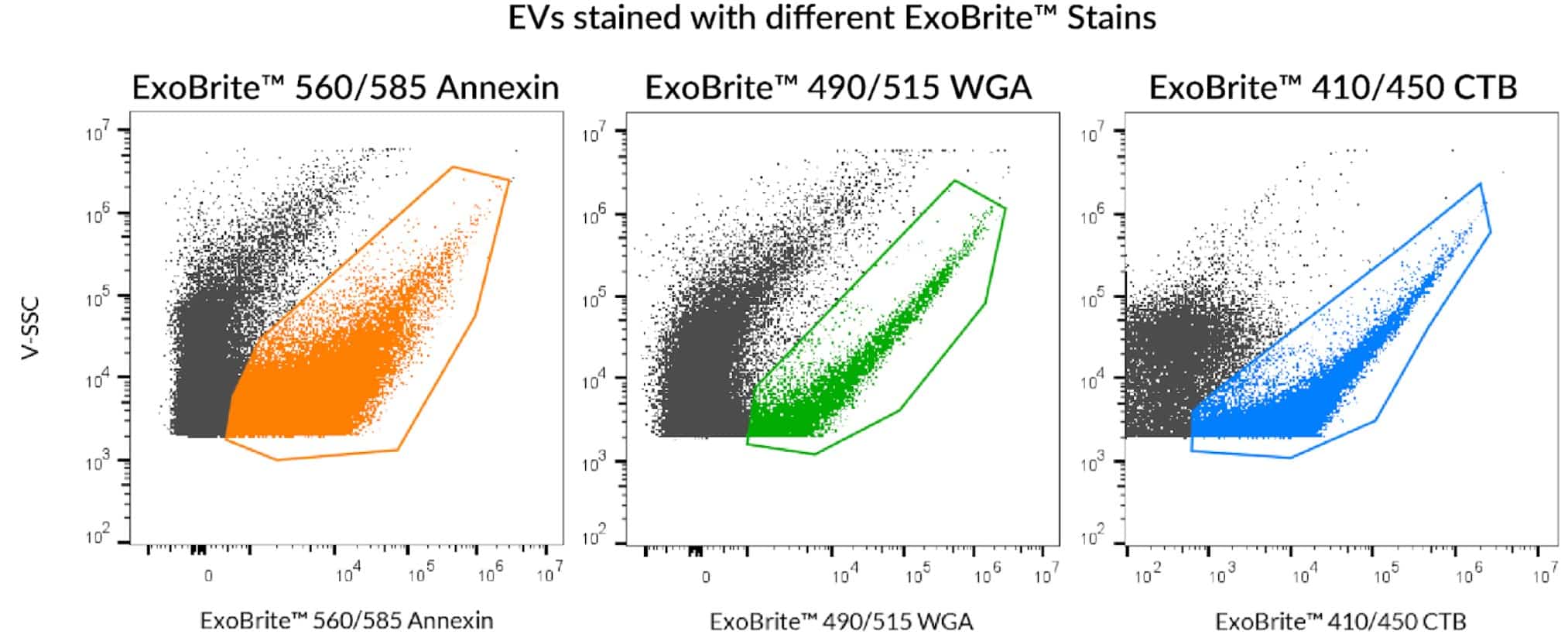
ExoBrite™ EV Surface Stains are conjugates of probes (Annexin V, WGA, and CTB) that are developed and validated for bright and specific fluorescent staining of EV surface targets with minimal aggregation in flow cytometry.
In addition, ExoBrite™ EV Surface Stains are compatible with antibody co-staining and do not bind non-specifically to polystyrene beads, and therefore unlike hydrophobic membrane dyes, they can be used to stain bead-bound EVs.
For flexible panel design, ExoBrite™ EV Surface Stains are available in four colors for the Pacific Blue™, FITC, PE, and APC channels.
Advantages of ExoBrite™ EV Surface Stains:
- Available as ExoBrite™ Annexin V, CTB, and WGA conjugates
- Optimized for bright and sensitive EV staining by flow cytometry
- Stain purified or bead-bound EVs
- Compatible with antibody co-staining
- Available in 4 colors for flexible panel design
- Sampler kit with Annexin V, CTB, and WGA stains available
ExoBrite™ EV Surface Stains
| Product | Conjugate | Detection channels | Size | Catalog Number |
|---|---|---|---|---|
| ExoBrite™ True EV Membrane Stains | N/A | Pacific Blue™, FITC, PE, APC | 100 Labelings, 500 Labelings | 30129, 30130 |
| ExoBrite™ Annexin EV Staining Kits | Annexin V | Pacific Blue™, FITC, PE, Cy®3, APC | 100 Labelings, 500 Labelings | 30119-30122 |
| ExoBrite™ WGA EV Staining Kits | Wheat Germ Agglutinin (WGA) | 30123-30126 | ||
| ExoBrite™ CTB EV Staining Kits | Cholera Toxin Subunit B (CTB) | 30111-30114 | ||
| ExoBrite™ EV Surface Stain Sampler Kit, Green | Annexin V, Wheat Germ Agglutinin (WGA), Cholera Toxin Subunit B (CTB) | FITC | 100 Labelings | 30127 |
| ExoBrite™ STORM CTB EV Staining Kits | Cholera Toxin Subunit B (CTB) | FITC, Texas Red®, Cy®5, Cy®5.5 | 100 Labelings, 500 Labelings | 30115-30118 |
Validated EV Sources for ExoBrite™ EV Surface Stains
| EV Source | ExoBrite™ True EV Membrane Stains | ExoBrite™ CTB Stains | ExoBrite™ WGA Stains | ExoBrite™ Annexin Stains |
|---|---|---|---|---|
| A549 cells | Yes | Yes | Yes | Yes |
| CHO cells | Yes | No | Yes | Yes |
| hASC (human adipose stem cells) | ND | No1 | ND | ND |
| HEK293 cells | Yes | Yes1 | Yes | Yes |
| HeLa cells | Yes | No | Yes | Yes |
| HUVEC (human umbilical vein endothelial cells) | ND | No1 | ND | ND |
| J774 cells | Yes | Yes | Yes | Yes |
| Jurkat cells | Yes | Yes | Yes | Yes |
| MCF-7 cells | Yes | Yes | Yes | Yes |
| Plasma | Yes | No | ND | Yes |
| Raji cells | ND | Yes | Yes | Yes |
| RAW 264.7 cells | Yes | Yes | Yes | Yes |
| Serum | Yes | No | ND | Yes |
| Skeletal myoblasts | ND | Yes1 | ND | ND |
| THP-1 cells | Yes | ND | ND | ND |
| U2OS cells | Yes | No | Yes | Yes |
| U937 cells | Yes | No | Yes | Yes |
| NIH3T3 cells | Yes | Yes | Yes | Yes |
| HepG2 cells | Yes | No | Yes | Yes |
| Yeast (S. cerevisiae) | Yes | No | Yes | Yes |
Value of “Yes” or “No” indicates coverage of EVs based on Biotium’s internal data or customer-reported data. Value of “ND” indicates no data.

TECHNOLOGY PAGE
Extracellular Vesicle Research
TECHNOLOGY PAGE
Validated Antibodies for EV & Exosome Detection: Against CD9, CD63, and CD81
Cytoplasm
ViaFluor® SE Cell Proliferation Dyes
Viafluor® SE dyes are amine-reactive dyes that covalently label proteins throughout the cell. They are membrane-permeable compounds that are non-fluorescent until they enter viable cells. In the cytoplasm, they are hydrolyzed by cytoplasmic esterase enzymes to release fluorescent amine-reactive dyes that react with proteins throughout the cytoplasm and intracellular compartments.
ViaFluor® SE dyes can be used for dye dilution of cell division by flow cytometry or as cell-filling cytoplasmic stains. The cellular staining can also withstand fixation and permeabilization for subsequent immunostaining. We offer blue fluorescent ViaFluor® 405, green fluorescent ViaFluor® CFSE, and red fluorescent ViaFluor® 650.
Calcein AM
Calcein AM is a cell-filling green fluorogenic cytoplasmic stain that uniformly labels the entire cell. It works by a similar mechanism as ViaFluor® CFSE, but the dye is not reactive, so labeling is not covalent.
Only viable cells generate and retain the dye. Calcein AM is widely used as a viability stain to quantitate live cells, detect cell lysis, or monitor dye efflux by transporter proteins.
ViaFluor® SE Dyes Features
- Membrane-permeable compound is hydrolyzed in live cells to release amine-reactive dye
- Covalent, fixable intracellular labeling
- Can be used for flow cytometry analysis of cell division by dye dilution
- For long-term labeling (days to weeks)
- Choice of blue, green, and far-red fluorescence
Calcein AM Features
- Membrane-permeable compound is hydrolyzed in live cells to release green fluorescent dye
- Uniform intracellular labeling Dead cells don’t retain dye, for true endpoint viability assay
- For short term labeling (≤24 hours)
Membrane-Permeant Cytoplasm Stains | Features |
|---|---|
| ViaFluor® SE Dyes | • Membrane-permeable compound is hydrolyzed in live cells to release amine-reactive dye • Covalent, fixable intracellular labeling • Can be used for flow cytometry analysis of cell division by dye dilution • For long-term labeling (days to weeks) • Choice of blue, green, or far-red fluorescence |
| Calcein AM | • Membrane-permeable compound is hydrolyzed in live cells to release green fluorescent dye • Uniform intracellular labeling • Dead cells don't retain dye, for true endpoint viability assay • For short term labeling (≤24 hours) |
Neuronal Stains
Anterograde & Retrograde Tracers
Hydroxystilbamidine (also called Fluoro-Gold™) has been used extensively as a retrograde tracer for neurons and also as a histochemical stain. Fluoro-Gold™ is used for retrograde tracing and dendrite filling. Biotin ethylenediamine is equivalent to Neurobiotin™, a useful anterograde and transneuronal tracer.
Cholera Toxin Conjugates
WGA Conjugates
Dextran Conjugates
| Product | Ex/Em | Catalog number | |
|---|---|---|---|
| Hydroxystilbamidine (Fluoro-Gold™) | 361/536 nm | 80014 | |
| Hydroxystilbamidine (Fluoro-Gold™), 4% in H2O | 361/536 nm | 80023 | |
| Biotin ethylenediamine, hydrobromide (Neurobiotin™) | N/A | 90057 | |
| Biotin ethylenediamine, hydrochloride | N/A | 90075 | |
| Cholera Toxin Conjugates | Choice of 10 colors | ||
| Wheat Germ Agglutinin Conjugates | Choice of 13 colors, plus HRP | ||
| Dextran Conjugates | Choice of 11 colors & 5 molecular weights | ||
Cytosolic Tracers
CF® Dye Hydrazides
We offer a wide selection of our bright and photostable CF® Dye Hydrazides. Water-soluble and aldehyde-fixable, hydrazides can be microinjected as neuronal morphology tracers. Learn more about our CF® Dyes.
Calcein, Calcein AM, and ViaFluor® SE Dyes
Calcein is a water-soluble green fluorescent FITC derivative widely used for the study of cell membrane integrity that can be introduced by microinjection.
Calcein-AM is a membrane-permeant AM ester of calcein that can be loaded into cells by incubation, where it is cleaved by esterases to release the dye. Calcein AM can also be used to quantitate cell viability, because dead cells will not hydrolyze the ester or retain the hydrolyzed dye.
ViaFluor® SE Dyes work similarly to Calcein AM, except the hydrolyzed dyes are amine-reactive and covalently label cytoplasmic proteins, so fluorescent staining can withstand fixation and permeabilization.
Other Classic Fluorescent Tracers
Lucifer Yellow CH and Lucifer Yellow Cadaverine are aldehyde-fixable fluorescent cytosolic and gap junction tracers.
Hydroxystilbamidine (equivalent to Fluoro-Gold) is a UV-excitable green fluorescent dye that has been used extensively as a retrograde tracer for neurons and as a histochemical stain. It is available in solid form and as a ready-to-use 4% solution.
A number of other classic hydrophilic dyes have also been used as micro injectable tracers.
Biotin Derivatives
Biocytin (e-biotinoyl-L-lysine) is a cellular tracer that can be introduced by microinjection; biocytin-hydrazide is an aldehyde-fixable analog.
We also offer bright, photostable, and pH-independent CF® Dye biotin and CF® Dye biocytin, as well as classic fluorescent biotin derivatives.
Cytosolic Tracers | Features |
|---|---|
| CF® Hydrazides | • Microinjectable, fixable fluorescent tracers • Wide selection of bright & photostable CF® dyes |
| Calcein | • Water soluble green fluorescent tracer for microinjection |
| Calcein AM | • Membrane-permeable compound is hydrolyzed in live cells to release water soluble green fluorescent dye • Uniform intracellular labeling • Dead cells don't retain dye, for true endpoint viability assay |
| ViaFluor® SE Dyes | • Membrane-permeable compound is hydrolyzed in live cells to release amine-reactive dye • Covalent, fixable intracellular labeling • Choice of blue or green fluorescence |
| Lucifer Yellow Derivatives | • Widely used green fluorescent tracers for neuronal morphology and gap junction studies • Fixable Lucifer Yellow CH and Lucifer Yellow Cadaverine • Lucifer Yellow Cadaverin Biotin-X for secondary detection with streptavidin |
| Hydroxystilbamidine (equivalent to Fluoro-Gold™) | • Widely used UV-excitable green fluorescent retrograde neuronal tracer • Available in solid and 4% solution in water |
| Biotin Derivatives | • Biotin-based tracers for secondary detection with streptavidin |
| CF® Dye Biotin | • Biotin conjugates of our bright & photostable CF® dyes |
| CF® Biocytin | • Aldehyde-fixable biocytin conjugated with bright & photostable CF® dyes. |
Synaptic Probes and Nerve Terminal Dyes
α-Bungarotoxin Conjugates
Fluorescent α-bungarotoxin can be used for labeling of nicotinic acetylcholine receptors in sections of neuromuscular junction.
We offer α-bungarotoxin conjugates with a selection of our CF® Dyes and other labels. CF® Dyes offer advantages in brightness and photostability compared to other commercially available fluorescent dyes. Learn more about our CF® Dyes.
- Fluorescent probe for nicotinic acetylcholine receptors
- Stains neuromuscular junction endplates
- Wide selection of bright & photostable CF® dyes

Nerve Terminal Dyes
SynaptoGreen™ and SynaptoRed™ nerve terminal probes (originally called FM® dyes) are membrane dyes used to trace endocytic vesicles. They are a series of fluorescent cationic styryl dyes developed to follow synaptic activity at neuromuscular junctions or synapses.
The dyes label synaptic vesicles in neuronal tissues and cultured neurons in an activity-dependent fashion. They can also be used to label endocytic vesicles in other cell types.
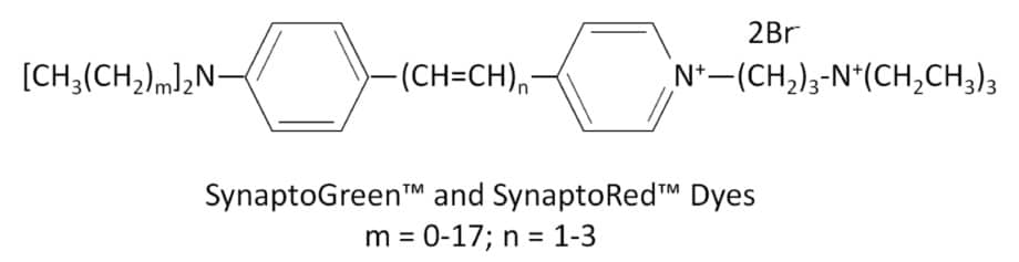
Nerve terminal dyes have highly hydrophilic, cationically charged head groups at one end with lipophilic tails at the other end. They are virtually non-fluorescent in aqueous solution, but become intensely fluorescent in membranes.
Following nerve stimulation, the dye molecules are internalized in newly formed endocytic vesicles. During exocytosis, the dyes are released from the vesicles along with neurotransmitters, causing a decrease in fluorescence signal. As a result, the change in fluorescent intensity reflects the amount of endocytosis/exocytosis or synaptic activity.
The rate of fluorescence increase during endocytosis (on-rate), and the rate of fluorescence decrease during exocytosis (off-rate) vary from dye to dye.
AM dyes and HM dyes are fixable nerve terminal dyes. After staining with these dyes, cells can be fixed and permeabilized for subsequent immunostaining.
See the table below for a list of nerve terminal dyes and their properties.
A common problem encountered with nerve terminal dyes is background fluorescence due to residual membrane staining, even after extensive washing. To reduce this background fluorescence, we offer three quencher or dye-clearing agents.
- ADVASEP-7, a sulfonated β-cyclodextrin, forms a water soluble inclusion complex with SynaptoGreen™ C4 that can be removed more effectively by washing.
- Biotium’s unique quencher, SCAS, reduces background fluorescence as soon as it is added to the preparation without the need for washing.
- Sulforhodamine 101 quenches SynaptoGreen™ background staining via fluorescent resonance energy transfer (FRET).
We offer these reagents as individual products and in kits with dyes and the quencher/dye-clearing agents.
Properties of Nerve Terminal Dyes
| Nerve Terminal Dye | m* | n* | Fixable? | Size | Catalog number | Features |
|---|---|---|---|---|---|---|
| SynaptGreen™ Dyes (Ex/Em ~480/660 nm in membranes) | ||||||
| SynaptoGreen™ C1 | 0 | 1 | No | 5 mg, 5 x 1 mg | 70042, 70043 | • Green nerve terminal probe • Shortest tail for slowest on-rate & fastest off-rate |
| SynaptoGreen™ C2 (equivalent to FM®2-10) | 1 | 1 | No | 70044, 70045 | • Equivalent to FM®2-10 | |
| SynaptoGreen™ C3 | 2 | 1 | No | 70023, 70026 | • Green nerve terminal probe | |
| SynaptoGreen™ C4 (equivalent to FM®1-43) | 3 | 1 | No | 70020, 70022 | • Equivalent to FM®1-43 | |
| SynaptoGreen™ C5 (equivalent to FM®1-84) | 4 | 1 | No | 70046, 70047 | • Equivalent to FM®1-84 | |
| SynaptoGreen™ C18 (equivalent to FM®3-25) | 17 | 1 | No | 70048, 70049 | • Equivalent to FM®3-25 | |
| AM1-43 | 3 | 1 | Yes | 1 mg | 70024 | • Fixable version of SynaptoGreen C4 • Equivalent to FM®1-43FX |
| AM1-44 | 4 | 1 | Yes | 70038 | • Improved fixability over AM1-43 | |
| AM2-10 | 1 | 1 | Yes | 70036 | • Fixable analog of SynaptoGreen™ C2 | |
| AM3-25 | 17 | 1 | Yes | 70051 | • Fixable far-red nerve terminal probe | |
| HM1-43 | 3 | 1 | Yes | 70053 | • Fixable red nerve terminal probe | |
| SynaptoRed Dyes™ (Ex/Em ~510/750 nm in membranes) | ||||||
| SynaptoRed™ C1 | 0 | 3 | No | 5 mg, 5 x 1 mg | 70040, 70041 | • One carbon shorter than SynaptoRed™ C2 |
| SynaptoRed™ C2 (equivalent to FM®4-64) | 1 | 3 | No | 70021, 70027 | • Equivalent to FM®4-64 | |
| SynaptoRed™ C2M** (equivalent to FM®5-95) | 1 | 3 | No | 70019, 70028 | • More water soluble than SynaptoRed™ C2 • Equivalent to FM®5-95 |
|
| AM4-64 | 1 | 3 | Yes | 1 mg | 70025 | • Fixable version of SynaptoRed™ C2 |
| AM4-65 | 3 | 3 | Yes | 70039 | • Fixable version of SynaptoRed™ C2 | |
| AM4-66 | 4 | 3 | Yes | 70050 | • Fixable and spectrally identical to SynaptoRed™ C2 | |
**The positively-charged end of SynaptoRed C2M is a trimethylammonium group.
FM is a registered trademark of Thermo Fisher Scientific.
Nerve Terminal Staining Kits
| Nerve Terminal Staining Kit | Nerve Terminal Dye | Background Reducer | Catalog number |
|---|---|---|---|
| Nerve Terminal Staining Kit I | SynaptoGreen™ C4 (5 x 1 mg ) | ADVASEP-7 (250 mg) | 70030 |
| Nerve Terminal Staining Kit II (A) | AM1-43 (1 mg) | ADVASEP-7 (100 mg) | 70031 |
| Nerve Terminal Staining Kit II (B) | AM1-43 (1 mg) | SCAS (100 mg) | 70031-1 |
| Nerve Terminal Staining Kit III | SynaptoGreen™ C4 (5 x 1 mg) | Sulforhodamine 101 (100 mg) | 70032 |
| Nerve Terminal Staining Kit V | SynaptoRed™ C2 (5 x 1 mg) | ADVASEP-7 (250 mg) | 70034 |
Transwell is a registered trademark of Corning Inc; Triton is a registered trademark of The Dow Chemical Company; MitoTracker and FM are registered trademarks of Thermo Fisher Scientific.


