 New Products
 Earth-Friendly Products
 Biotium Choice Antibodies
 Special Offers


Content #1
Content #1
Content #1
ViaFluor® SE Cell Proliferation Kits use amine-reactive dyes to covalently label cells throughout the cytoplasm and intracellular compartments. ViaFluor® SE dyes are initially membrane-permeable and non-fluorescent. After the compound enters viable cells, it is hydrolyzed by cytoplasmic esterases to releases the fluorescent amine-reactive dye. The dye covalently reacts with amine groups on intracellular proteins, forming stable fluorescent conjugates that are retained in the cell.
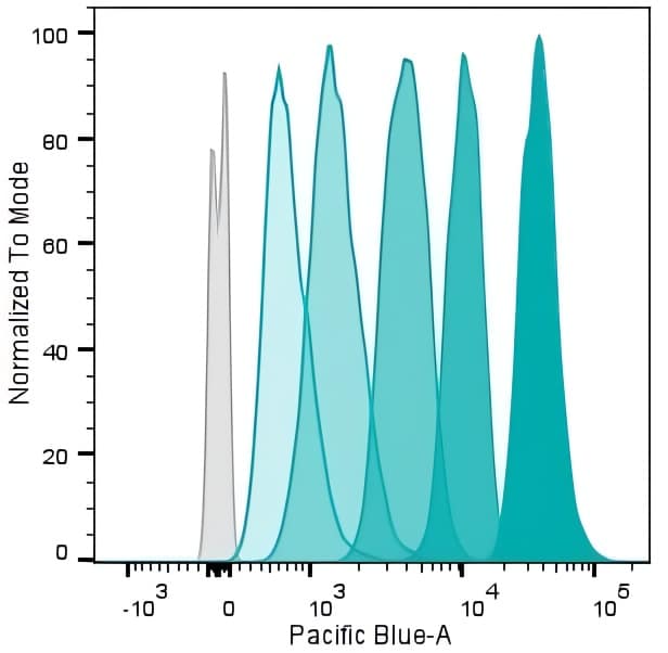
Cell proliferation dyes like ViaFluor® SE were developed to monitor cell division by flow cytometry. Immediately after staining cells with the dye, a single, bright fluorescent population is detected. With each cell division, daughter cells inherit roughly half of the fluorescent label, resulting in the appearance of a successively dimmer peak on a flow cytometry histogram for each cell division. Thus, flow analysis can be used to count multiple divisions of cells grown in culture or injected in vivo after labeling with the proliferation dye. ViaFluor® SE dyes are bright, highly reactive, and have low toxicity, features that are critical for cell division tracking.
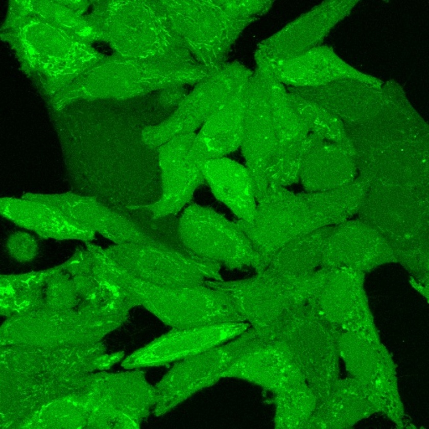
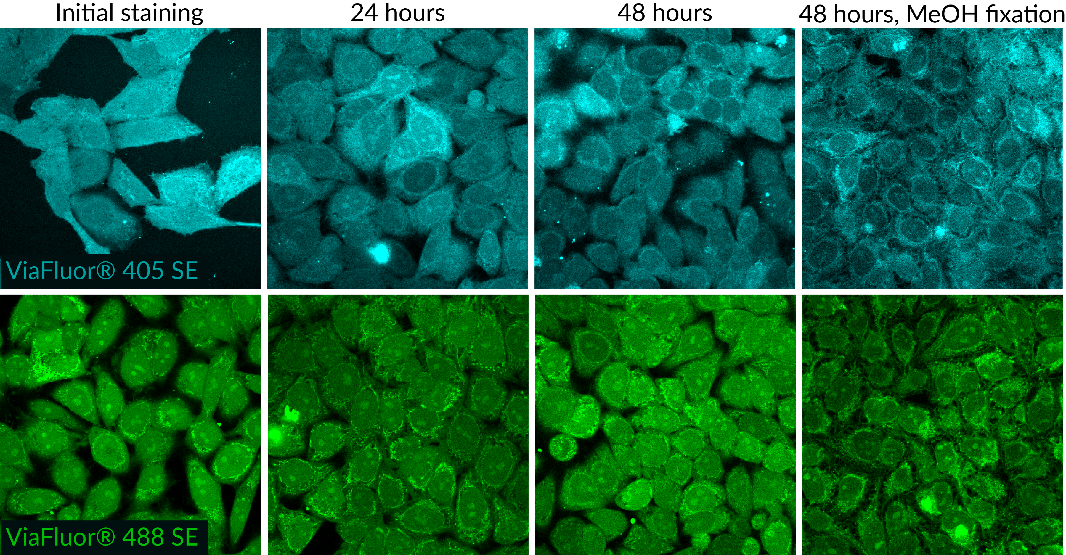
Another useful application of ViaFluor® SE is for cell tracing and tracking by microscopy. Traditionally, lipophilic membrane dyes like our CellBrite™ Dyes (DiO, DiI, and related dyes) have been used for long term cell tracking, because they are stable, non-toxic, and do not readily transfer between cells. But lipophilic dyes tend to label cells unevenly due to poor solubility. The dyes initially stain the cell surface, but become internalized by endocytosis, making it difficult to view cell morphology over time. While the staining can be fixed, it has poor tolerance for permeabilization.
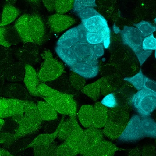
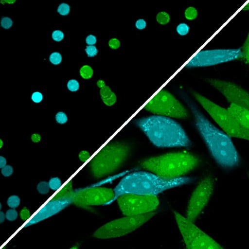
Like lipophilic membrane dyes, ViaFluor® SE labeling is also non-toxic, stable, and does not readily transfer between cells. But because ViaFluor® SE dyes uniformly label the cell interior, they are more useful for imaging morphology over time than lipophilic membrane dyes. Two different cell populations can be labeled with different ViaFluor® SE dyes, mixed together, and tracked in co-culture over time. Because labeling is covalent, the dyes have excellent tolerance for fixation and permeabilization. See four example workflows for different ViaFluor® SE applications below, and download the complete ViaFluor® SE Protocol.
1. Stain cells with ViaFluor® SE1
2. Culture cells to allow division2
3. If needed: Detach adherent cells
4. Optional: Fix, permeabilize cells
5. Optional: Stain other markers
6. Analyze by flow cytometry
B. Imaging Cell Morphology by Microscopy
1. Stain cells with ViaFluor® SE1
2. Optional: Culture cells2
3. Optional: Fix, permeabilize cells
4. Optional: Stain other markers
5. Image by microscopy
1. Stain cells with ViaFluor® SE1,3
2. Detach cells
3. Mix different cell populations
4. Culture cells2
5. Optional: Fix/permeabilize, stain other markers
6. Analyze by microscopy or flow4
D. Labeling Suspension Cells for Co-culture
1. If necessary: Detach cells
2. Stain cells with ViaFluor® SE1,3
3. Mix different cell populations.
4. Culture cells2
5. Optional: Fix/permeabilize, stain other markers
6. Analyze by microscopy or flow4
Notes:

Content #1
Content #1
Content #1
Content #2
Content #3