 New Products
 Earth-Friendly Products
 Biotium Choice Antibodies
 Special Offers


Content #1
Content #1
Content #1
GPRC5D-targeted T cell engagers (TCEs), such as talquetamab and forimtamig, have demonstrated promising efficacy in relapsed and refractory multiple myeloma. GPRC5D is a G protein–coupled receptor enriched on malignant plasma cells and is minimally expressed across normal hematopoietic tissues, making it an ideal surface antigen for targeted immunotherapy. However, more information on how tumor cells evade antigen-directed immunotherapies at the genetic, epigenetic, and protein localization levels is needed.
In a 2026 Nature Medicine publication, Lee, H. et al. characterized mechanisms of resistance to TCEs in multiple myeloma using cytotoxicity assays, structural modeling, flow cytometry, and confocal imaging. Functional studies showed that truncating and frameshift GPRC5D mutations eliminated surface expression of the transmembrane protein and conferred complete resistance to talquetamab and forimtamig. In contrast, the missense p.Tyr257Ser variant retained partial, dose-dependent sensitivity despite appearing negative by standard flow cytometry and confocal microscopy.
The researchers used Biotium’s CytoLiner™ 570/590 Fixed Cell Membrane Stain to assess membrane colocalization of wildtype and missense variant p.Tyr257Ser GPRC5D transduced in K562 cells. Using optimized non-permeabilized staining conditions, low-level membrane localization of p.Tyr257Ser GPRC5D was detected, explaining its residual responsiveness to high-avidity, bivalent TCEs. These findings advance understanding of antigen escape in immunotherapy-treated myeloma, and CytoLiner™ 570/590 Fixed Cell Membrane Stain supported the multiplex imaging workflows essential to translational cancer research.
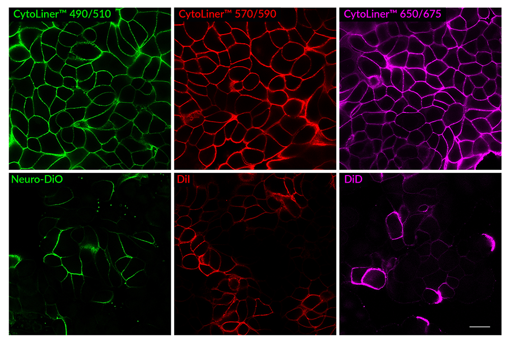
Comparison of fixed cell staining with CytoLiner™ Fixed Cell Membrane Stains versus classic lipophilic carbocyanine membrane dyes. In comparison to the traditional lipophilic carbocyanine dyes, CytoLiner™ dyes show more uniform and reliable staining of PFA-fixed, mildly-permeabilized cell membranes. PFA-fixed MCF7 cells were permeabilized for 10 minutes at RT with PBS/0.1% Triton® X-100, then rinsed with PBS and stained with carbocyanine dyes in PBS or CytoLiner™ Stains in 1X CytoLiner™ Buffer for 10 minutes at RT, then rinsed with PBS. Imaged by confocal microscopy; scale bar: 20 um.
Biotium offers a wide selection of bright, photostable CF® Dye–labeled primary and secondary antibodies optimized for confocal microscopy and flow cytometry, as well as CytoLiner™ Fixed Cell Membrane Stains for precise membrane delineation in formaldehyde-fixed cells. Additional tools for workflows requiring staining live cells before fixation include CellBrite® Fix Membrane Stains or MemBrite® Fix Cell Surface Staining Kits. View Biotium’s Membrane and Cell Surface Stains Comparison to find the right membrane stain for your application.
Full Citation
Lee, H., Ahn, S., Gonzales, G.A. et al. Multimodal antigenic escape to GPRC5D-targeted T cell engagers in multiple myeloma. Nat Med (2026). https://doi.org/10.1038/s41591-025-04175-8
