 New Products
 Earth-Friendly Products
 Biotium Choice Antibodies
 Special Offers


Content #1
Content #1
Content #1
Although resting CD4+ T cells are commonly infected by HIV-1 in vivo, they are highly resistant to infection with cell-free virus in vitro without artificial activation. This paradox has challenged researchers’ understanding of how HIV-1 establishes infection in physiologically resting immune cells. Cell-cell spread (CCS) through virological synapses is known to be far more efficient than cell-free infection, though it has not been clear how direct contact between infected and uninfected T cells altered the target cells’ permissivity to infection.
In a 2026 Nature publication, Mesner, Whelan et al. developed a virological synapse (VS)-priming assay using donor T cells expressing fusion-defective HIV-1 Env-F522Y, allowing CD4 engagement and synapse formation without productive viral transfer. In live-cell instant Structured Illumination Microscopy (iSIM) experiments, Biotium’s NucSpot® Live 650 Nuclear Stain enabled high-resolution tracking of HIV-1 capsid movement into the nucleus. During dynamic imaging of infected T cells, CellBrite® Steady 550 Membrane Stain provided clear plasma membrane visualization during dynamic imaging of infected T cells. Combining these stains with fluorescently-tagged HIV-1 capsids, the researchers continuously tracked individual viral particles from cell entry through cytoplasmic trafficking, nuclear envelope arrest, and nuclear translocation in living primary human T cells (Figure 1). Additional dSTORM imaging of primary labelled nucleoporins (Nups) using a Biotium CF®568-conjugated secondary antibody further revealed increased Nup54 puncta at the nuclear envelope following HIV-1 envelope glycoprotein (Env) stimulation, consistent with nuclear pore complex remodeling (Figure 2).
The researchers demonstrated that cell–cell contact alone activates a CD4–LCK signaling cascade that triggers CDK1-dependent phosphorylation of nucleoporins, remodeling the nuclear pore complex (NPC) to promote capsid nuclear import without inducing classical T-cell activation or cell-cycle progression. VS-primed cells exhibited significantly increased capsid localization at the nuclear envelope, elevated 2-LTR circle formation (a surrogate for nuclear import), enhanced proviral integration, and compared with non-primed controls (Figure 2) .
These findings provide a mechanistic explanation for how viruses manipulate nuclear transport pathways during infection and why physiologically resting T cells resist cell-free HIV-1 infection in vitro yet are infected efficiently in vivo. This work not only advances understanding of HIV-1 pathogenesis and host–virus interactions, but also highlights Biotium’s CellBrite® Steady Membrane Stains, NucSpot® Live Cell Nuclear Stains, and CF® Dyes as powerful tools for super-resolution virology and live-cell imaging.
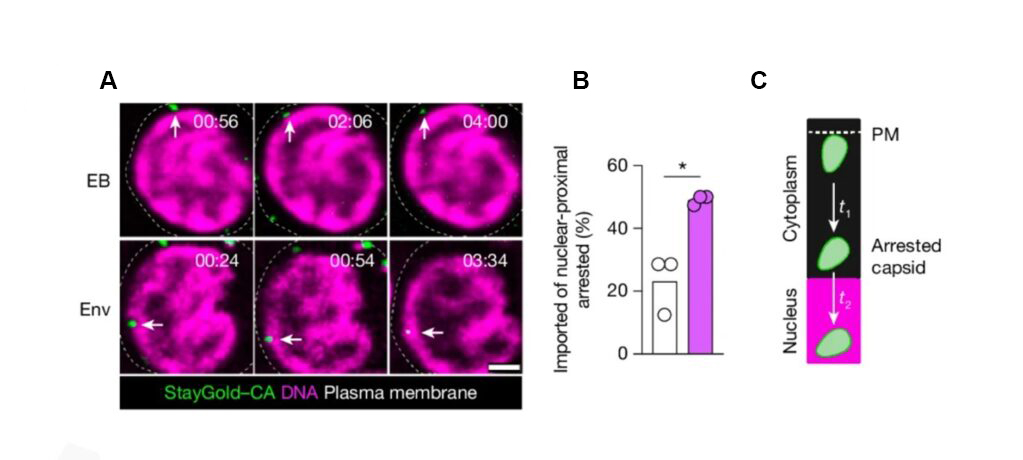
Figure 1. NucSpot® 650, CellBrite® Steady 550, and CF®568 secondary antibody enable visualization of CD4 signaling-driven HIV-1 capsid nuclear import. (a) Live-cell iSIM imaging of mStayGold–CA HIV-1 in resting T cells stimulated with empty beads (EB, top) or Env-coated beads (bottom). CellBrite® Steady 550 membrane stain (dashed white line) and NucSpot® 650 nuclear stain (magenta) provide subcellular landmarks for the plasma membrane and nucleus, respectively. Timepoints (min:s) indicate elapsed time after virus–cell association. Scale bar, 200 nm. (b) Percentage of nuclear-proximal arrested capsids that successfully entered the nucleoplasm in EB- vs Env-bead-stimulated cells. n = 3 donors. (c) Time from capsid entry at the plasma membrane to nuclear-proximal arrest (t1) and from arrest to nuclear import (t2) in EB and Env conditions. n = 21 virions (t1); n = 5 (EB) and n = 13 (Env) virions (t2). Figure adapted from Mesner et al. 2026. Open access — CC BY 4.0.
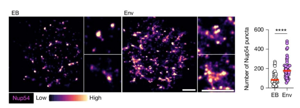
Figure 2. CF®568 secondary antibody enable visualization of NPC remodeling in primary resting T cells. dSTORM imaging of Nup54 puncta in resting T cells stimulated with EB or Env beads for 6 h. Nup54 was visualized using a rabbit primary antibody and CF®568-conjugated secondary antibody (Biotium). Localization maps are pseudo-colored from lowest (black) to highest (yellow) intensity. Quantification shows the number of Nup54 puncta per cell; n = 46 cells from 6 donors. Scale bar, 1 µm. Data in all figures are mean ± SEM. Statistical analysis: color-coded paired two-tailed t-tests (b); paired two-tailed t-tests (d); two-tailed Mann–Whitney U-tests (e, f). Figure adapted from Mesner et al. 2026. Open access — CC BY 4.0.
Learn more about Biotium’s NucSpot® Live Cell Nuclear Stains, no-wash nuclear dyes with low toxicity that are ideal for long-term live cell labeling. CellBrite® Steady Membrane Stains allow fluorescence imaging of live cell surfaces for up to several days in culture. To find the right stain for your application, view Membrane & Cell Surface Stains Comparison.
Full Citation
Mesner D*, Whelan MVX*, Shivkumar M, Reuschl A-K, Zenezini Chiozzi R, Thalassinos K, de Bruin RAM & Jolly C. HIV-1 signalling remodels nuclear pores to licence infection. Nature (2026). https://doi.org/10.1038/s41586-026-10453-3 (*equal contribution).
