New Products
 Earth-Friendly Products
 Biotium Choice Antibodies
 Special Offers
 Quick Order Tool
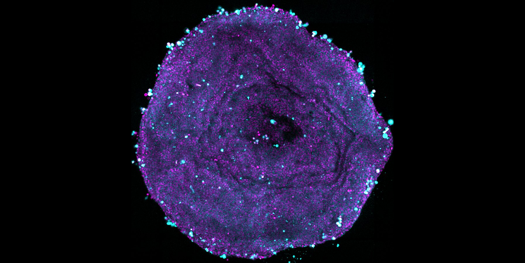
Bright Stains Engineered for Deep Penetration
Biotium offers a selection of unique stains engineered to meet the demands of 3D cell culture, delivering the brightness, photostability, and deep-tissue penetration required for spheroids, organoids, and other complex models. Whether you're staining nuclei, cell surfaces, or performing viability analysis, Biotium's application-tested stains give researchers the confidence to generate publication-ready data for complex 3D models.
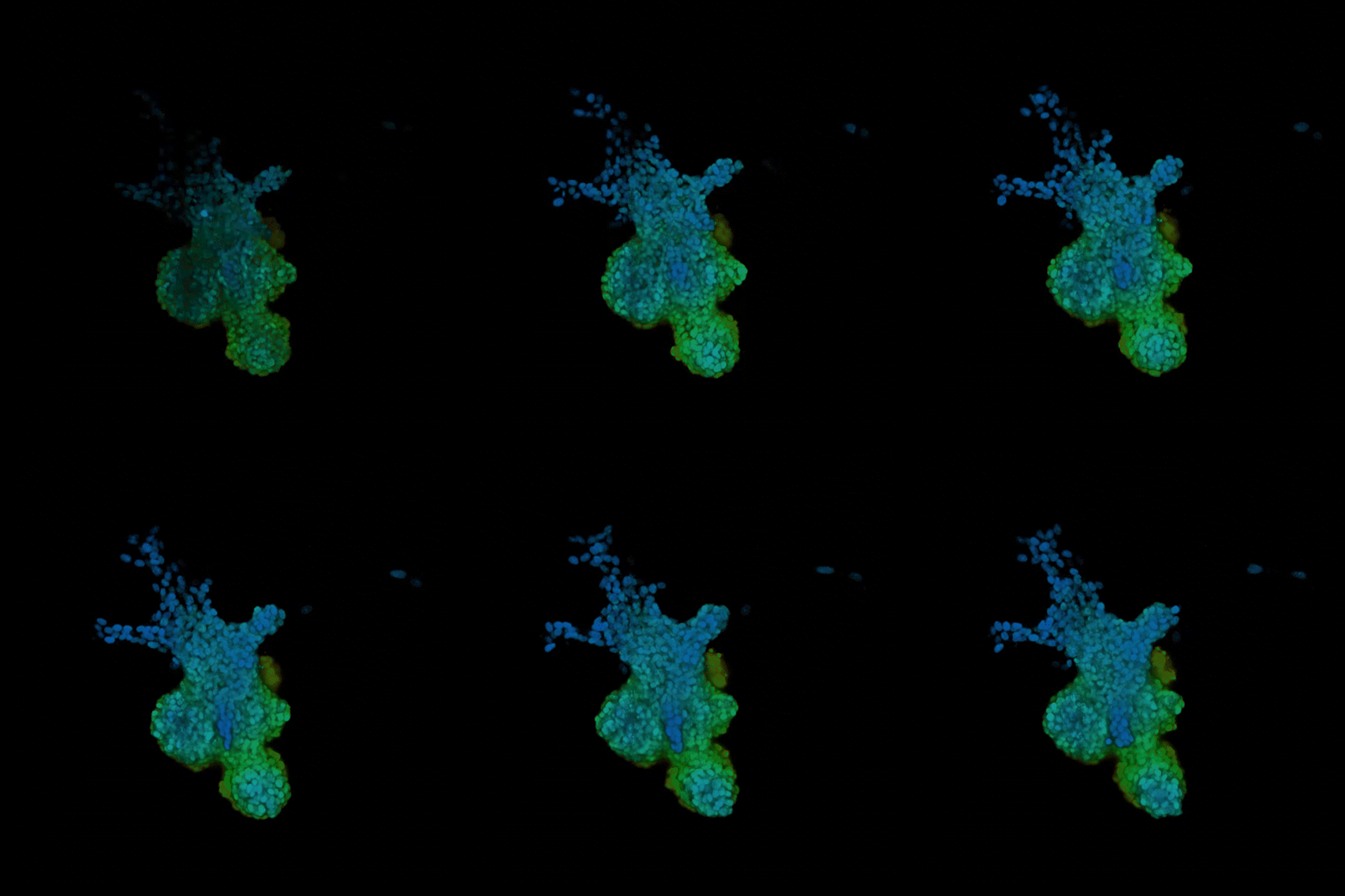
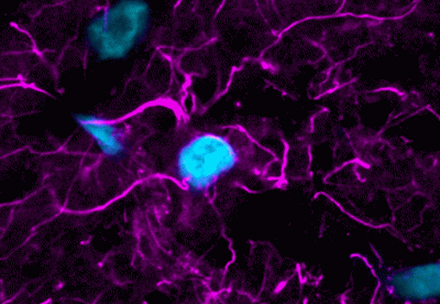
Real-Time Imaging of Nuclei in Organoids with NucSpot® Live
NucSpot® Live Cell Nuclear Stains are cell-permeant stains designed for bright nuclear-specific staining in long-term imaging. The stains have low toxicity and can be added before or after fixation. Staining with NucSpot® Live 650 is validated on the Leica Viventis SCAPE light sheet microscope to monitor 3D tissue dynamics of branching mouse mammary gland organoids.
- No-wash, nuclear-specific stains validated for spheroids and organoids
- Low-toxicity for real-time live cell imaging
- Fix before or after labeling
- Available in green or far-red colors
View Product Page

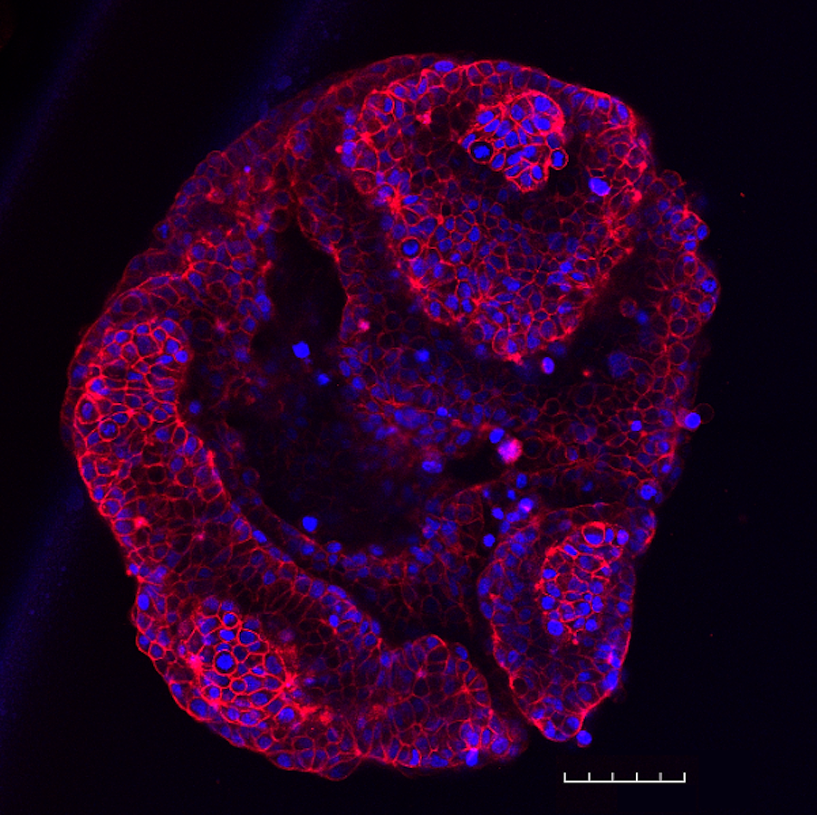
Sharp Cell Membrane Imaging in Spheroids with CellBrite® Steady
CellBrite® Steady Membrane Staining Kits are unique membrane dyes that distribute between the cell surface and intracellular compartments, allowing stable cell surface staining for long-term imaging in live cells. CellBrite® Steady Dyes are validated for imaging in both organoid and spheroid cultures.
- Validated for use in spheroids and organoids
- Retained on cell surface, unlike other membrane dyes
- Optional Enhancer masks intracellular signal
- 9 color options from blue to near-IR
- Rapid, even staining in complete cell culture medium
View Product Page
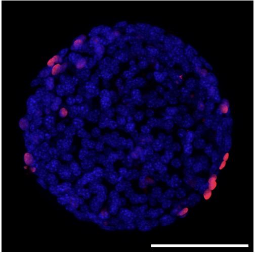
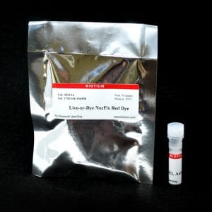
Viability Assessment of Spheroids with NucFix™ Red
Live-or-Dye NucFix™ Red is a reactive, cell membrane impermeant dye that specifically stains the nuclei of dead cells. The dye is able to enter into dead cells that have compromised membrane integrity and covalently label the cell nucleus, allowing for clear differentiation of live and dead cells. Live-or-Dye NucFix™ Red has been validated for fixable viability staining in spheroids, but staining in 3D cultures needs to be validated and optimized empirically for each cell type.
- Validated as a nuclear-specific dead cell stain in spheroids
- Highly dead-cell specific
- Covalent labeling with reactive dye
- Withstands fixation and permeabilization
View Product Page
Read Peer-Reviewed Studies Validating Biotium Stains for 3D Cultures

Literature Digest
Literature Digest: Validated Stains for Spheroids, Organoids, and Other 3D Cultures
Unlock Deeper Insights with MiniMab™ SdAbs
MiniMab™ single-domain antibodies (SdAbs) from Biotium are engineered for optimal performance in immunofluorescence and other applications. At ~15 kDa, they are significantly smaller than conventional IgG antibodies, enabling deeper tissue penetration, faster staining, and better access to masked epitopes. Combined with availability in bright near-IR CF® Dyes, MiniMab™ SdAbs are ideal for thick tissue sections, whole mount tissue, organoids, and other 3D cultures.
The utility of MiniMab™ SdAbs in whole tissue was validated with CF®568 GFAP rVHH (SdAb2409.GFAP) staining in whole mouse brain on the SmartSPIM light sheet microscope from LifeCanvas Technologies.
Features of MiniMab™ single-domain antibodies
- Deeper tissue penetration and better access to masked epitopes than IgG antibodies, ideal for 3D cultures
- Improved solubility and faster staining
- Developed and optimized for immunofluorescence
- Labeled with bright, photostable CF® Dyes, including near-infrared CF®740
A whole mouse brain was fixed in paraformaldehyde (PFA) and processed using the standard SHIELD protocol from LifeCanvas Technologies. The brain was then delipidated and immunolabeled with CF®568 GFAP rVHH (SdAb2409.GFAP) MiniMab™. Imaging was performed on a SmartSPIM light-sheet microscope using a 3.6x objective. Video courtesy of LifeCanvas Technologies.
MiniMab™ SdAb Catalog
Stains Validated in 3D Culture
| Product | Target | Fixable? | Tolerates optical clearing? | Applications / Notes |
|---|---|---|---|---|
| Hoechst 33342 Hoechst 33258 | All cell nuclei | Yes | Yes | • Commonly used nuclear stain |
| NucSpot® Live Nuclear Stains | All cell nuclei | Yes | ND | • Validated in-house and publications for staining spheroids • Validated by publications and collaborators for live imaging of organoids |
| Live-or-Dye NucFix™ Red | Dead cell nuclei | Yes | Yes | • Validated in publications for staining spheroids |
| CellBrite® Cytoplasmic Membrane Dyes | Cell membranes | Yes1 | No | • Lipophilic carbocyanine dyes have been used for labeling cells before or after spheroid formation |
| CellBrite® Steady Membrane Staining Kits | Cell membranes | No | ND | • Validated in-house for staining spheroids • Validated in publications for staining organoids |
| MitoView™ Dyes | Mitochondria | No | No | • MitoView™ Green validated for staining organoids |
| NucView® 488 Caspase-3 Substrate | Apoptotic cells | Yes | Yes | • Validated in publications for staining spheroids and organoids |
| Calcein-AM | Viable cells (whole cell interior) | No | No | • Commonly used to stain cells in Matrigel® or spheroids |
| Cell-Membrane Impermeant Nucleic Acid Dyes | Necrotic cells2 | No | No | • Wide selection of dead-cell specific stains that could be used for 3D cultures |
| ViaFluor® SE Cell Proliferation Dyes | Whole cell interior | Yes | Yes | • Validated in publications for staining spheroids • Can be used to label cells before seeding in Matrigel® or spheroids |
| LipidSpot™ Lipid Droplet Stains | Lipid droplets | Yes1 | ND | • LipidSpot™ 488 has been validated by publications for staining spheroids |
| LysoView™ Dyes | Lysosomes | No | ND | • May be suitable for staining spheroids, 3D cultures, or Matrigel® cultures, but have not yet been validated |
2 Necrotic cell stains also stain late apoptotic cells with leaky membranes; staining is not dead cell-selective in fixed samples.
Matrigel is a registered trademark of Corning, Inc.