New Products
 Earth-Friendly Products
 Biotium Choice Antibodies
 Special Offers

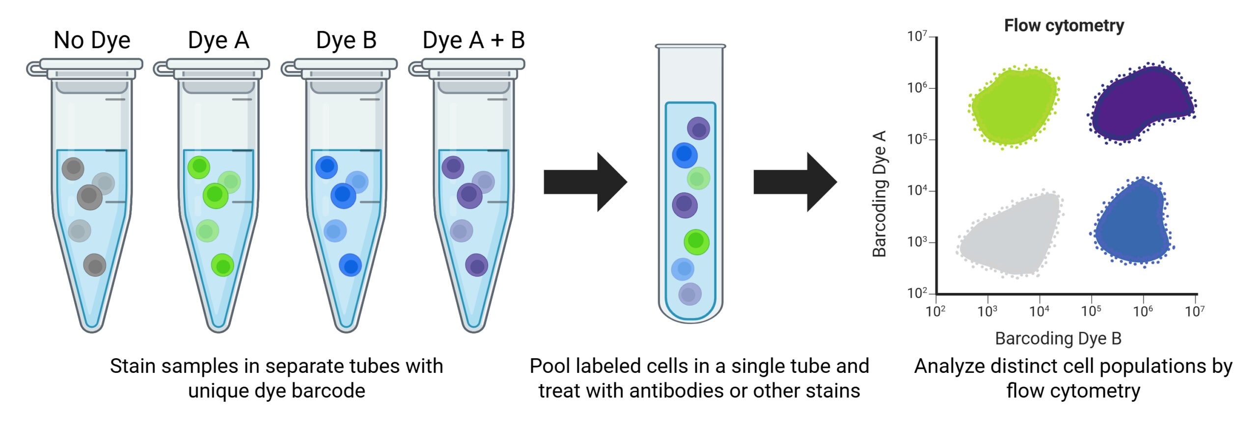
What is Fluorescent Cell Barcoding for Flow Cytometry?
Fluorescent cell barcoding (FCB) is a multiplexing technique that labels cell samples with unique fluorescent signatures, enabling them to be pooled and digitally deconvoluted in downstream analysis. Pooled samples can be treated collectively in a single tube with antibodies, viability dyes, or other stains and analyzed in a single run, ensuring uniform labeling across all samples and eliminating inter-sample technical variability. This approach significantly reduces both the time and reagent costs associated with processing large numbers of samples.
Several methods for FCB have been developed for flow cytometry, this includes the use of amine-reactive NHS ester dyes or fluorescent antibody conjugates. NHS ester dyes covalently label cells at varying concentrations, allowing multiple unique barcodes to be generated from a small number of dyes. However, traditional NHS ester dyes are typically only compatible with fixed cells and intracellular staining. Fluorescent antibody conjugates can be used for both live and fixed cell barcoding, but this approach can suffer from non-specific binding, epitope competition, and antibody detachment and re-attachment to neighboring cells.
Advantages of Fluorescent Cell Barcoding
| Traditional Staining | Fluorescent Cell Barcoding |
|---|---|
| Each sample run separately | All samples analyzed in a single run |
| Variability between samples | Technical variability minimized |
| More reagent consumption | Significantly fewer reagents needed |
| Longer acquisition time | Significantly faster data collection |
Flexible Live Cell Barcoding with ViaPlex™
The ViaPlex™ Cell Barcoding Kit enables convenient and flexible cell barcoding of up to 15 cell populations in a single tube, saving time and reagents. Cells are labeled using covalently bound fluorescent dyes with up to three concentrations per dye.
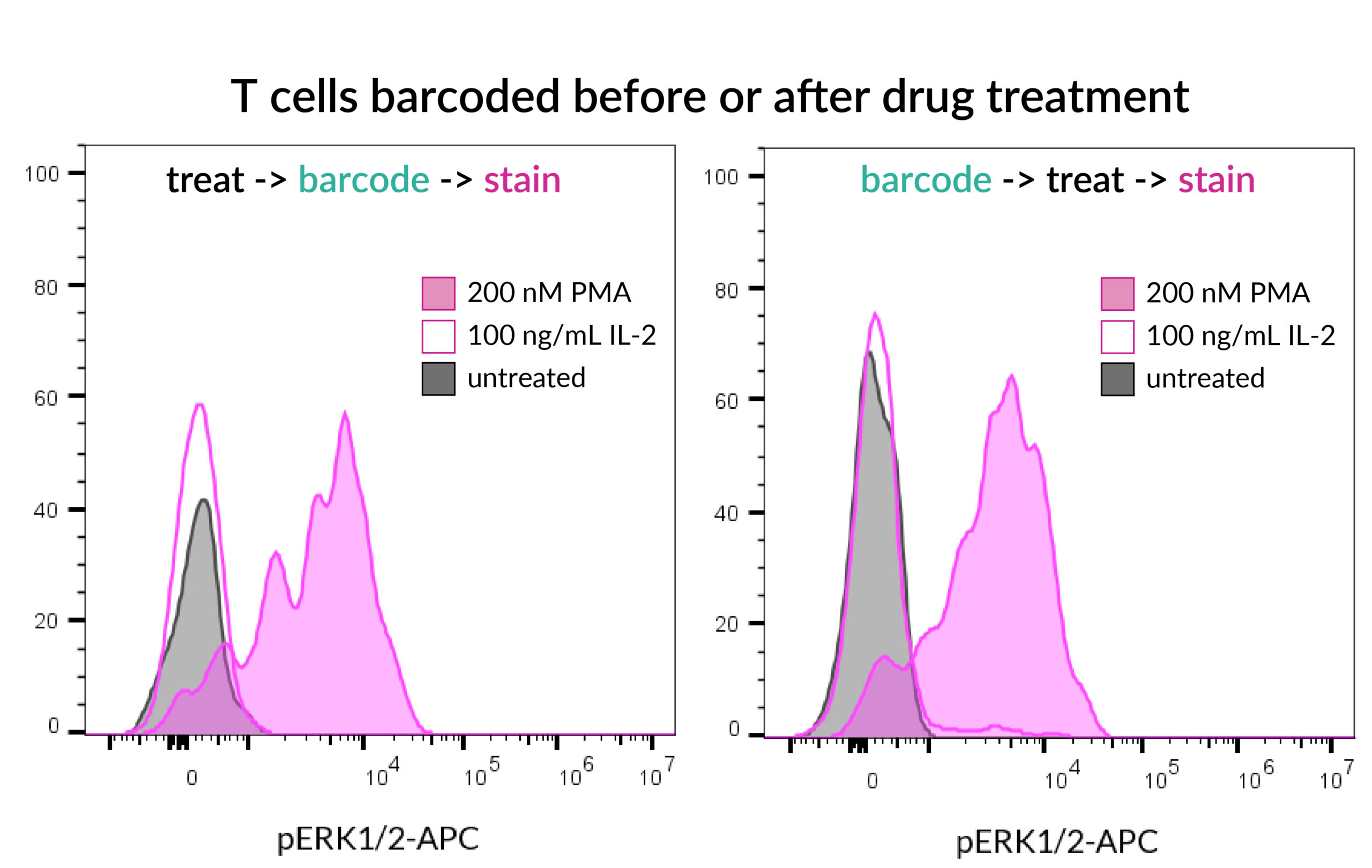
Unlike traditional NHS-ester labeling, such as the BD Phosflow™ Violet Kit, ViaPlex™ barcoding is compatible with surface and intracellular antibody staining workflows, and barcoding can be performed either before or after cell treatment to accommodate a wide range of experimental designs. ViaPlex™ labeling does not require fixation or permeabilization, enabling assays on live cells. However, the covalent dye labeling also allows barcoded cells to be fixed and permeabilized for intracellular detection.
ViaPlex™ Features
- Saves time and reagent cost
- Multiplex up to 15 samples in one staining reaction; optional 16th barcode via compensation
- Compatible with surface and/or intracellular staining
- Works with live cells, fixation optional after barcoding
- Covalent dyes stably label cells for clean sample separation
- Uses Pacific Blue® and FITC channels
Advantages of ViaPlex™ Cell Barcoding
| Traditional NHS-ester barcoding | ViaPlex™ Cell Barcoding |
|---|---|
| Does not work with live cells, requires fixation before barcoding | Works with live cells, fixation optional after barcoding |
| Not suitable for surface staining, intracellular only | Can be used for both surface and intracellular staining |
| Cell treatment must be done before barcoding | Cell treatment can be done before or after barcoding |
View Product Page

ViaPlex™ Barcoding Workflow Overview
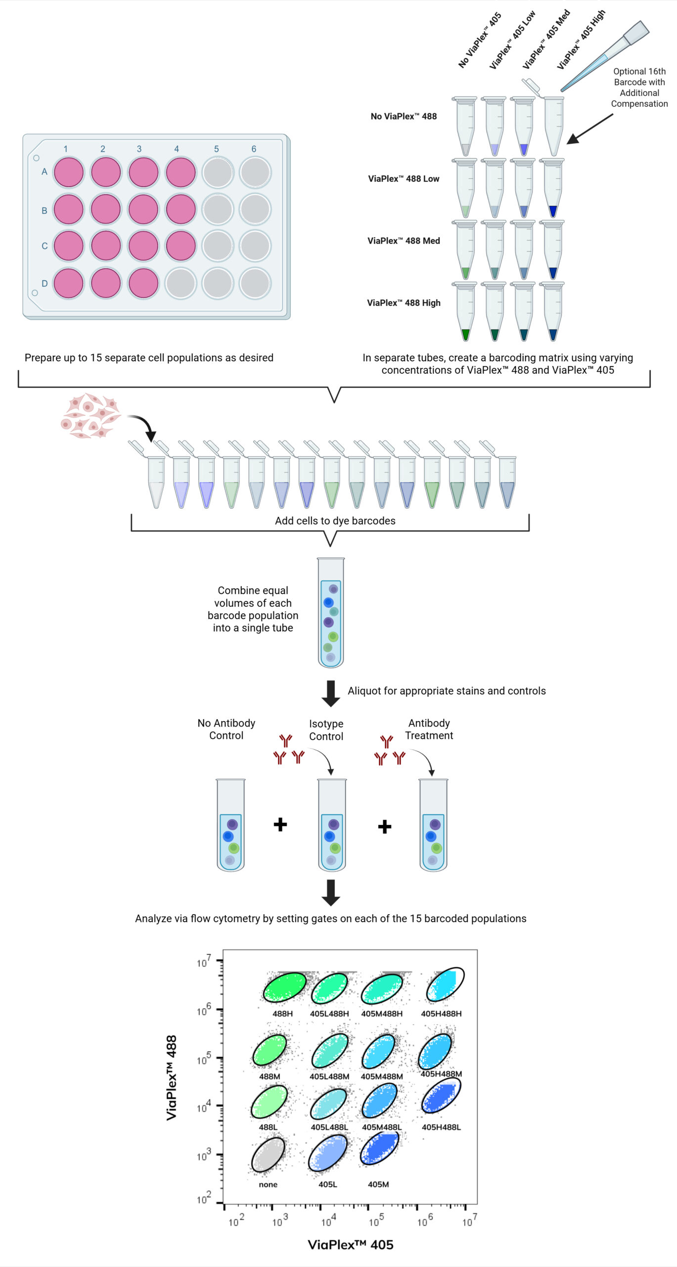
Prepare Cells
Obtain up to 15 distinct cell populations. Cells can be treated as desired (e.g., add any drugs or cytokines for the desired amount of time).
Setup the Barcoding Matrix
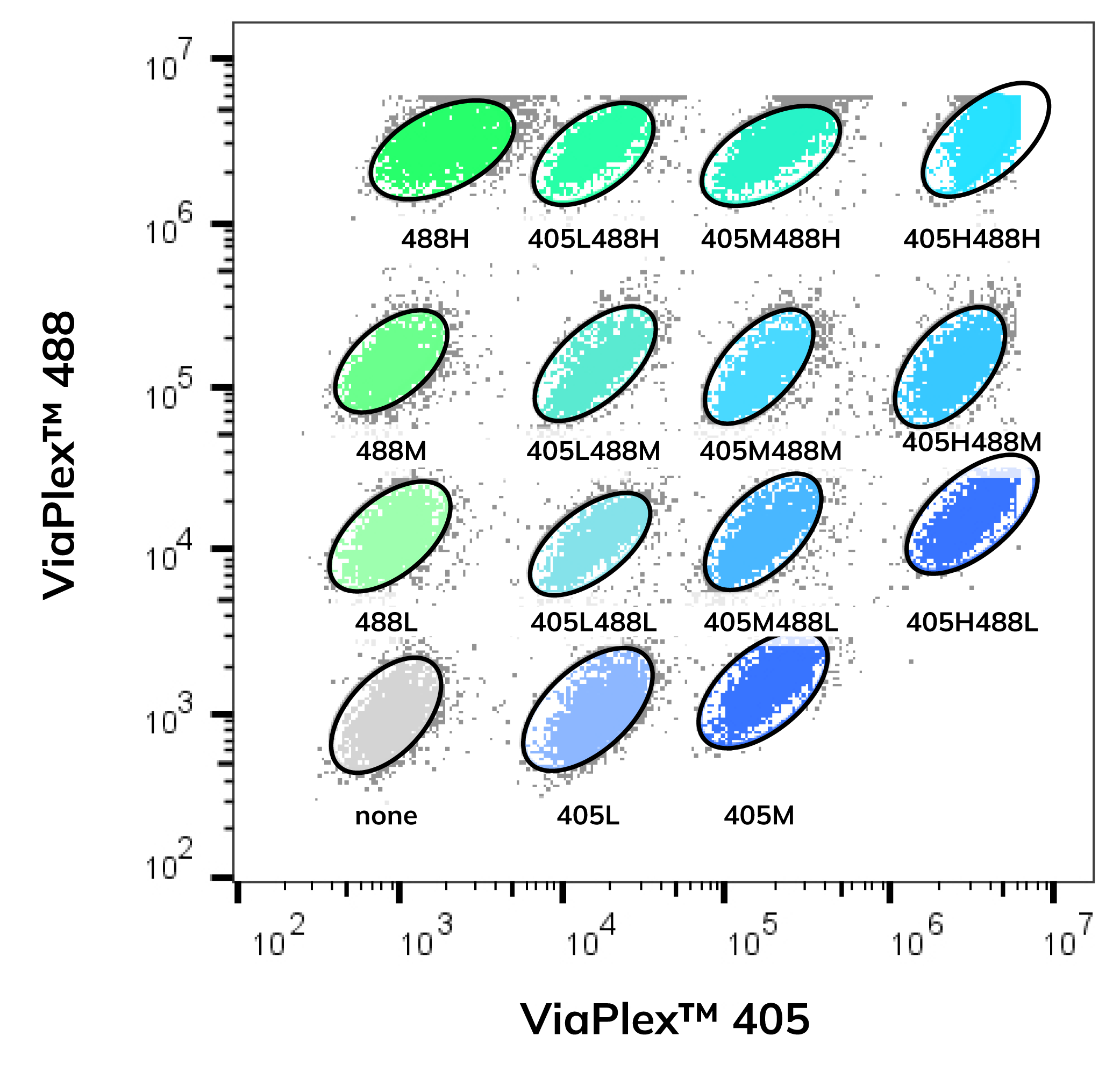
Using just two ViaPlex™ Dyes at three intensity levels (negative, mid, high), you can generate up to 15 unique combinations. An optional 16th barcode can be added but will require compensation during analysis. Each dye combination is then used to stain each individual cell population.
Stain Cells with Dye Barcodes
Stain each cell population with a unique dye barcode prepared from the barcode matrix, then pool samples in a single tube for downstream staining and analysis.
Aliquot Barcoded Cells for Staining
Aliquot the combined barcoded cells into separate tubes for staining your targets of interest and appropriate controls. Use stains that are complementary to ViaPlex™ 405 and ViaPlex™ 488.
Analyze by Flow Cytometry
Analyze the sample by setting gates on each of the 15 individual barcoded populations. An optional 16th barcode can be added if compensation is performed. Detect your antibodies or other stains of interest in the appropriate channels.
Overview of ViaPlex™ Barcoding Workflow
Phosflow is a trademark of BD Biosciences.