 New Products
 Earth-Friendly Products
 Biotium Choice Antibodies
 Special Offers


Content #1
Content #1
Content #1
Targeted activation of immune checkpoints is an emerging strategy for treating Type 1 Diabetes (T1D), where autoreactive T cells destroy insulin-producing beta cells. Autoimmune diabetes has been linked to disruption of the PD-1/PD-L1 pathway, which normally suppresses T cell activity to maintain immune tolerance. To address the limitations of systemic immunosuppression, an immune modulating monoclonal TCR against autoimmunity (ImmTAAI) molecule was created to selectively activate PD-1 signaling at the beta cell surface. This is a bispecific molecule that combines a T cell receptor targeting a preproinsulin peptide presented by HLA-A2 with a PD-1 agonist domain, enabling localized suppression of autoreactive T cells.
In a 2026 Science Advances publication, Becker et al. evaluated ImmTAAI function in live human pancreas tissue slices through confocal imaging, binding assays, and functional coculture systems using engineered T cell “avatars” designed to mimic the behavior of autoreactive T cells in T1D. Biotium’s CF®647 succinimidyl ester dye was used to label and visualize ImmTAAI molecules in live and fixed human pancreas slices. This labeling enabled precise tracking of ImmTAAI localization and quantification of its binding to beta cells via confocal microscopy without affecting binding affinity, specificity, or functional potency of ImmTAAI.
The researchers found that ImmTAAI binds specifically and dose-dependently to beta cells in a human leukocyte antigen (HLA)-dependent manner, with increased binding under inflammatory conditions. ImmTAAI treatment increased T cell movement, reduced T cell–beta cell interactions, and suppressed cytotoxic activity of target beta cells. Furthermore, ImmTAAI conferred protection of beta cells from immune attack in cell culture and in tissue slices, and helped preserve insulin secretion in live pancreas slices from a donor recently diagnosed with type 1 diabetes.
CF®647 enabled colocalization studies in a PD/PD-L1 therapeutic model and confirmed selective targeting within complex tissue environments. These findings highlight the value of bright, photostable far-red dyes like CF®647 for imaging-driven validation of targeted immunotherapies.
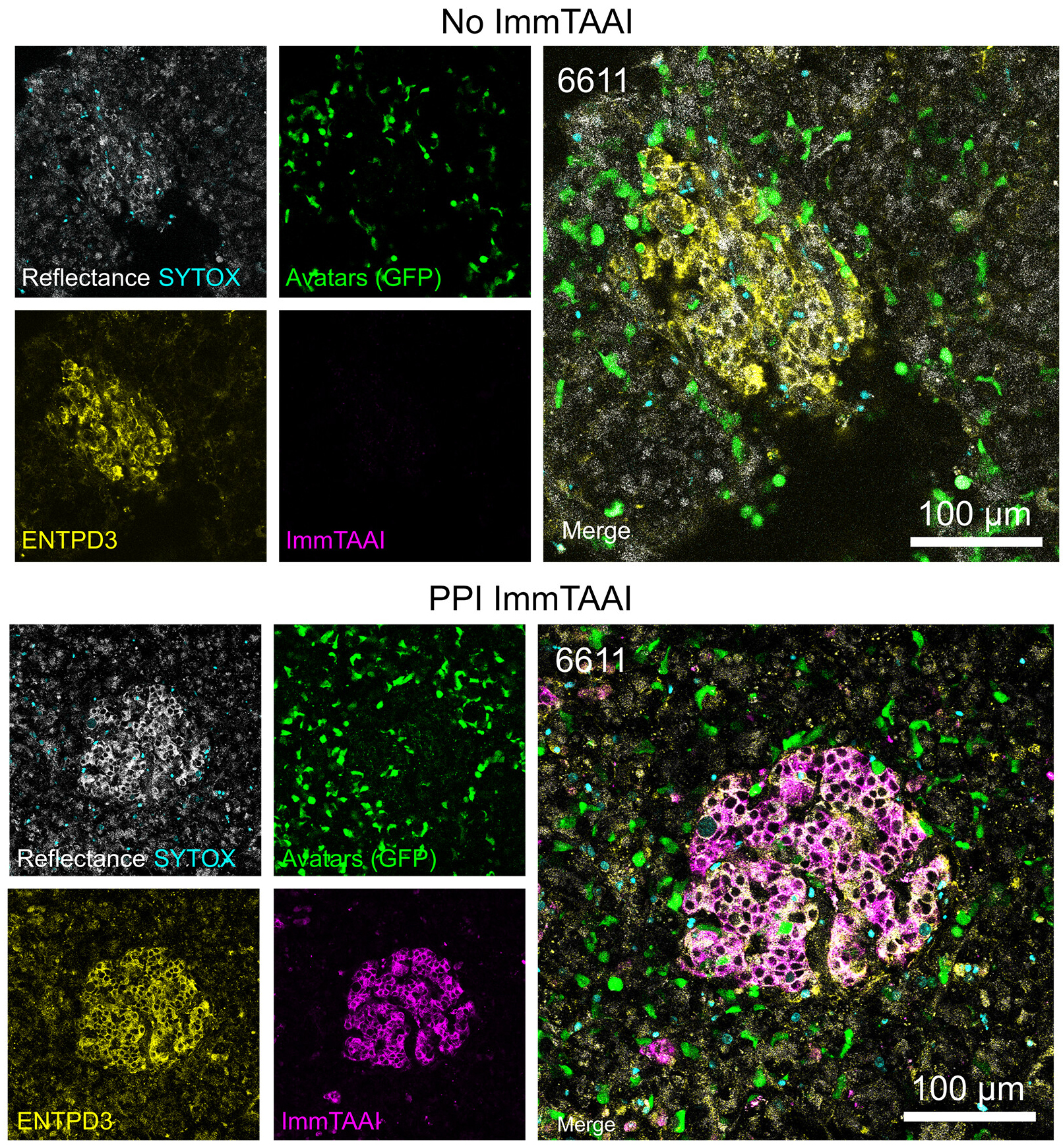
Live-cell confocal imaging (18 h) of pancreas slices treated with or without PPI ImmTAAI after addition of 200,000 IGRP-specific T cells per slice shows CF®647-labeled ImmTAAI colocalization with ENTPD3. Adapted from Becker et. al. Reproduced under CC BY 4.0.
Learn more about Biotium’s next-generation CF® Dye probes featuring exceptional brightness, photostability, and signal-to-noise, available in over 40 colors from blue to near-IR. CF® Dyes are also available in convenient Mix-n-Stain™ Labeling Kits for quick and efficient antibody labeling.
Full Citation
Matthew W. Becker et al. Beta cell–targeted PD-1 agonist inhibits cell-mediated autoimmunity in pancreas tissue slices. Sci. Adv. 12, eaec9029(2026). DOI:10.1126/sciadv.aec9029
