 New Products
 Earth-Friendly Products
 Biotium Choice Antibodies
 Special Offers


Content #1
Content #1
Content #1
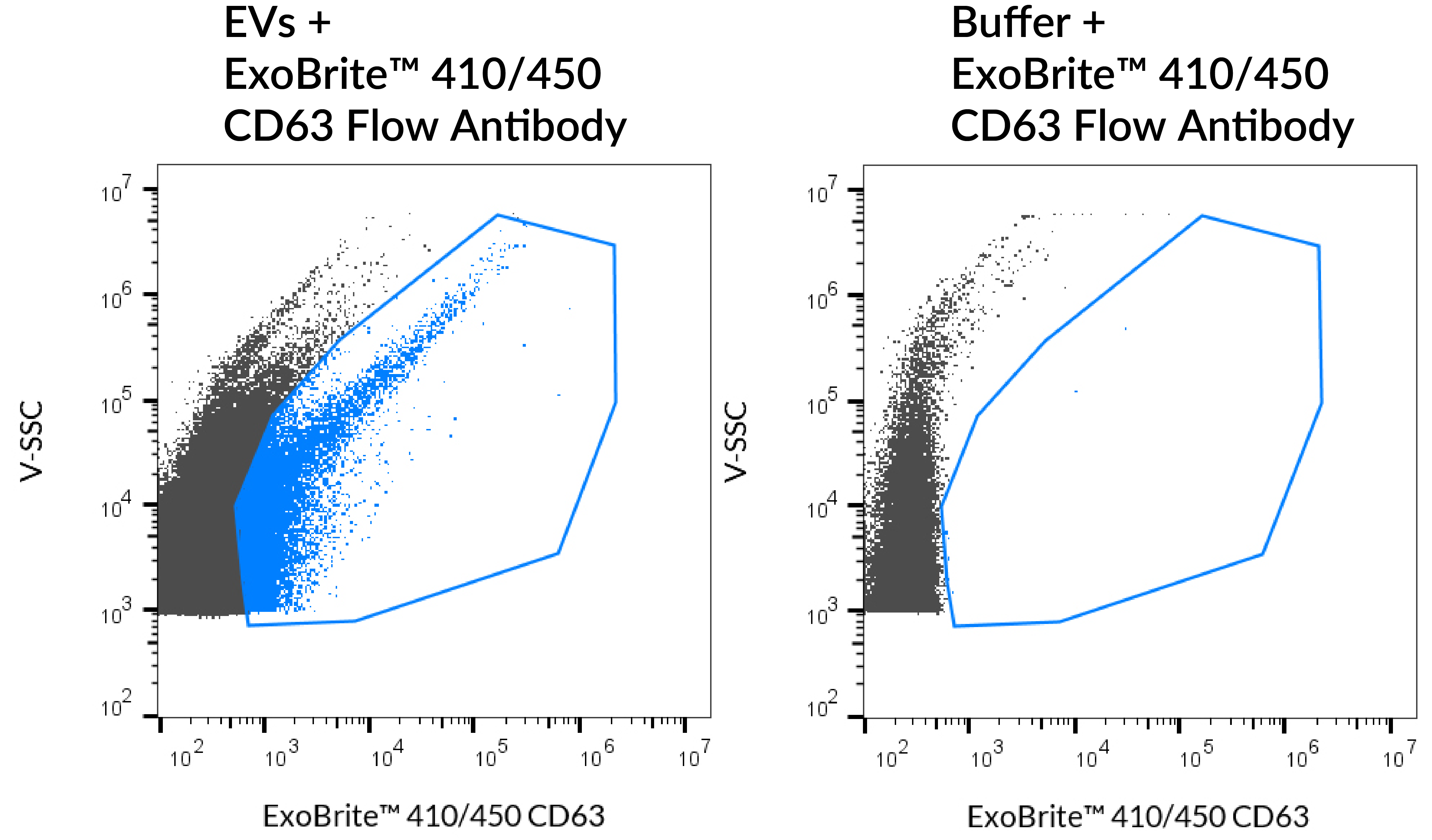
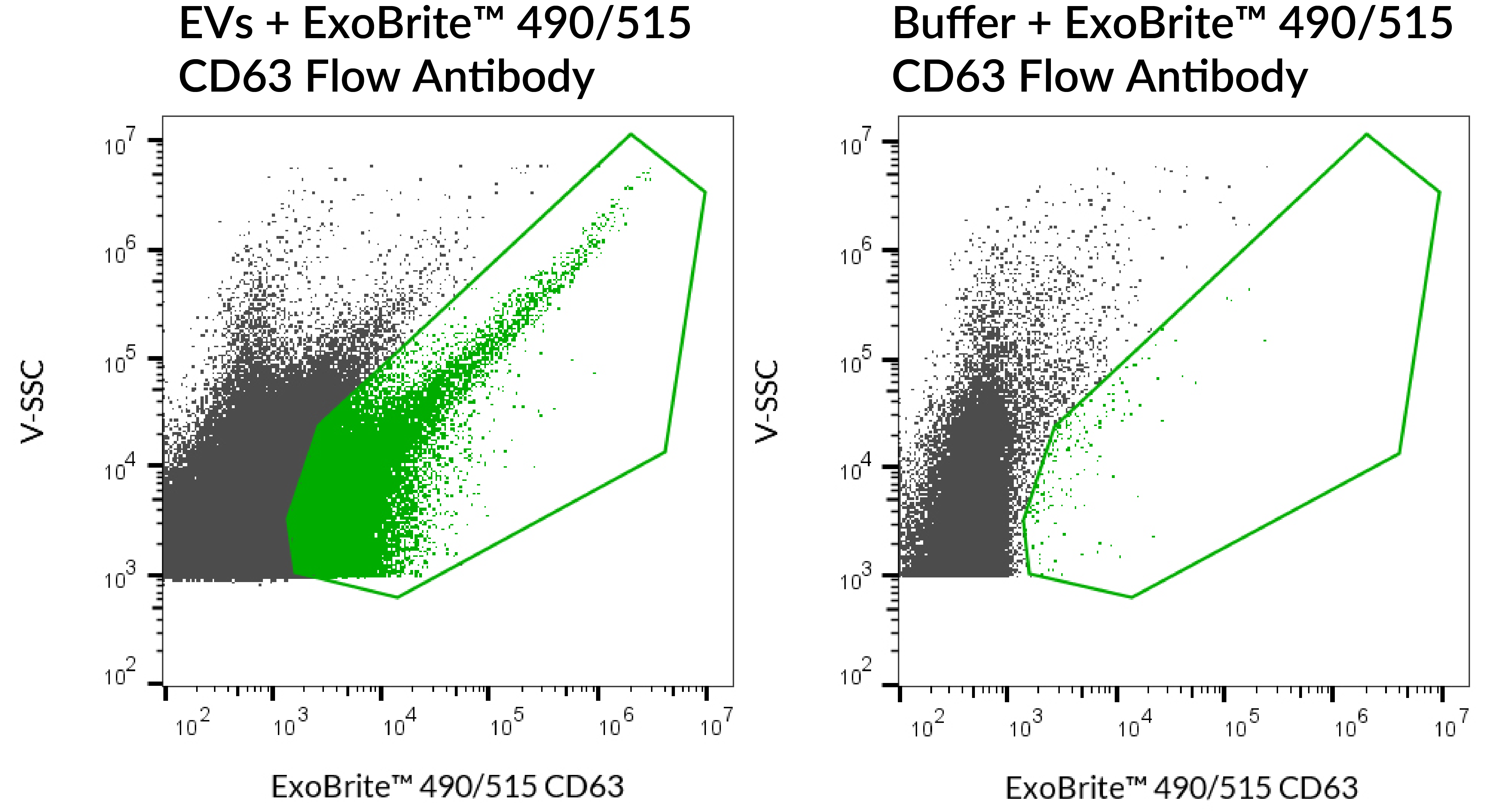
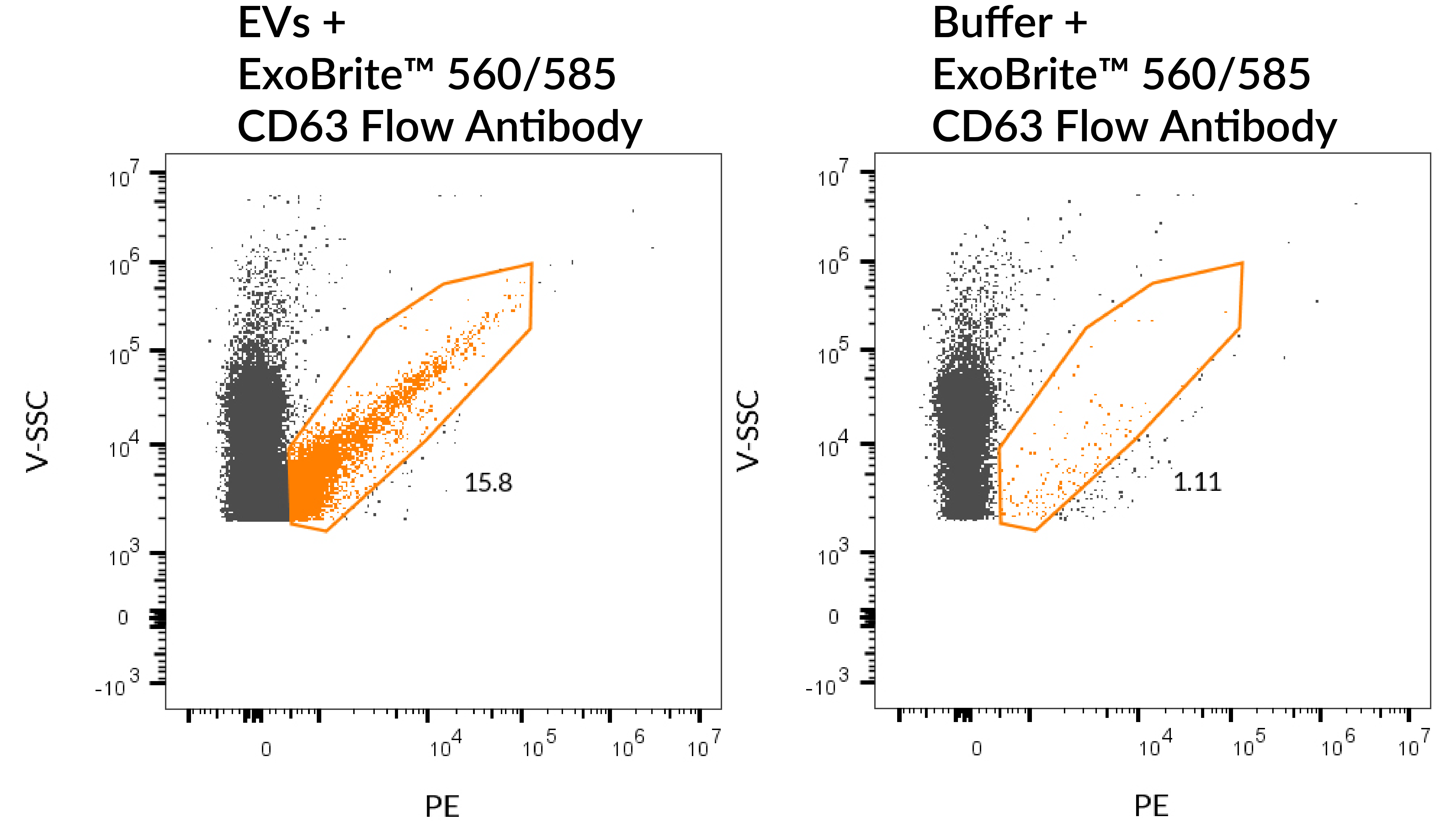
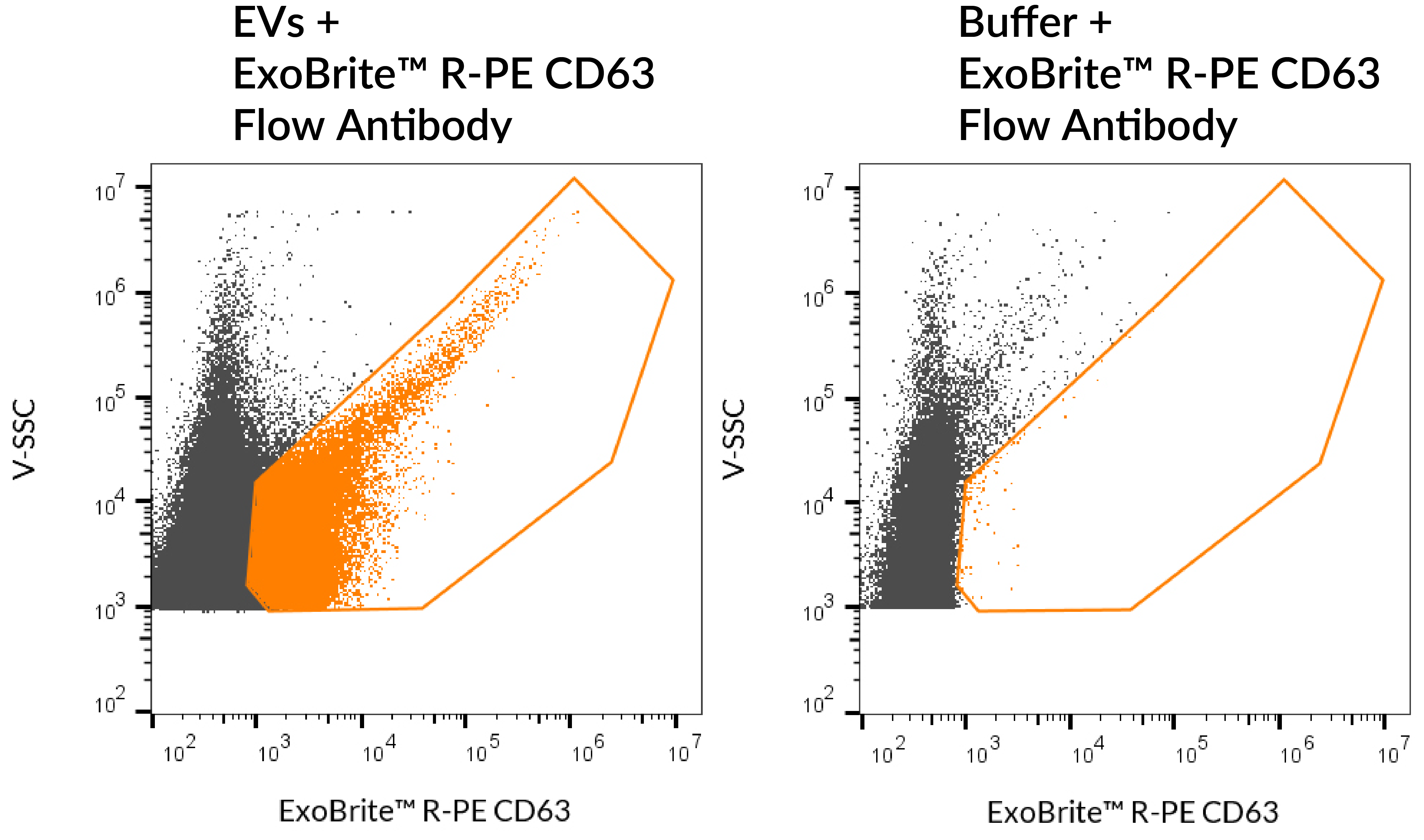
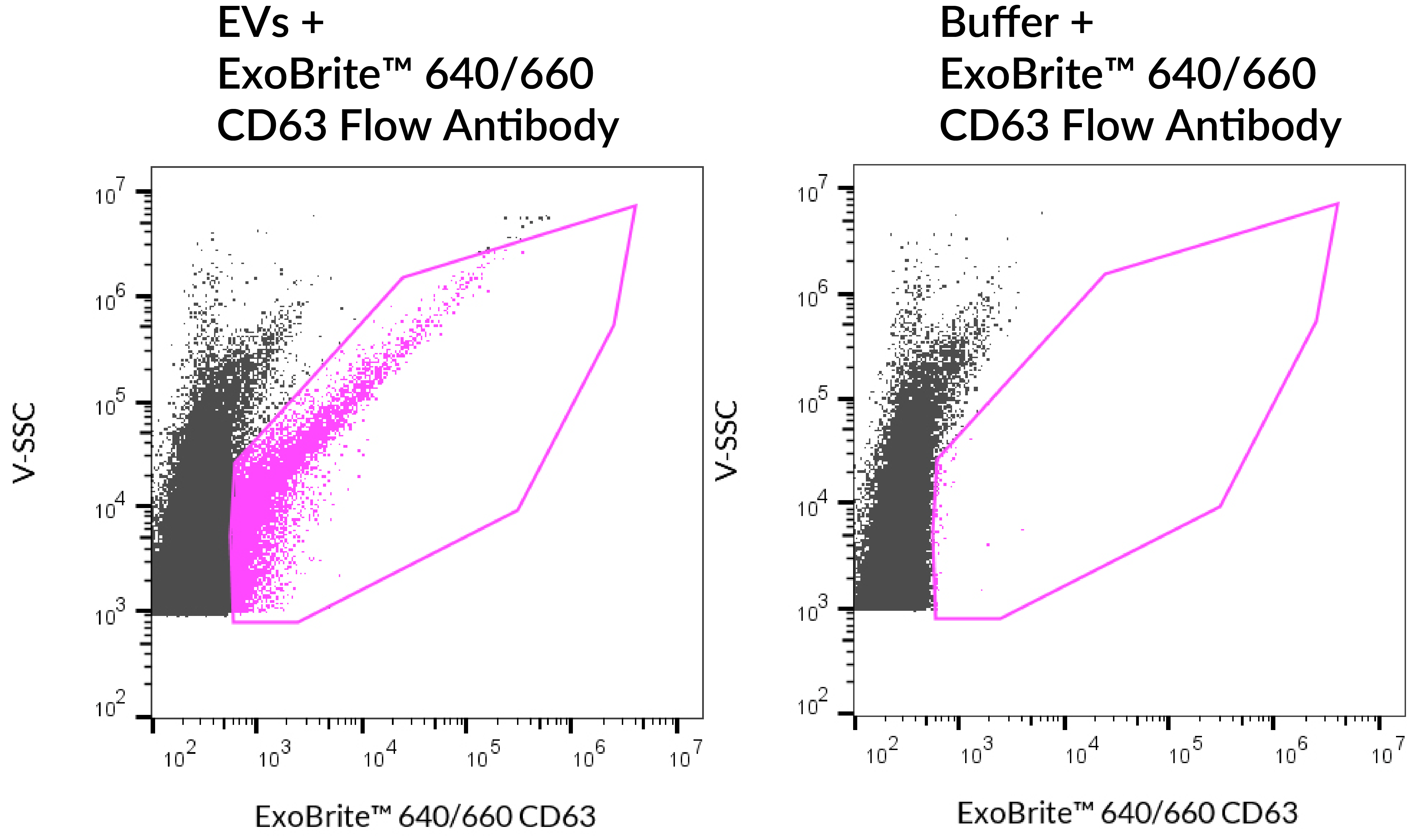
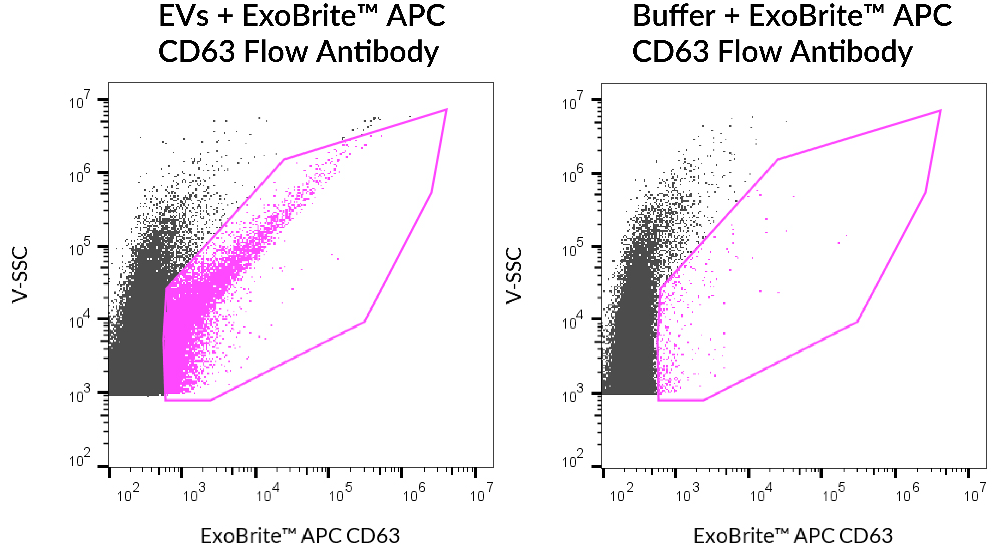
Validated antibody for optimal detection of EV marker CD63 in purified or bead-bound EVs by flow cytometry.
ExoBrite™ CD63 Flow Antibody is validated by Biotium for optimal detection of EV marker CD63 in purified or bead-bound EVs by flow cytometry. The antibody conjugates offer exceptional signal-to-noise and include color options for the Pacific Blue™, FITC, PE, and APC channels.
Biotium also offers validated ExoBrite™ Flow Antibodies for tetraspanin proteins CD9, CD81, and other EV markers.
EV antibodies you can trust
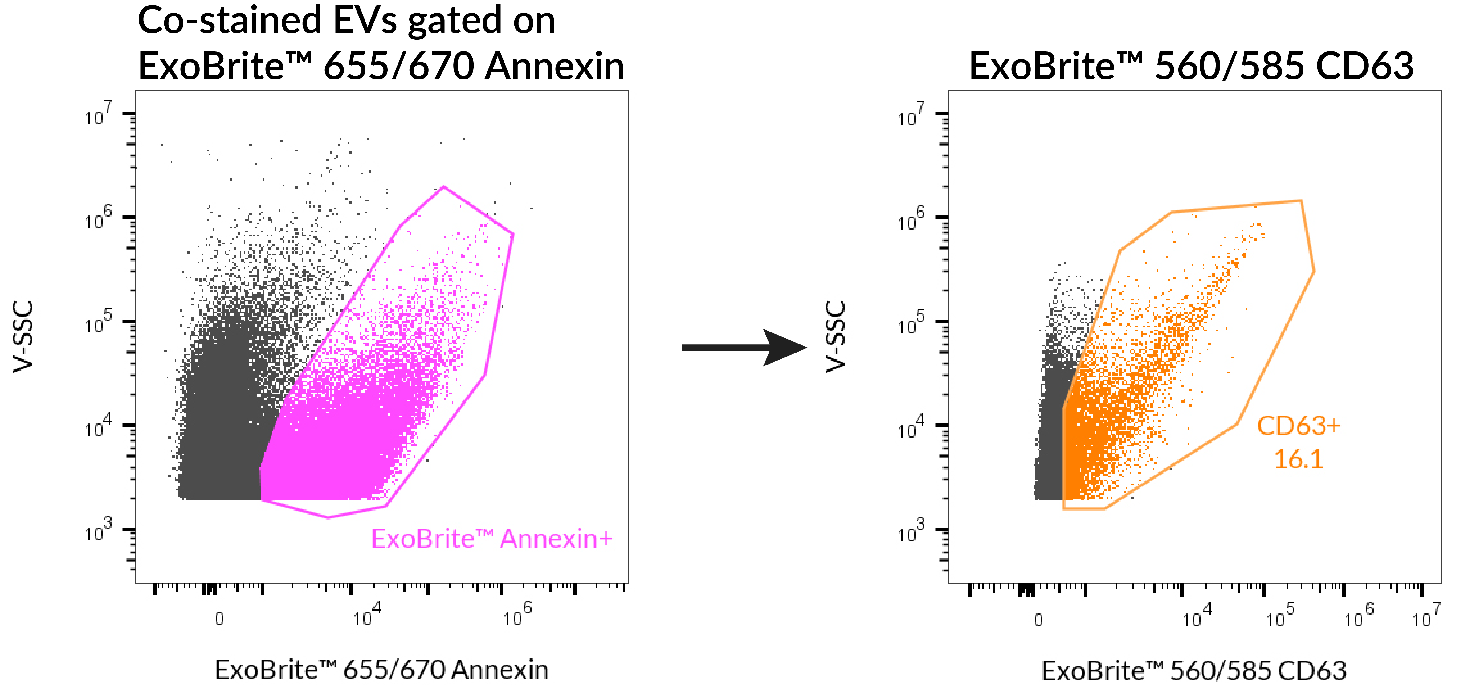
Other commercially available antibodies against mouse and human tetraspanin proteins CD9, CD63, and CD81 are generally not validated for isolated EVs and may require tedious optimization for your EV prep and staining protocol. The antibodies and dye labels of ExoBrite™ Flow Antibody Conjugates were carefully selected and validated for robust detection of isolated EVs. In addition, the antibodies are provided in a proprietary buffer formulation for reduced antibody aggregation and brighter EV staining for optimal accuracy and signal-to-noise. Single-color and 3-color ExoBrite™ CD9/CD63/CD81 Antibody Cocktails for human tetraspanin proteins are also available for high-coverage EV staining or EV phenotyping, respectively.
For general EV staining, Biotium's ExoBrite™ True EV Membrane Stains offer unparalleled coverage of EVs in a sample and address issues of dye aggregation often seen with PKH and other common membrane dyes. Biotium also offers optimized ExoBrite™ EV Surface stains conjugated to cholera toxin B (CTB), wheat germ agglutinin (WGA), and Annexin V. These stains are specially formulated for bright and specific detection of isolated EVs by flow cytometry. These ExoBrite™ EV Surface stains may also be combined with antibody staining, for multi-parameter analysis.
Biotium offers conjugated ExoBrite™ Western Antibodies against human CD9, CD63, and CD81 designed for optimal detection in EV extracts by fluorescent western blot.
Note: In our testing, we have found that ExoBrite™ 490/515 dye may bind to streptavidin coated surfaces or beads if free biotin binding sites are not blocked. We recommend performing a biotin blocking step after binding your biotinylated capture antibody to streptavidin beads or surfaces when using ExoBrite™ 490/515 conjugates. Alternatively, consider using a different ExoBrite™ dye for staining EVs captured on streptavidin beads or surfaces.
| Cocktail | Ex/Em | Detection Channel | Size | Catalog No. |
|---|---|---|---|---|
| ExoBrite™ CD9/CD63/CD81 Single-Color Antibody Cocktail (Green) | 490/516 nm | FITC | 25 tests | P030-125 |
| 100 tests | P030-500 | |||
| ExoBrite™ CD9/CD63/CD81 Single-Color Antibody Cocktail (Orange) | 562/584 nm | PE | 25 tests | P031-125 |
| 100 tests | P031-500 | |||
| ExoBrite™ CD9/CD63/CD81 Single-Color Antibody Cocktail (Far-Red) | 642/663 nm | APC | 25 tests | P032-125 |
| 100 tests | P032-500 | |||
| ExoBrite™ CD9/CD63/CD81 3-Color Antibody Cocktail (Blue/Green/Far-Red) | 411/452 nm 490/516 nm 642/663 nm | Pacific Blue® FITC APC | 25 tests | P028-125 |
| 100 tests | P028-500 | |||
| ExoBrite™ CD9/CD63/CD81 3-Color Antibody Cocktail (Green/Orange/Far-Red) | 490/516 nm 562/584 nm 642/663 nm | FITC PE APC | 25 tests | P029-125 |
| 100 tests | P029-500 |
Extracellular vesicles (EVs) derived from mesenchymal stem cells (MSCs) are emerging as powerful, cell-free immunomodulatory therapies for inflammatory diseases such as COVID-19. However, because the mechanism is poorly understood, optimizing EV-based therapies remains challenging.
In a 2025 Springer Nature study, Infante et al. investigated how COVID-19 patient serum reshapes the transcriptome and paracrine activity of Wharton’s jelly–derived MSC stem cells (WJ-MSCs). WJ-MCSs exposed to serum from hospitalized COVID patients showed downregulation of NEAT1 and MALAT1, two pro-inflammatory two long noncoding RNAs (lncRNAs). Furthermore, the researchers found that EVs derived from the treated cells had enhanced immunosuppressive activity when administered to T-cells.
The researchers isolated EVs from WJ-MSC cells after NEAT1 and/or MALAT1 knockdown, and tested whether there was an effect on T-cell proliferation. A Western blot of EVs derived from control and lncRNA-knockdown MSCs were probed with ExoBrite™ 680/700 CD81 Western Antibody. ExoBrite™ 770/800 Calnexin Western Antibody was also used as an endoplasmic reticulum marker to assess cellular contamination.
EV enriched samples in control, NEAT1 knockdown, MALAT1 knockdown, and NEAT1/MALAT1-double knockdown were confirmed by bright CD81 detection and the absence of Calnexin. They found that the MALAT1 knockdown EVs were found to have an inhibitory effect on T-cell proliferation. These results illustrate the importance of EV characterization using tools like Biotium’s ExoBrite™ antibodies in translational EV research.
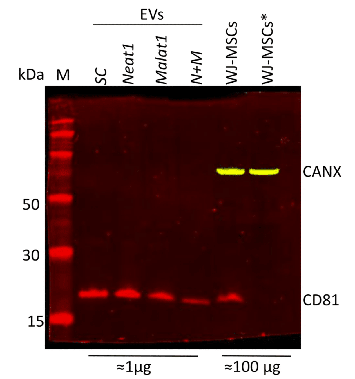
Isolation and characterization of EVs from various lncRNA knock-down WJ-MSCs. Western blot analysis using ExoBrite™ 680/700 CD81 and ExoBrite™ 770/800 Calnexin in EV and MSC lysates. Asterisk (*) indicates reduced conditions used in the MSCs lysate. Modified from Infante et. al. Reproduced under CC BY 4.0.
Learn more about Biotium’s many stains and antibodies for EV research, including ExoBrite™ CD9/CD63/CD81 Antibody Cocktails for flexible and bright multiplexing detection by flow cytometry. Biotium also offers ExoBrite™ stains for pan-EV labeling, optimized fluorescent conjugates of CTB, WGA, and Annexin V for EV detection, ExoBrite™ antibodies for STORM imaging, and more.
Full Citation:
Infante, A., Cabodevilla, L., Gener, B. et al. Modulation of NEAT1 and MALAT1 expression in WJ-MSCs by Covid-19 serum: a foundation for EVs-mediated therapy. Respir Res 26, 313 (2025). https://doi.org/10.1186/s12931-025-03394-4
While early studies of EVs attempted to use first-generation membrane dyes like DiI or PKH to stain EVs, more recently this class of dyes has been found to be largely unsuitable for EV staining due to their high degree of aggregation. Dye aggregation not only generates nonspecific particles that are indistinguishable from EVs in flow cytometry, but also results in poor EV labeling efficiency. Biotium developed the ExoBrite™ True EV Membrane Stains in response to our customers difficulties with using traditional membrane dyes to stain EVs. See our Literature Digest for more information.
We strongly recommend our ExoBrite™ Flow Antibody Conjugates for staining both purified or bead-bound EVs. The antibodies are validated and optimized to offer bright signal and low background. They are available against human or mouse CD9, CD63, and CD81 tetraspanin proteins.
