 New Products
 Earth-Friendly Products
 Biotium Choice Antibodies
 Special Offers


Content #1
Content #1
Content #1
A unique additive that can be added to extracellular vesicle (EV) stain reactions to improve the staining specificity for applications like flow cytometry.
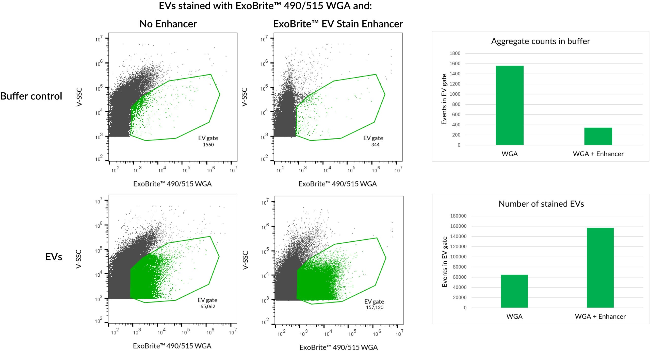
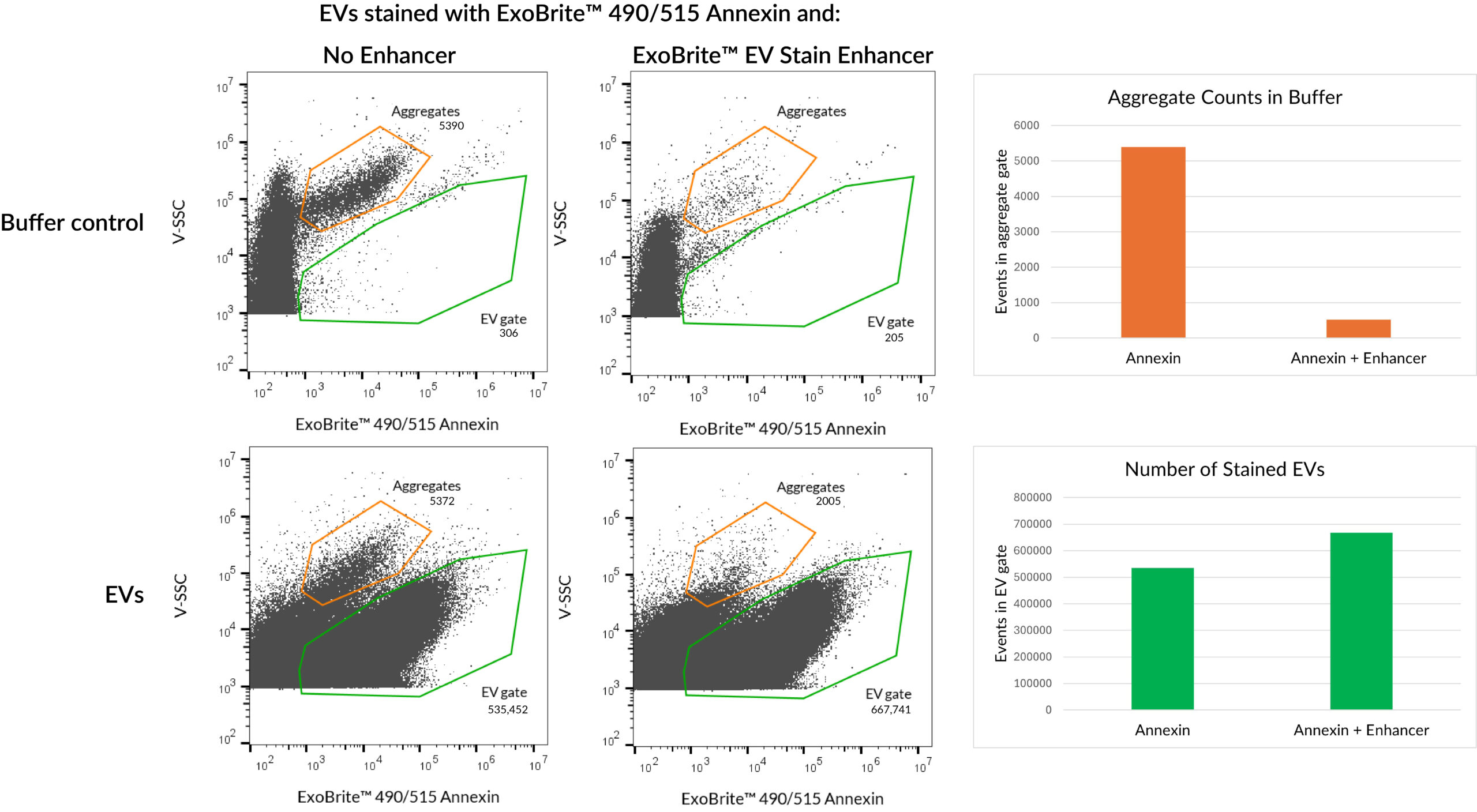
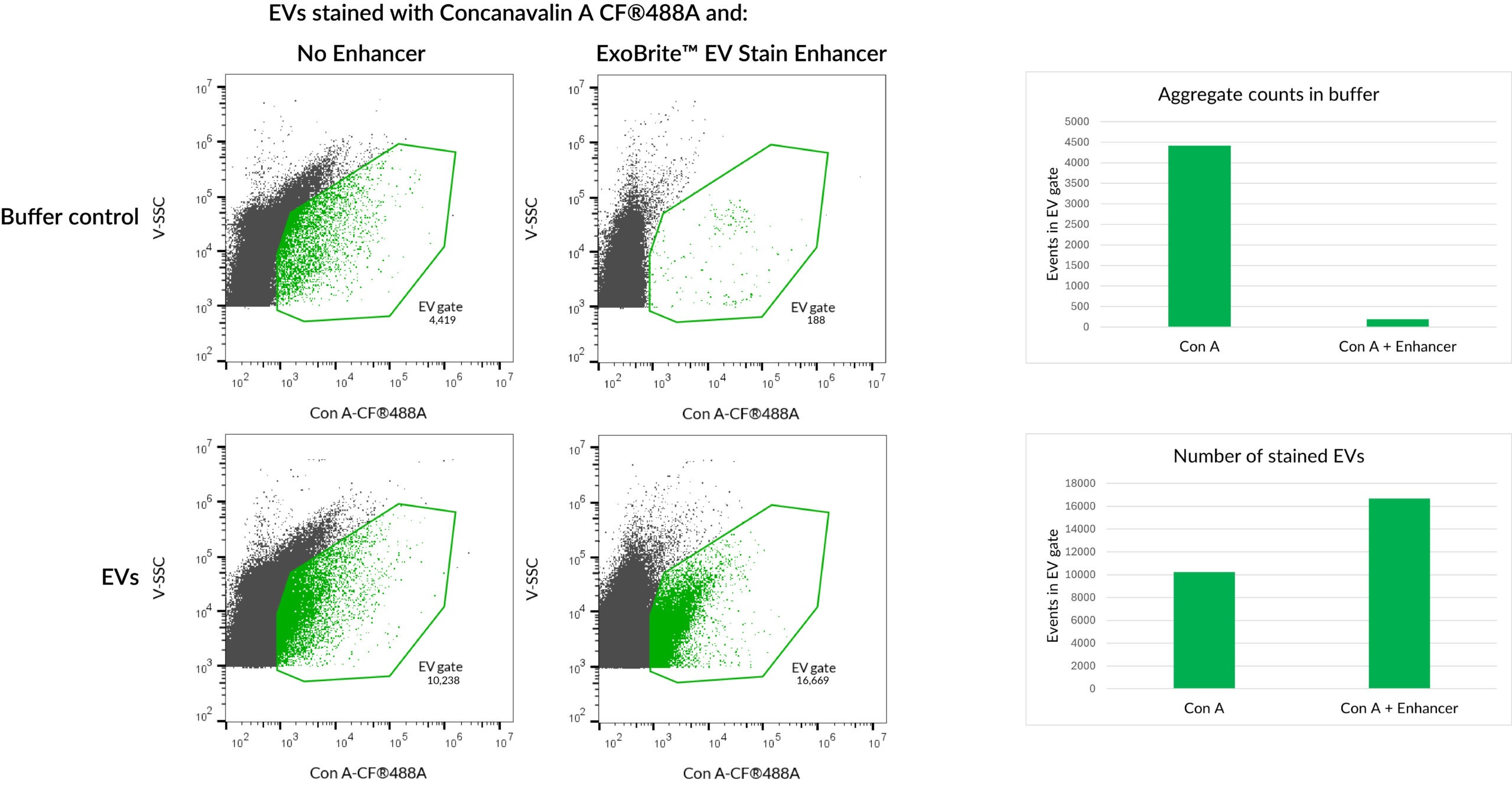
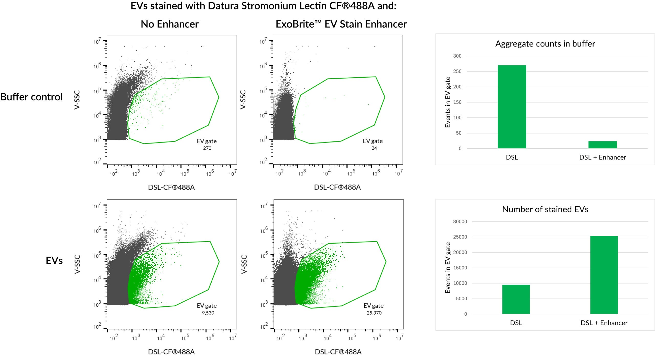
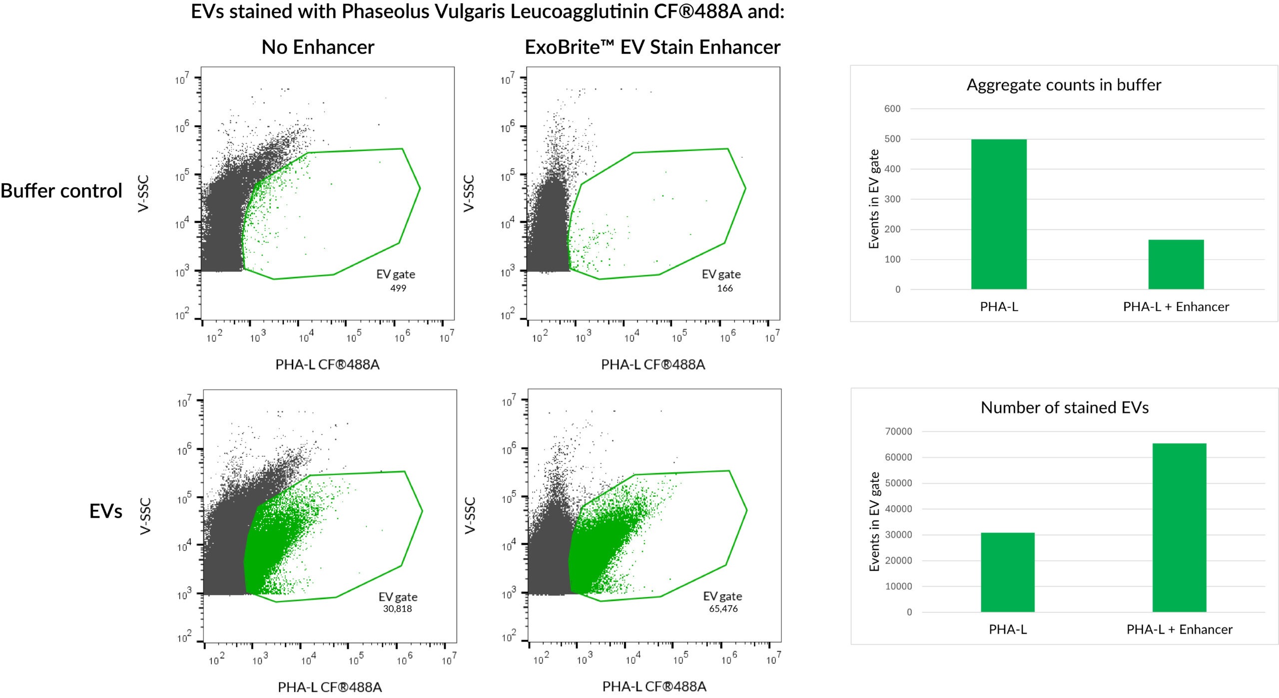
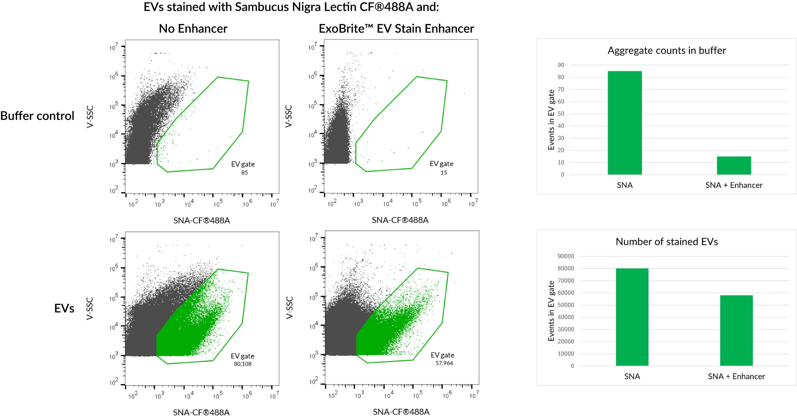
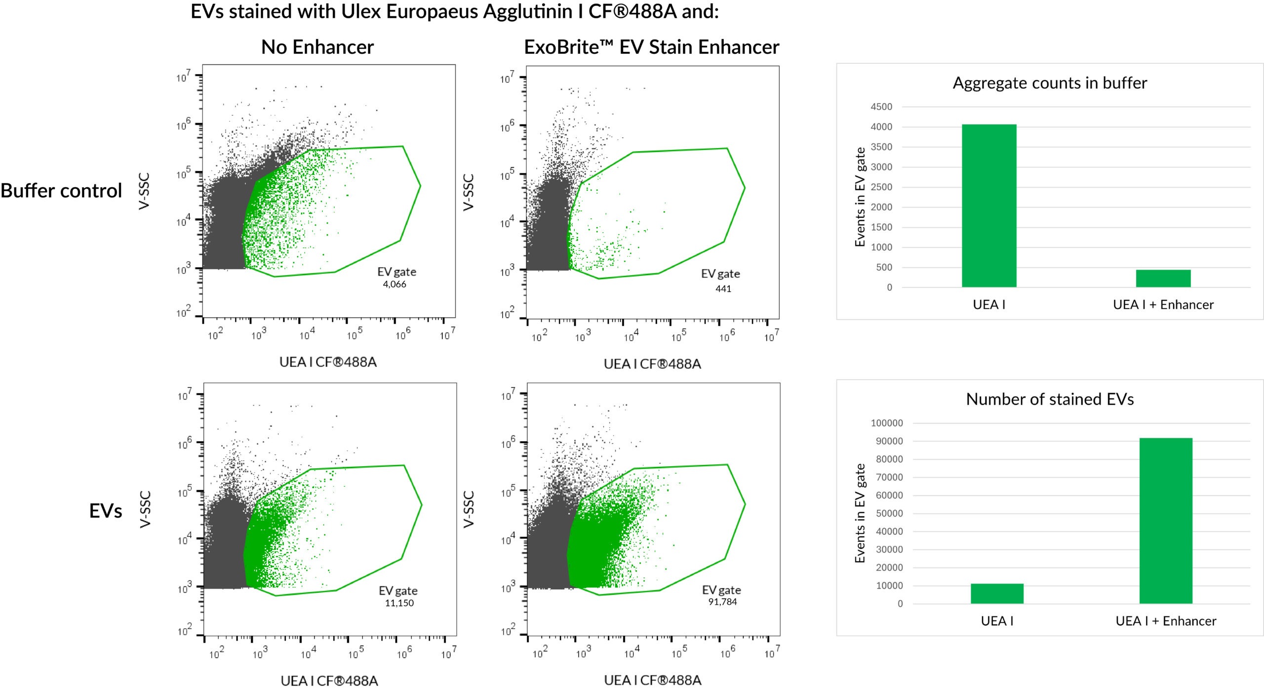
The ExoBrite™ EV Stain Enhancer is a unique additive that can be added to extracellular vesicle (EV) stain reactions to improve the staining specificity for applications like flow cytometry. The ExoBrite™ Stain Enhancer works by reducing the aggregation of certain EV stains, which allows the conjugate to stain the EVs more efficiently, resulting in a better signal-to-noise ratio and fewer false positives.
Features
Enhancer has been shown to be beneficial for staining EVs with WGA, other lectins, and Annexin V, but it is not recommended for lipophilic EV stains like ExoBrite™ True EV Membrane Stains or PKH dyes. Enhancer can be used to decrease aggregation of Cholera Toxin B (CTB), but generally is not required due to the intrinsically low aggregation of CTB conjugates. Enhancer is generally not required for use with ExoBrite™ antibody conjugates because they are already formulated to reduce aggregation, however, it may provide benefits for certain antibodies that do show aggregates in flow.

HeLa EVs were stained with ExoBrite™ 490/515 WGA Stain with or without ExoBrite™ EV Stain Enhancer. The Enhancer reduces dye aggregates and increases the number of detected stained EVs, for improved signal-to-noise.
Biotium’s ExoBrite™ True EV Membrane Stains are genuine lipophilic membrane dyes are designed for superior pan-EV labeling over other membrane dyes including PKH, DiO, DiI, and DiD. Biotium also offers
ExoBrite™ EV Surface Stains which are fluorescent conjugates of probes for labeling EV membrane surface targets. ExoBrite™ EV Surface Stains are available as cholera toxin subunit B (CTB), wheat germ agglutinin (WGA), or Annexin V conjugates. A convenient ExoBrite™ EV Surface Stain Sampler Kit is also available, the kit includes each ExoBrite™ EV Surface Stains (CTB, WGA, and Annexin V) for assessing which stain offers the best coverage for the EV samples of interest.
ExoBrite™ Antibody Conjugates are optimized for detection of CD9, CD63, and CD81 EV markers by flow cytometry and western blotting. For super-resolution imaging by STORM, learn about our ExoBrite™ STORM CTB EV Staining Kits available in four CF® Dyes validated for STORM.
| ExoBrite™ EV Surface Stain | Pros | Cons |
|---|---|---|
| ExoBrite™ True EV Membrane Stains | • Near-complete staining of EVs in a sample • Broad compatibility with different EV sources • Validated for flow and fNTA | • Can't be used to stain bead-bound EVs • May have more aggregation than CTB & Annexin |
| ExoBrite™ Annexin EV Staining Kits | • Broad compatibility with different EV sources • Validated for flow and fNTA • Low background aggregates | • May not stain every EV in a sample • Doesn't work well on bead-bound EVs |
| ExoBrite™ WGA EV Staining Kits | • Broad compatibility with different EV sources • Can be used with bead-bound EVs | • May not stain every EV in a sample • Doesn't work well for fNTA |
| ExoBrite™ CTB EV Staining Kits | • Validated for flow and fNTA • Extremely low background • Can be used with bead-bound EVs | • May not stain every EV in a sample • Does not stain EVs from every source |
| ExoBrite™ Antibodies | • Highly specific for human tetraspanins CD9, CD63, CD81, and other EV markers • Validated for EV flow • Broad compatibility for different EV sources • Can be used with bead-bound EVs • Can be used for WB | • Depends on the expression level of the target protein on the EVs |
| ExoBrite™ EV Stain Enhancer | • Improves signal-to-noise by reducing or eliminating aggregates of certain EV stains • Validated with several different lectins and Annexin V • Does not interfere with antibody staining of EVs • Easy to use, just add directly to the staining reaction | • Not recommended for use with lipophilic EV stains |
Extracellular vesicles (EVs) derived from mesenchymal stem cells (MSCs) are emerging as powerful, cell-free immunomodulatory therapies for inflammatory diseases such as COVID-19. However, because the mechanism is poorly understood, optimizing EV-based therapies remains challenging.
In a 2025 Springer Nature study, Infante et al. investigated how COVID-19 patient serum reshapes the transcriptome and paracrine activity of Wharton’s jelly–derived MSC stem cells (WJ-MSCs). WJ-MCSs exposed to serum from hospitalized COVID patients showed downregulation of NEAT1 and MALAT1, two pro-inflammatory two long noncoding RNAs (lncRNAs). Furthermore, the researchers found that EVs derived from the treated cells had enhanced immunosuppressive activity when administered to T-cells.
The researchers isolated EVs from WJ-MSC cells after NEAT1 and/or MALAT1 knockdown, and tested whether there was an effect on T-cell proliferation. A Western blot of EVs derived from control and lncRNA-knockdown MSCs were probed with ExoBrite™ 680/700 CD81 Western Antibody. ExoBrite™ 770/800 Calnexin Western Antibody was also used as an endoplasmic reticulum marker to assess cellular contamination.
EV enriched samples in control, NEAT1 knockdown, MALAT1 knockdown, and NEAT1/MALAT1-double knockdown were confirmed by bright CD81 detection and the absence of Calnexin. They found that the MALAT1 knockdown EVs were found to have an inhibitory effect on T-cell proliferation. These results illustrate the importance of EV characterization using tools like Biotium’s ExoBrite™ antibodies in translational EV research.
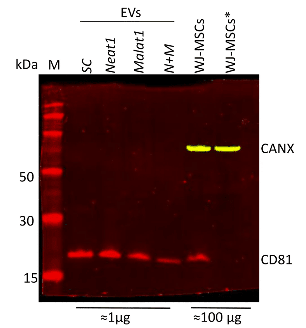
Isolation and characterization of EVs from various lncRNA knock-down WJ-MSCs. Western blot analysis using ExoBrite™ 680/700 CD81 and ExoBrite™ 770/800 Calnexin in EV and MSC lysates. Asterisk (*) indicates reduced conditions used in the MSCs lysate. Modified from Infante et. al. Reproduced under CC BY 4.0.
Learn more about Biotium’s many stains and antibodies for EV research, including ExoBrite™ CD9/CD63/CD81 Antibody Cocktails for flexible and bright multiplexing detection by flow cytometry. Biotium also offers ExoBrite™ stains for pan-EV labeling, optimized fluorescent conjugates of CTB, WGA, and Annexin V for EV detection, ExoBrite™ antibodies for STORM imaging, and more.
Full Citation:
Infante, A., Cabodevilla, L., Gener, B. et al. Modulation of NEAT1 and MALAT1 expression in WJ-MSCs by Covid-19 serum: a foundation for EVs-mediated therapy. Respir Res 26, 313 (2025). https://doi.org/10.1186/s12931-025-03394-4
While early studies of EVs attempted to use first-generation membrane dyes like DiI or PKH to stain EVs, more recently this class of dyes has been found to be largely unsuitable for EV staining due to their high degree of aggregation. Dye aggregation not only generates nonspecific particles that are indistinguishable from EVs in flow cytometry, but also results in poor EV labeling efficiency. Biotium developed the ExoBrite™ True EV Membrane Stains in response to our customers difficulties with using traditional membrane dyes to stain EVs. See our Literature Digest for more information.
We strongly recommend our ExoBrite™ Flow Antibody Conjugates for staining both purified or bead-bound EVs. The antibodies are validated and optimized to offer bright signal and low background. They are available against human or mouse CD9, CD63, and CD81 tetraspanin proteins.
