 New Products
 Earth-Friendly Products
 Biotium Choice Antibodies
 Special Offers


Content #1
Content #1
Content #1

Jump to a section:
Hoechst & DAPI Everything You Need to Know
How to Stain Live Cells
How to Stain Fixed Cells or Tissue Sections
DAPI and Hoechst Technical Information
Interested in Ordering?
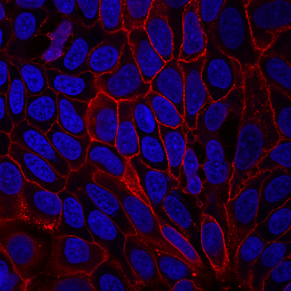
Hoechst and DAPI are popular blue fluorescent, nuclear-specific dyes that can be used to stain live or fixed cells. Both DAPI and Hoechst are minor-groove binding dyes with a preference for A/T-rich regions of DNA over G/C-rich DNA. The dyes have minimal fluorescence in solution, but become brightly fluorescent upon binding to DNA. Therefore, they can be used to stain cells without a wash step. The staining is very stable and the dyes have low toxicity in most cell types. Hoechst and DAPI are extremely stable in water at 10 mg/mL, and can be stored at 4°C for years as long as they are protected from light. However, there are some notable differences between DAPI and Hoechst that are important to highlight.
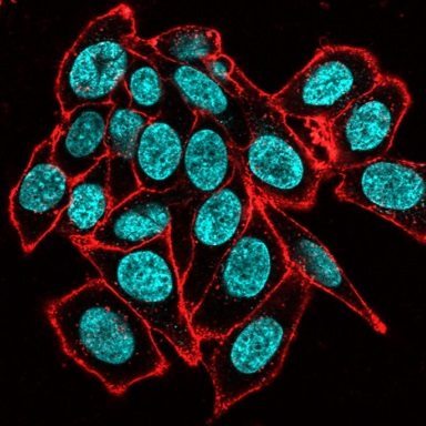
Hoechst dyes are generally preferred for live cell staining over DAPI because they are less toxic and more cell permeant. Biotium sells both Hoechst 33342 and Hoechst 33258, structurally similar dyes that are widely used in cell cycle studies and as nuclear counterstains for live or fixed cells. Hoechst 33528 is slightly more water soluble and less cell permeant than Hoechst 33342. There have been some reports that Hoechst 33342 induces apoptosis or shows more toxicity in some cell types (see for example Zhang et al., 1997 or Zhang et al., 1999). There are also some reports that note differences in their quantitative staining in some cell types. Both Hoechst dyes are typically used for staining at 1 ug/mL. When working with Hoechst, it is not recommended to store dilute solutions of Hoechst dye, because the dye will be lost to precipitation or adsorption to the container over time. Biotium offers stable concentrated stock solutions of Hoechst in water at 10 mg/mL.
DAPI is somewhat less cell membrane permeant and more toxic than Hoechst dyes, and is therefore preferred for fixed cell staining over live cell staining. As a fixed cell stain we recommend a DAPI concentration at 1 ug/mL, though live cell staining with DAPI can be performed at higher concentrations (usually 10 ug/mL). DAPI is stable in dilute solutions, and can be added directly to antifade mounting medium for long-term use. Biotium offers DAPI dilactate, a more soluble DAPI salt, which is useful for making stock solutions for cell staining. We also offer a ready-to-use stock solution of DAPI in water, and antifade EverBriteTM Mounting Medium with DAPI (see ordering information).
Hoechst and DAPI stain bacteria more dimly than mammalian cells. Live or killed bacteria (gram-negative or gram-positive) can be stained with 12-15 ug/mL Hoechst or DAPI in PBS or 150 mM NaCl for 30 minutes at room temperature. Dead cells tend to stain more brightly than live cells. In S. cerevisiae, DAPI and Hoechst preferentially stain dead cells with nuclear and cytoplasmic localization. In live yeast, Hoechst shows dim nuclear and cytoplasmic staining, while DAPI shows dim mitochondrial staining. The dyes can be used to stain yeast at 12-15 ug/mL in PBS.
A less familiar issue with DAPI and Hoechst is photoconversion by UV light, which causes the dyes to fluoresce in other channels. Some strategies to avoid this include imaging green fluorescence before switching to the DAPI channel, or moving to an unexposed field of view before imaging the green channel after UV exposure of the sample (Roberts, 2019). Using hardset mounting medium like EverBriteTM Hardset instead of glycerol-based wet-set medium can reduce photoconversion.
Biotium also offers NucSpot® Nuclear Stains which resolves the issue of photoconversion by offering bright and specific nuclear staining in 7 colors from green to near-IR. Learn more about how to avoid issues with UV photoconversion of DAPI and Hoechst.
Be sure to also check out our protocols for IF staining of cells and other helpful tech tips.
For live cell staining, morphology or viability of some cell types may be affected by medium exchange. In addition, floating dead cells may be lost during medium removal, and suspension cells must be collected by centrifugation to exchange the medium. Direct addition of 10X dye solution is a convenient staining method that doesn't require medium exchange, but care must be taken to mix immediately yet gently to avoid high transient dye concentration or disruption of cells by pipetting. Note that we do not recommend adding highly concentrated dye directly to cells in culture, as this will result in local areas of high dye exposure. Biotium also offers low-toxicity NucSpot® Live Stains stains for long-term live cell imaging, available in green and far-red fluorescence. For short-term imaging, Biotium offers RedDotTM1 far-red stain, which serves as an alternative to Draq5TM and may also be used for cell cycle analysis by flow cytometry.
Live cell staining by medium exchange
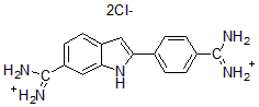
DAPI, dihydrochloride salt
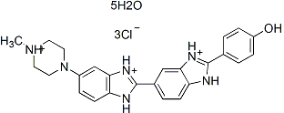
Hoechst 33258, pentahydrate
Hoechst 33342, trihydrochloride trihydrate
| Product | Unit Size | Catalog Number |
|---|---|---|
| Hoechst 33258, 10 mg/mL in H2O | 10 mL | 40044 |
| Hoechst 33258, pentahydrate | 100 mg | 40045 |
| Hoechst 33342, 10 mg/mL in H2O | 10 mL | 40046 |
| Hoechst 33342, trihydrochloride trihydrate | 100 mg | 40047 |
| DAPI in H2O, 10 mg/mL | 1 mL | 40043 |
| DAPI, dilactate | 10 mg | 40009 |
| DAPI, dihydrochloride | 10 mg | 40011 |
| EverBrite™ Mounting Medium with DAPI | 10 mL | 23002 |
| Drop-n-Stain EverBrite™ Mounting Medium with DAPI | 10 mL | 23009 |
| EverBrite™ Hardset Mounting Medium with DAPI | 10 mL | 23004 |

Content #1
Content #1
Content #1
Content #2
Content #3