 New Products
 Earth-Friendly Products
 Biotium Choice Antibodies
 Special Offers


Content #1
Content #1
Content #1
Cell membrane-impermeant, nuclear-specific counterstains. Suitable for fixed cells or staining dead cells in live cultures.
NucSpot® Nuclear Stains are cell membrane-impermeant, nuclear-specific counterstains available in a variety of colors from green to near-infrared (near-IR).
Commonly used blue nuclear stains, such as DAPI and Hoechst, undergo photoconversion after exposure to UV wavelengths and cause cross-talk in other channels. NucSpot® Nuclear Stains were developed to address these photoconversion issues by offering bright and nuclear-specific counterstaining in a wide selection of colors from green to near-IR. The have minimal fluorescence until they bind to DNA and can be used for no-wash nuclear staining. Unlike other nucleic acid dyes such as propidium iodide (PI), TOTO®, TO-PRO®, and similar dyes that stain both the nucleus and cytoplasm, NucSpot® Nuclear Stains selectively stain the nucleus in fixed and permeabilized cells without the need for RNase treatment. NucSpot® Nuclear Stains also can be used for selective staining of dead cells in unfixed cell cultures for analysis by flow cytometry or fluorescence imaging. Several of the dyes can be continuously incubated with cells for multi-day imaging.
| Product | Ex/Em | Detection Channel | Size | Catalog No. |
|---|---|---|---|---|
| NucSpot® 500/515 | 497/513 nm | FITC* | 20 uL | 41040-T |
| 100 uL | 41040 | |||
| NucSpot® 555/570 | 559/566 nm | Cy®3 or PE* | 20 uL | 41033-T |
| 100 uL | 41033 | |||
| NucSpot® 568/580 | 572/583 nm | Cy®3 or PE* | 20 uL | 41036-T |
| 100 uL | 41036 | |||
| NucSpot® 594/615 | 603/613 nm | Texas Red® or PE-Texas Red®* | 20 uL | 41037-T |
| 100 uL | 41037 | |||
| NucSpot® 650/665 | 653/671 nm | Cy®5 or APC* | 20 uL | 41034-T |
| 100 uL | 41034 | |||
| NucSpot® 680/700 | 683/707 nm | Cy®5.5* | 20 uL | 41035-T |
| 100 uL | 41035 | |||
| NucSpot® 750/780 | 757/780 nm | Cy®7 or APC-Cy®7* | 20 uL | 41038-T |
| 100 uL | 41038 |
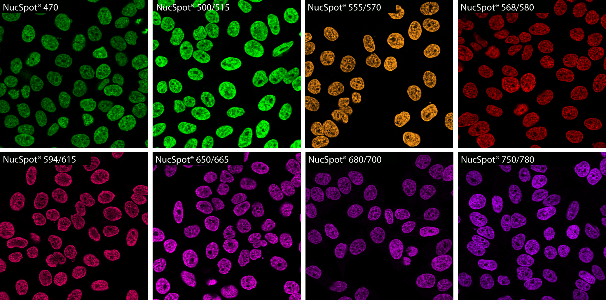
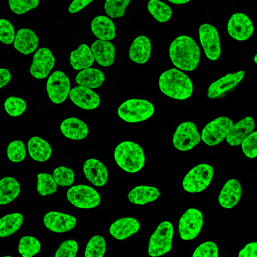
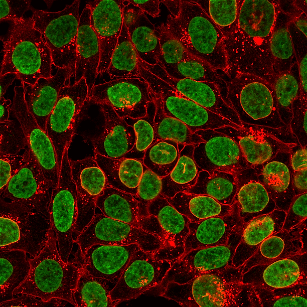
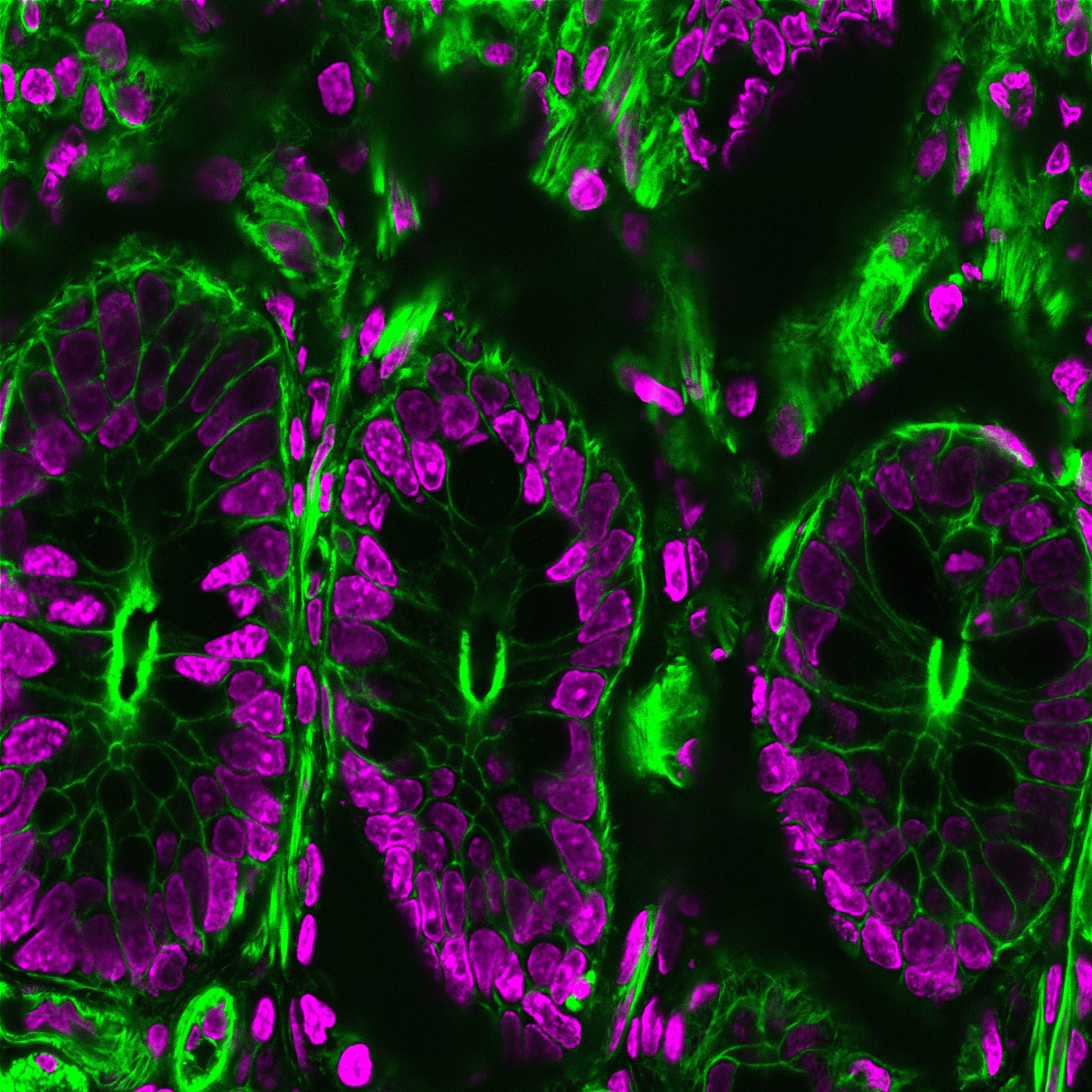
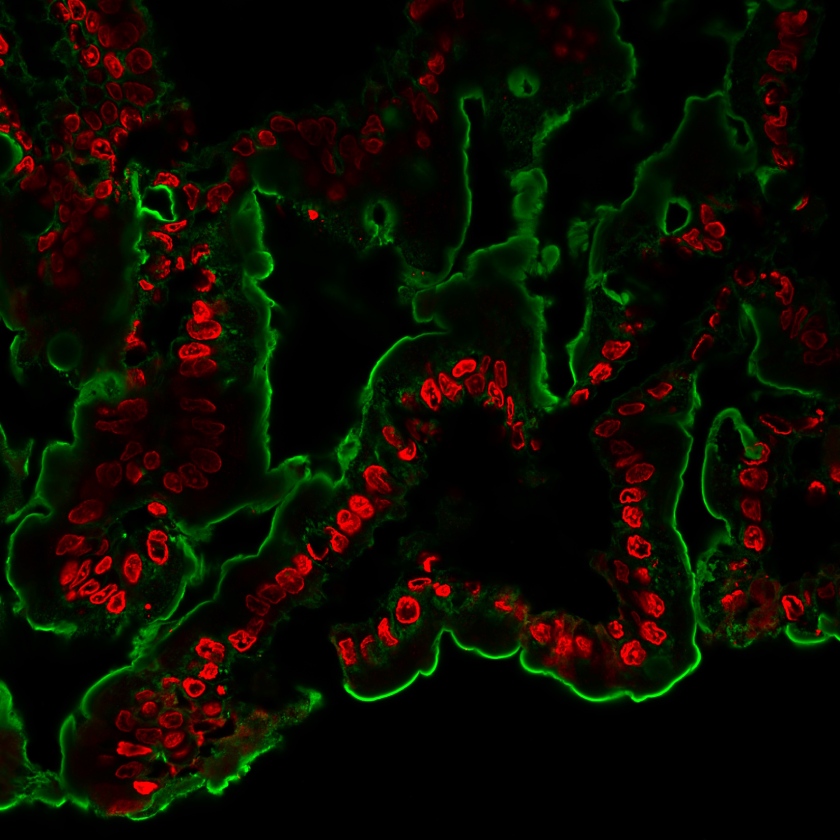
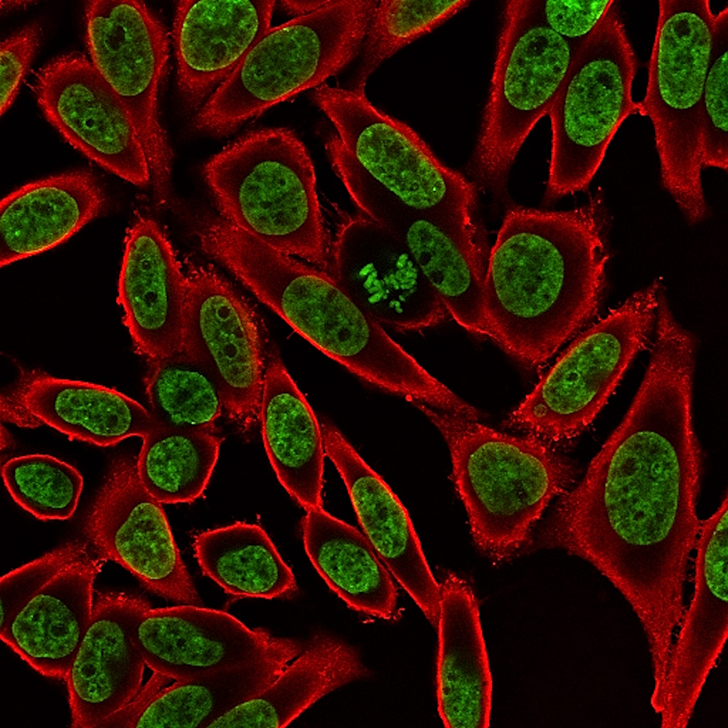
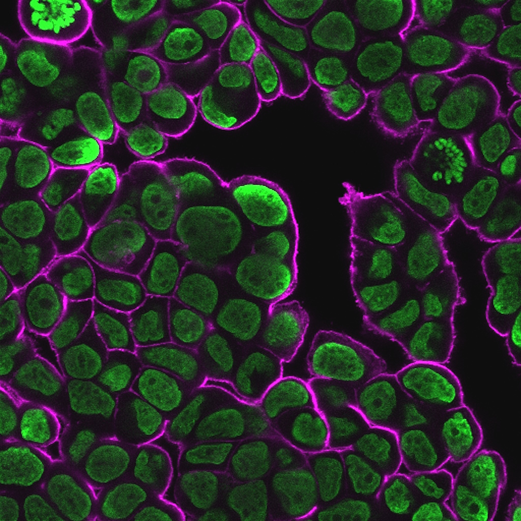
PFA-fixed, Triton® X-100 permeabilized HeLa cells stained with NucSpot® Nuclear Stains in PBS and imaged by confocal microscopy without a wash step. All stains were used at 1X concentration except for NucSpot® 750/780, which was used at 5X concentration in order to image using 640 nm excitation.
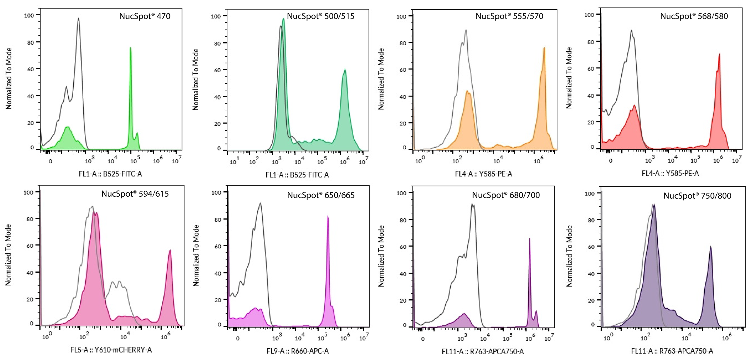
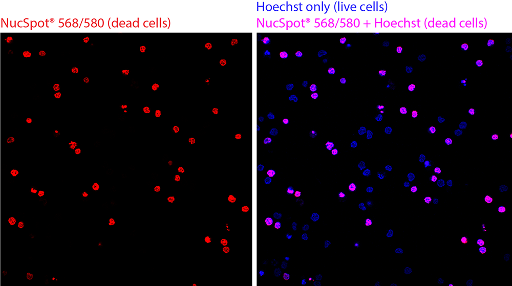
Live dead discrimination by flow cytometry using NucSpot® Nuclear Stains. A mixture of untreated and heat-killed Jurkat cells was stained with each NucSpot® Nuclear Stain and analyzed by flow cytometry on a BD CytoFLEX flow cytometerin the detection channel shown on the histogram x-axis. Solid peaks represent stained cells; open gray peaks represent unstained cells. The concentrations of each dye used were: 1X NucSpot® 470, 0.1X NucSpot® 500/515, 0.5X NucSpot® 555/565, 1X NucSpot® 568/580; 0.5X NucSpot® 594/615, 0.05X NucSpot® 650/665, 0.2X NucSpot® 680/700, 0.5X NucSpot® 750/780.
NucSpot® Nuclear Stains offer improved photostability over commonly used cyanine-based nuclear stains. The figure below shows how NucSpot® 650/665 is significantly more photostable than Thiazole Red Homodimer (TOTO® -3).
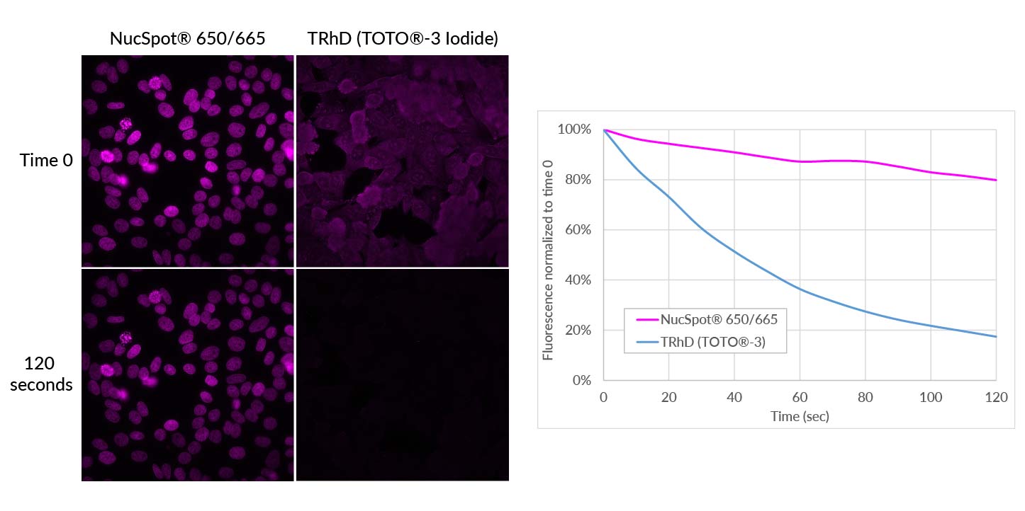
Photostability of NucSpot® 650/665 compared to Thiazole Red Homodimer. PFA-fixed, permeabilized HeLa cells were stained with 1X NucSpot® 650/665 or 200 nM Thiazole Red Homodimer (equivalent to TOTO®-3 Iodide). Cells were continuously imaged for 120 seconds by epifluorescence microscopy through a Cy®5 filter set, and images were captured every 10 seconds. Mean fluorescence normalized to time 0 for each image is plotted on the right. NucSpot® 650/665 fluorescence was well-preserved, while Thiazole Red/TOTO®3 fluorescence photobleached rapidly. In addition, NucSpot® 650/665 specifically stained the cell nucleus, while in the absence of RNase-pretreatment, TOTO®-3 stained both cytoplasm and nucleus.
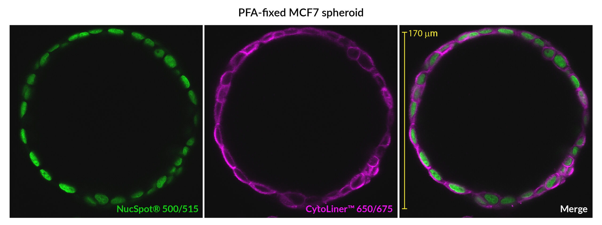
NucSpot® 500/515 is a green fluorescent nuclear stain for the FITC channel. In addition to bright nuclear counterstaining of fixed cells, it can also be used for multi-day live/dead staining of mammalian cells in culture. NucSpot® 500/515 is designed as an improved alternative to NucSpot® 470 which has limited use for cell viability tracking due to its instability in culture medium during long incubation periods.
NucSpot® 555/570 and NucSpot® 568/580 have orange and visible red fluorescence, respectively, and are nuclear-specific alternatives to PI and similar dyes.
NucSpot® 594/615 has deep red fluorescence for the Texas Red® channel.
NucSpot® 650/665 has far-red fluorescence with superior nuclear specificity compared to first-generation far-red nuclear stains such as RedDot™2 and Draq7™.
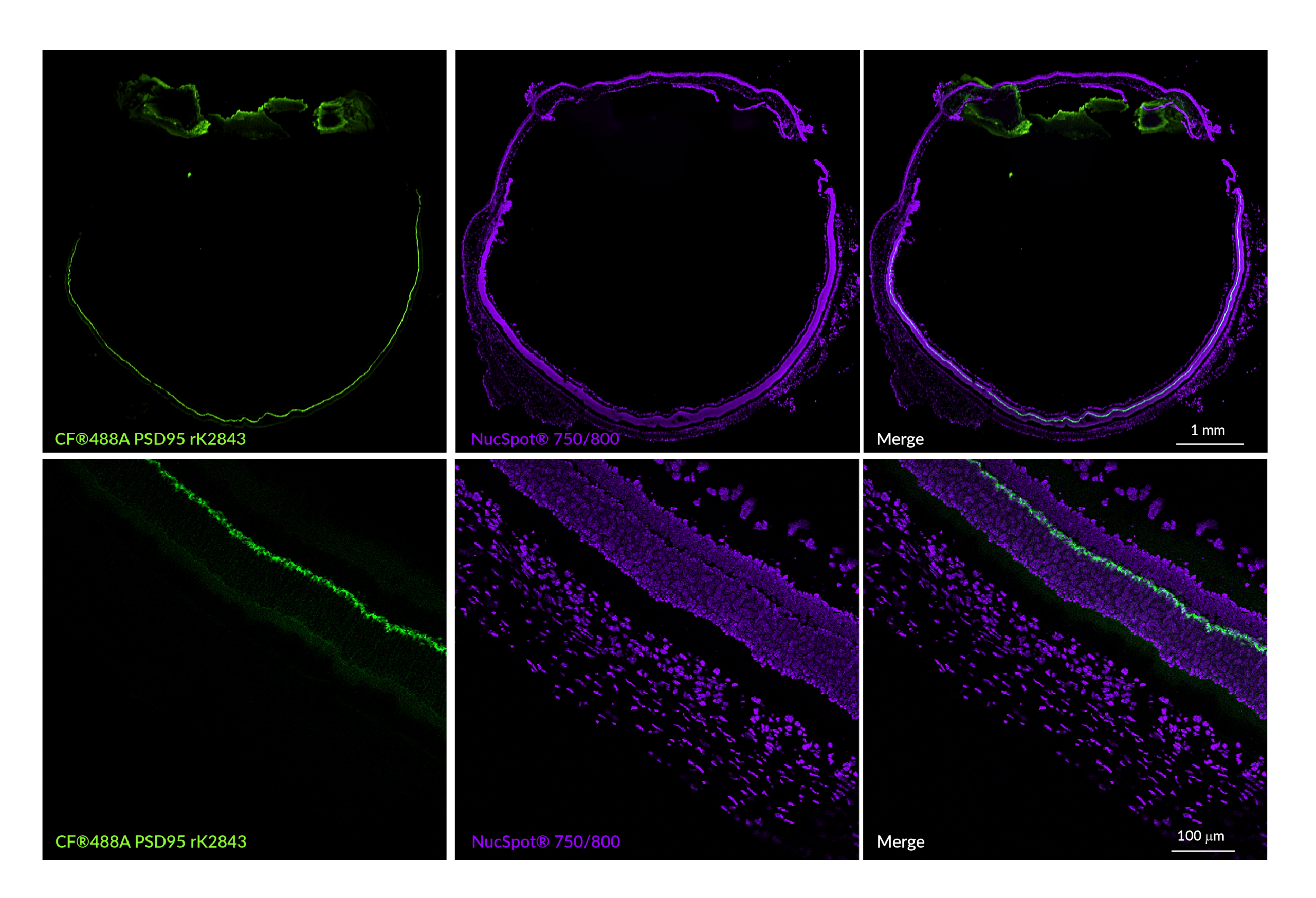
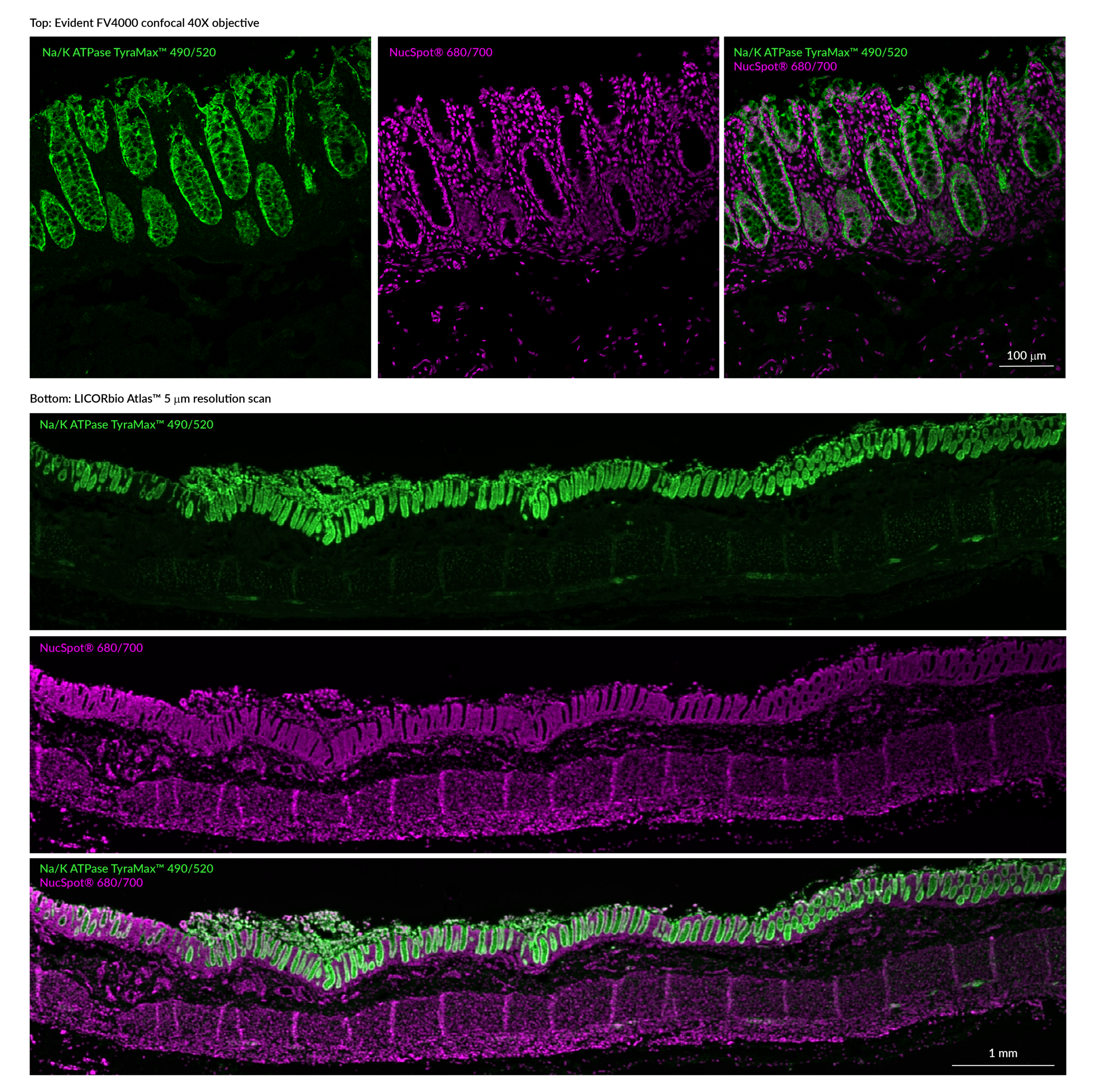
NucSpot® 680/700 and NucSpot® 750/780 are spectrally unique DNA stains for far-red and near-IR detection.
Biotium also offers NucSpot® Far-Red, a flow cytometry stain developed as an improved alternative to 7-AAD. It shows less bleed-through fluorescence in the PE-Texas Red® channel compared to 7-AAD and is ideal for selective detection of dead cells or DNA content analysis by flow cytometry without RNase treatment.
Learn more about our full selection of Cellular Stains for the nucleus and other organelles.
It has been reported in publications that concentrations of serum above 10% in the assay may affect the results.
See the following publications for more information
Our ViaFluor® SE Cell Proliferation assay is a dye dilution assay for cell division, like CFSE and CellTrace™ Violet from Thermo. This type of assay is commonly used to measure lymphocyte proliferative responses in culture and in vivo (if the labeled cells are injected back into mice). It requires flow cytometry to analyze and allows you to count how many cell divisions have occurred in the labeled cells.
For more information and a typical procedure for using fluorescent ViaFluor® SE Dyes with PMBCs, which can easily be adapted for use with other cell types, please see our Tech Tip: Measuring Cell Division in PMBCs by Flow Cytometry
If flow cytometry is not an option, we offer absorbance-based and fluorescence-based microplate assays for quantitating cell numbers. These measure mitochondrial activity (resazurin/MTT/XTT) or intracellular esterase activity (calcein AM) as a readout of viable cell numbers. Please visit the Cell Viability and Apoptosis technology page for more information.
The ATP-Glo™ assay is a luminescence assay for cellular ATP levels, which are proportional to the number of live cells. This assay requires a luminometer.
CellTrace is a trademark of Thermo Fisher Scientific.
Our Resazurin Cell Viability Assay (Cat. No. 30025) has red fluorescence (Ex/Em 530-560/590 nm), and is specifically designed for microplate reader. It is an economical, easy-to-use, and homogeneous (no-wash) assay for quantifying live cells. It is similar to alamarBlue®, PrestoBlue®, and CellTiter-Blue®.
The Calcein AM Cell Viability Assay (Cat. No. 30026) has green fluorescence (Ex/Em 485/530 nm), and also works well for microplate reader. This assay requires culture medium to be removed from cells before adding the viability dye in buffer. We also offer the Viability/Cytotoxicity Assay for Animal Live & Dead Cells, which combines calcein-AM with the fluorescent dead cell stain EthD-III, and is compatible with microplate reader.
Bioscience kits
The guaranteed shelf life from date of receipt for bioscience kits is listed on the product information sheet. Some kits have an expiration date printed on the kit box label, this is the guaranteed shelf life date calculated from the day that the product shipped from our facility. Kits often are functional for significantly longer than the guaranteed shelf life. If you have an older kit in storage that you wish to use, we recommend performing a small scale positive control experiment to confirm that the kit still works for your application before processing a large number of samples or precious samples.
Antibodies and other conjugates
The guaranteed shelf life from date of receipt for antibodies and conjugates is listed on the datasheet sheet which can be downloaded on the product page. Antibodies and other conjugates often are functional for significantly longer than the guaranteed shelf life. If you have an older conjugate in storage that you wish to use, we recommend performing a small scale positive control experiment to confirm that the product still works for your application before processing a large number of samples or precious samples.
For lyophilized antibodies, we recommend reconstituting the antibody with glycerol and antimicrobial preservative like sodium azide for the longest shelf life (note that sodium azide is not compatible with HRP-conjugates).
Chemicals, dyes, and gel stains
Biotium guarantees the stability of chemicals, dyes, and gel stains for at least a year from the date you receive the product. However, the majority of these products are highly stable for many years, as long as they are stored as recommended. Storage conditions can be found on the product information sheet or product safety and data sheet, material safety data sheet, and on the product label. Fluorescent compounds should be protected from light for long term storage.
If you have a Biotium compound that has been in storage for longer than one year that you wish to use, we recommend performing a small scale positive control experiment to confirm that the compound still works for your application before processing a large number of samples or precious samples.
Expiration date based on date of manufacture (DOM)
If your institution requires you to document expiration date based on date of manufacture for reagents, please contact [email protected] for assistance.
Chemical products with special stability considerations:
Esters
Ester compounds include the following:
Ester dyes are stable in solid form as long as they are protected from light and moisture. Esters are not stable in aqueous solution. Concentrated stock solutions should be prepared in anhydrous DMSO (see Biotium catalog no. 90082). Stock solutions in anhydrous DMSO can be stored desiccated at -20°C for one month or longer. Esters should be diluted in aqueous solution immediately before use. Succinimidyl esters (SE) should be dissolved in a solution that is free of amine-containing compounds like Tris, glycine, or protein, which will react with the SE functional group. AM esters and diacetate compounds should be dissolved in a solution that is free of serum, because serum could contain esterases that would hydrolyze the compound.
A note on CF® Dye succinimidyl ester stability
Succinimidyl esters (SE) are generally susceptible to hydrolysis, which can result in lower labeling efficiency. Many commercially available fluorescent dyes used for life science research are heavily sulfonated dyes which makes them particularly hygroscopic, worsening the hydrolysis problem. In addition, for several commercially available SE reactive dyes, the SE group is derived from an aromatic carboxylic acid, while the SE group in all of Biotium’s CF® Dyes is prepared from an aliphatic carboxylic acid. This structural difference reduces the susceptibility of CF® Dye SE reactive groups to hydrolysis, resulting in relatively stable reactive dyes with consistently higher labeling efficiency compared to other SE derivatives of other fluorescent dyes.
Maleimides, MTS and thiosulfate dyes
Like the succinimidyl ester dyes, these dyes are also susceptible to hydrolysis, although generally to a much lower degree. Thus, for long term storage, anhydrous DMSO is recommended for making stock solutions.
Other reactive dyes
Amines, aminooxy (also known as oxylamine), hydrazide, azide, alkyne, BCN, and tyramide reactive dyes, as well as dye free acids, are generally stable in aqueous solution when stored at -20°C for 6-12 months or longer, as long as no compounds are present that may react with the dye’s functional group. See the product information sheets for specific reactive dyes more information.
Coelenterazines and D-luciferin
Coelenterazines are stable in solid form when stored as recommended; they are not stable in aqueous solution. Concentrated coelenterazine stock solutions (typically 1-100 mg/mL) should be prepared in ethanol or methanol; do not use DMSO or DMF to dissolve coelenterazines, because these solvents will oxidize the compounds. Ethanol or methanol stocks of coelenterazine can be stored at -20°C or below for six months or longer; alcohol stocks may evaporate during storage, so use tightly sealing screw cap vials and wrap the vials with Parafilm for long term storage. Propylene glycol also can be used as a solvent to minimize evaporation. If the solvent evaporates, the coelenterazine will still be present in the vial, so note the volume in the vial prior to storage so that you can adjust the solvent volume to correct for evaporation if needed. Prepare working solutions in aqueous buffers immediately before use. Coelenterazines are stable for up to five hours in aqueous solution.
Aquaphile™ coelenterazines are water soluble formulations of coelenterazines. They are stable in solid form when stored as recommended. Aquaphile™ coelenterazines should be dissolved in aqueous solution immediately before use. They are stable for up to five hours in aqueous solution.
Note that coelenterazines are predominantly yellow solids, but may contain dark red or brown flecks. This does not affect product stability or performance. If your coelenterazine is uniformly brown, then it is oxidized and needs to be replaced.
D-luciferin is stable in solid form and as a concentrated stock solution when stored as recommended; it is not stable at dilute working concentrations in aqueous solution. Prepare concentrated D-luciferin stock solutions (typically 1-100 mg/mL) in water, and store in aliquots at -20°C or below for six months or longer. Prepare working solutions immediately before use.
For dyes or reagents that are supplied lyophilized (as solids), it is hard to compare quantities based on appearance of the dye in the tube, because during the lyophilization process the dye can dry down in different ways, either spread out all over the tube, clumped together, or coating the sides or bottom of the tube. Centrifugation of the tube may not help in collecting the dye solid to the bottom of the tube as this generally works for solutions. However, lyophilized solids are packaged based on highly accurate absorbance measurement of the reagent solution prior to drying, so the vial will contain the correct amount of dye.
Biotium ships all antibodies (primary, secondary and conjugates) at room temperature. We guarantee their quality and performance under these conditions based upon our stability testing. Antibodies were subjected to accelerated stability testing by storing them at various temperatures (4°C, room temperature, or 37°C) for 1 week to mimic simulated shipping conditions and tested in immunostaining experiments. All antibodies showed the expected brightness and specificity, even after storage at sub-optimal temperatures for a week or longer. You can also download our Product Storage Statement here.
In line with our goal to be more environmentally friendly by reducing the use of excess packaging, and lowering shipping costs for our customers, products that have passed our stability testing are shipped at room temperature.
Once you have received the antibody vial, please follow the long-term storage instructions on the product information (PI) sheet.
