Lori Roberts holds a PhD in Biomedical Science from the University of California, San Francisco. She currently serves as the Director of Bioscience at Biotium.
EvaEZ™ is a fluorescence-based assay that allows you to quantitate DNA polymerase activity without using radioisotopes. While the assay is simple to use, there are many factors that can affect the quality of your data. In this article, we've outlined our advice for getting the best results using our EvaEZ™ Assay with your enzyme. The […]
Jump to a section: Hoechst & DAPI Everything You Need to Know How to Stain Live Cells How to Stain Fixed Cells or Tissue Sections DAPI and Hoechst Technical Information Interested in Ordering? Hoechst & DAPI - Everything You Need to Know Hoechst and DAPI are popular blue fluorescent, nuclear-specific dyes that can be used […]
Cross-talk and specificity in multi-color imaging experiments Cross-talk between fluorescence channels is an important concern for multi-color imaging experiments. The detection of fluorescence from one dye in multiple detection channels can result in artifactual co-localization of signals. Cross-talk usually occurs because of the overlap of dye excitation and emission spectra, and can be managed by […]
The most common use of GelRed® and GelGreen® nucleic acid stains is in pre-cast agarose gels, where the dyes are added to molten agarose during gel preparation. However, due to their larger size designed to improve safety, band migration of DNA in pre-cast gels may be affected. Some samples, such as restriction digested DNA can […]
ViaFluor® SE Cell Proliferation Kits use amine-reactive dyes to covalently label cells throughout the cytoplasm and intracellular compartments. ViaFluor® SE dyes are initially membrane-permeable and non-fluorescent. After the compound enters viable cells, it is hydrolyzed by cytoplasmic esterases to releases the fluorescent amine-reactive dye. The dye covalently reacts with amine groups on intracellular proteins, forming […]
Dyes functionalized with succinimidyl ester (NHS Ester or SE) groups can be used to covalently label proteins on primary amines (typically lysine residues) to form a stable amide linkage. This is a common method for preparing fluorescently-labeled antibody conjugates. The protocol below is a typical procedure for labeling IgG antibodies in bicarbonate buffer. The protocol […]
This protocol can be adapted to use dUTP or dCTP labeled with other dyes, biotin, or haptens like digoxigenin. See our full selection of labeled nucleotides. * When using dUTP conjugates for labeling, use Taq DNA polymerase; dUTP inhibits archaeal polymerases such as Pfu and Vent®. Procedure: 1 - Set up labeling reactions 1.1 For […]
Membrane Stains for Different Cell Types, Organisms, and Sample Preparations For staining mammalian cells in 2D culture, please see our Membrane & Cell Surface Stains Comparison Guide and our Membrane & Surface Stains Brochure to select the right dye for your experiment. If you wish to use a membrane dye for a different cell type, […]
CellBrite® Cytoplasmic Membrane Dyes are membrane dyes that are useful for visualizing cell outlines. However, because they are lipophilic, the dyes tend to redistribute after detergent permeabilization (Fig. 1). For membrane staining of live cells followed by fixation and immunofluorescence, we recommend our newer CellBrite® Fix and MemBrite® Fix fixable cell surface stains. However, these […]
Problem: No Staining or Low Signal Primary antibody is not validated for application Check the supplier information to find out if your primary antibody is recommended for your application. If possible, validate the antibody using a positive control cell line or tissue that expresses the target. Confirm that the species reactivity of the antibody is […]
There are many variations on IF protocols, and steps may need to be optimized for different targets or applications. Some epitopes may require specific fixation conditions for detection. This is our basic protocol for staining adherent cells in dishes or cells grown on coverslips. Procedure: Rinse cells twice with PBS or HBSS to remove cell […]
Immunofluorescence staining (IF) can be performed in two ways, by direct or indirect detection. Direct IF uses a dye-conjugated antibody to stain the target protein. Indirect IF involves first binding the primary antibody to the target, then detecting the primary antibody using a conjugated secondary antibody. Indirect IF offers the advantage of higher sensitivity. Each […]
Immunofluorescence staining offers a variety of advantages compared to colorimetric immunocytochemistry, most importantly, simultaneous multicolor detection of several targets. However, fluorescence detection in tissue samples can be hindered by strong background fluorescence. Autofluorescence can arise from exogenous and endogenous sources, including aldehyde fixation, extracellular matrix components, red blood cells, other pigmented cell types, and lipofuscin. […]
Lectin conjugates, such as WGA (wheat germ agglutinin) or Con A (concanavalin A), bind with a high specificity to sugar groups on glycoproteins. WGA lectins bind to sialic acid and N-acetylglucosamine residues, while Con A lectins bind to a-mannopyranosyl and a-glucopyranosyl residues. Due to the expression of glycoproteins on cell membranes, conjugated lectins are widely […]
If you're struggling to find suitable antibodies for your experiment, directly labeling your own antibodies can be a great option that offers more flexibility. Direct labeling allows you to choose any combination of antibody and label, including combinations that are not commercially available. Direct labeling also allows you to only label the amount of antibody […]
To learn more about western blotting, including the advantages of near-infrared fluorescence detection, see our webinar: Fundamentals of Western Immunoblotting: Chemiluminescence and NIR Multiplex Imaging (49:44). General considerations for fluorescent western detection: Multiplex fluorescence western detection requires an imaging system capable of detecting multiple fluorescent conjugates. For best results, use a gel imager or scanner […]
Dyes functionalized with an aminooxy reactive group can be used to label molecules with a carbonyl group (aldehyde or ketone) to form a stable oxime bond. Aminooxy labeling can be used to conjugate glycoproteins after oxidation of the carbohydrates to generate aldhehyde groups. Aminooxy labeling of antibody glycosylation sites can be used as an alternative […]
Dyes functionalized with a maleimide group can react with thiol groups to form thioether-coupled products. The reaction can take place at neutral pH, and is not affected by the presence of amines. At neutral pH, the maleimide group does not react with histidine or arginine. Maleimide labeling of antibodies can be used as an alternative […]
Pluronic® F-127 is a nonionic detergent useful for solubilizing hydrophobic molecules in aqueous solutions. Pluronic® F-127 is commonly used to solubilize hydrophobic AM ester forms of fluorescent indicator dyes for calcium and other ions for loading of the dyes into cells. It also can be used to solubilize other hydrophobic dyes or compounds to facilitate […]
Bored waiting for your cells to grow? Kill time and test your knowledge with this Microbe Acrostic puzzle!
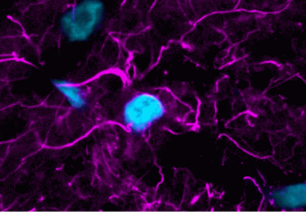
Photoconversion and cross-talk from blue nuclear dyes can ruin your image. Check out these tips on how to avoid it.
Content #1
Content #2
Content #3

 New Products
 Earth-Friendly Products
 Biotium Choice Antibodies
 Special Offers
 Quick Order Tool