New Products
 Earth-Friendly Products
 Biotium Choice Antibodies
 Special Offers
 Quick Order Tool


Content #1
Content #1
Content #1
This protocol can be adapted to use dUTP or dCTP labeled with other dyes, biotin, or haptens like digoxigenin. See our full selection of labeled nucleotides.
* When using dUTP conjugates for labeling, use Taq DNA polymerase; dUTP inhibits archaeal polymerases such as Pfu and Vent®.
1.1 For each labeling reaction, set up the PCR reaction mix as shown below:
| Component | Volume per reaction | Final concentration* |
|---|---|---|
| 10X Taq reaction buffer | 2 uL | 1X |
| 25 mM MgCl2 | 2 uL | 5 mM |
| 1 mM dATP | 2 uL | 100 uM |
| 1 mM dCTP | 2 uL | 100 uM |
| 1 mM dGTP | 2 uL | 100 uM |
| 1 mM dTTP | 1 uL | 50 uM |
| 10 uM forward primer | 1 uL | 500 nM |
| 10 uM reverse primer | 1 uL | 500 nM |
| Template DNA | 1 ng | 50 pg/uL |
| Taq | 1 U | 0.05 U/uL |
| Molecular grade dH20 | to 19 uL total |
1.2 Add 1 uL of 1 mM CF® dye dUTP to the reaction tube.
Amplify the reactions in a thermocycler using the following cycling protocol:
| Step | # Cycles |
|---|---|
| Denaturing/Taq activation 94°C, 2 min.1 | Hold |
| Denaturing 94°C 30 sec. | Cycle 30X |
| Annealing 30 sec.2 | |
| Extension 72°C 1 min. 3 | |
| Final extension 72°C 5 min. | Hold |
Use a PCR clean-up kit or G50 Sephadex® microspin column to remove unincorporated nucleotides.
4-1 Run 10% of the labeled product on an agarose gel along with a DNA ladder. Do not add fluorescent DNA dye to the agarose before casting. After electrophoresis, image the CF® dye fluorescence of the labeled probes on a UV gel transilluminator or laser-based gel scanner as appropriate for the wavelengths of the specific dye used.
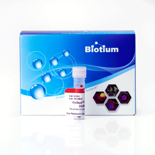
4-2 After imaging the probe fluorescence, post-stain the gel with a nucleic acid gel stain like GelRed® or GelGreen® to image unstained DNA ladder and unlabeled control PCR product.

Lori Roberts holds a PhD in Biomedical Science from the University of California, San Francisco. She currently serves as the Director of Bioscience at Biotium.