 New Products
 Earth-Friendly Products
 Biotium Choice Antibodies
 Special Offers


 Powered by Bioz


Content #1
Content #1
Content #1
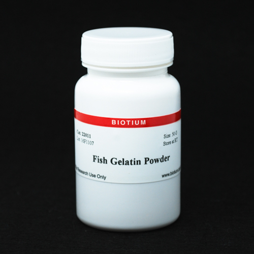
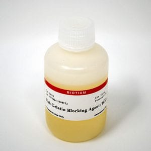
Gelatin from fish skin that can be added to blocking buffers to minimize non-specific antibody binding in immuno-detection procedures such as western blotting and cell staining.
Fish Gelatin Powder is purified from cold water fish skin. It can be used to make blocking buffers to minimize non-specific antibody binding in immunodetection procedures such as western blotting, immunofluorescence, and IHC.
Fish gelatin can be used to block non-specific binding sites on positively charged nylon or PVDF membranes or used for blocking and dilution of antibodies during immunostaining procedures. Unlike BSA or milk, fish gelatin does not contain IgG or serum proteins that could cross-react with mammalian antibodies.
1. Oncotarget (2017) Sep 28;8(49):86657-86670 doi:10.18632/oncotarget.21364
2. Heliyon (2018) Nov 16;4(11):e00917 doi:10.1016/j.heliyon.2018.e00917
3. Exp Eye Res (2019) Mar;180:122-128 doi:10.1016/j.exer.2018.12.016
4. Environ Mol Mutagen (2020) Feb;61(2):235-245 doi:10.1002/em.22331
1. Oncotarget (2017) Sep 28;8(49):86657-86670 doi:10.18632/oncotarget.21364
2. Heliyon (2018) Nov 16;4(11):e00917 doi:10.1016/j.heliyon.2018.e00917
3. Exp Eye Res (2019) Mar;180:122-128 doi:10.1016/j.exer.2018.12.016
4. Environ Mol Mutagen (2020) Feb;61(2):235-245 doi:10.1002/em.22331
Even though AccuOrange™ buffer does contain SDS, which is required for the dye to bind proteins, the assay is very sensitive to small changes in SDS concentration, and also cannot tolerate non-ionic detergents that form mixed micelles with SDS, like Triton®. Therefore we don't recommend using the kit for cell lysates or other samples with significant amounts of detergents.
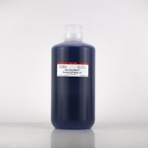
Gels stained with One-Step Blue® can be dried just like gels stained with Coomassie. The stain will not interfere with the detection of radiolabeled proteins.
The AccuOrange™ assay is a fluorescent dye-based assay. The dye binds to proteins primarily through hydrophobic interactions. Proteins denature upon heating; the dye binds to the exposed hydrophobic pockets of the protein after cooling. The free AccuOrange™ dye is fluorogenic due to non-radioactive decay but becomes highly fluorescent due to the rigid conformation inside the pocket.
The AccuOrange™ assay more sensitive than traditional protein quantitation assays such as BCA, Bradford and Lowry, and shows superior linearity and reproducibility than the NanoOrange® protein quantitation assay (Thermo Fisher Sci.), but has low tolerance for detergents like SDS and Triton® X-100.
