 New Products
 Earth-Friendly Products
 Biotium Choice Antibodies
 Special Offers


 Powered by Bioz


Content #1
Content #1
Content #1
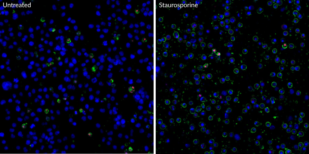
A convenient assay for quantifying apoptotic (green), necrotic (red), and total (blue) cells within the same cell population by flow cytometry or fluorescence microscopy.
The Apoptotic, Necrotic & Healthy Cells Quantitation Kit Plus provides a convenient assay for quantifying apoptotic (green), necrotic (red), and total (blue) cells within the same cell population by fluorescence microscopy.
Apoptosis and necrosis are two major processes by which cells die. Apoptosis is an active, genetically regulated disassembly of the cell from within. During apoptosis, phosphatidylserine (PS) is translocated from the inner to the outer leaflet of the plasma membrane for phagocytic cell recognition. The human anticoagulant Annexin V is a 35 kDa, Ca2 -dependent phospholipid-binding protein with high affinity for PS. Annexin V labeled with CF®488A stains apoptotic cells green by binding to PS exposed on the cell surface. CF®488A is spectrally similar to fluorescein (FITC), with much brighter and more photostable fluorescence.
Necrosis normally results from severe cellular insult. Both internal organelle and plasma membrane integrity are lost, resulting in the release of cellular contents into the surrounding environment. Ethidium Homodimer III (EthD-III) is a highly positively charged nucleic acid probe, which is impermeant to live cells, but stains cells with compromised membrane integrity with red fluorescence. EthD-III is a superior alternative to Propidium Iodide (PI) or Ethidium Homodimer I because of its significantly higher affinity for DNA and higher fluorescence quantum yield.
The kit also includes the cell membrane-permeant DNA dye Hoechst 33342 to stain all cell nuclei blue.
1. Cell Death Dis (2012) 3, e414. https://doi.org/10.1038/cddis.2012.154
2. JBC (2019) 294, 11259-11275. doi: 10.1074/jbc.RA119.007851
1. Cell Death Dis (2012) 3, e414. https://doi.org/10.1038/cddis.2012.154
2. JBC (2019) 294, 11259-11275. doi: 10.1074/jbc.RA119.007851
It has been reported in publications that concentrations of serum above 10% in the assay may affect the results.
See the following publications for more information
Our ViaFluor® SE Cell Proliferation assay is a dye dilution assay for cell division, like CFSE and CellTrace™ Violet from Thermo. This type of assay is commonly used to measure lymphocyte proliferative responses in culture and in vivo (if the labeled cells are injected back into mice). It requires flow cytometry to analyze and allows you to count how many cell divisions have occurred in the labeled cells.
For more information and a typical procedure for using fluorescent ViaFluor® SE Dyes with PMBCs, which can easily be adapted for use with other cell types, please see our Tech Tip: Measuring Cell Division in PMBCs by Flow Cytometry
If flow cytometry is not an option, we offer absorbance-based and fluorescence-based microplate assays for quantitating cell numbers. These measure mitochondrial activity (resazurin/MTT/XTT) or intracellular esterase activity (calcein AM) as a readout of viable cell numbers. Please visit the Cell Viability and Apoptosis technology page for more information.
The ATP-Glo™ assay is a luminescence assay for cellular ATP levels, which are proportional to the number of live cells. This assay requires a luminometer.
CellTrace is a trademark of Thermo Fisher Scientific.
Our Resazurin Cell Viability Assay (Cat. No. 30025) has red fluorescence (Ex/Em 530-560/590 nm), and is specifically designed for microplate reader. It is an economical, easy-to-use, and homogeneous (no-wash) assay for quantifying live cells. It is similar to alamarBlue®, PrestoBlue®, and CellTiter-Blue®.
The Calcein AM Cell Viability Assay (Cat. No. 30026) has green fluorescence (Ex/Em 485/530 nm), and also works well for microplate reader. This assay requires culture medium to be removed from cells before adding the viability dye in buffer. We also offer the Viability/Cytotoxicity Assay for Animal Live & Dead Cells, which combines calcein-AM with the fluorescent dead cell stain EthD-III, and is compatible with microplate reader.
