New Products
 Earth-Friendly Products
 Biotium Choice Antibodies
 Special Offers
 Quick Order Tool


Content #1
Content #1
Content #1
Extracellular vesicles (EVs) are nanoscale, membrane-bound particles released by cells that play important roles in intercellular communication by transporting proteins, lipids, and nucleic acids. Bovine milk is an abundant and accessible source of extracellular vesicles with potential applications in therapeutic delivery, diagnostics, and biomedical research. However, efficiently isolating these vesicles from milk remains challenging due to the complexity of the biological matrix and limitations of conventional isolation methods. Conventional ultracentrifugation or size exclusion chromatography approaches can require expensive, specialized equipment and may produce lower yields or affect vesicle integrity. Developing simpler, scalable, and cost-effective isolation methods could make EV research more accessible to laboratories with limited resources.
In a 2026 BioTechniques publication, Whitney, L. et al. investigated a modified aqueous two-phase system (ATPS) for isolating and characterizing EVs from raw bovine milk. Biotium’s ExoBrite™ 645/675 True EV Membrane Stain was used to fluorescently label the isolated EV membranes for flow cytometry analysis. The researchers combined the membrane stain with antibodies against EV-associated proteins CD63 and TSG101 to confirm the presence and identity of the isolated vesicles. Flow cytometry detected substantial populations of CD63- and TSG101-positive EVs above background, supporting successful EV recovery using the ATPS approach. Additional characterization found that the isolated particles exhibited typical EV morphology and size, as well as protein, lipid, and nucleic acid profiles consistent with extracellular vesicles.
Together, these findings demonstrated that the modified ATPS method can recover intact, biologically relevant EVs from raw bovine milk while requiring less specialized equipment than conventional isolation approaches. These findings are promising toward expanding access to extracellular vesicle research and unlocking the therapeutic and biomedical potential of milk-derived EVs.
By providing reliable fluorescent tools for identifying and characterizing EV populations, Biotium’s ExoBrite™ True EV Membrane Stains can help researchers visualize and distinguish extracellular vesicles during flow cytometry and other analytical workflows, supporting research into EV biology, isolation methods, and potential applications in drug delivery and diagnostics.
Learn more about Biotium’s wide range of EV stains, including ExoBrite™ WGA, CTB, and Annexin V conjugates for bright and sensitive EV surface staining. Biotium also offers ExoBrite™ Flow Antibody Conjugates and ExoBrite™ Antibody Cocktails for optimized detection of EV Markers by flow and fNTA.
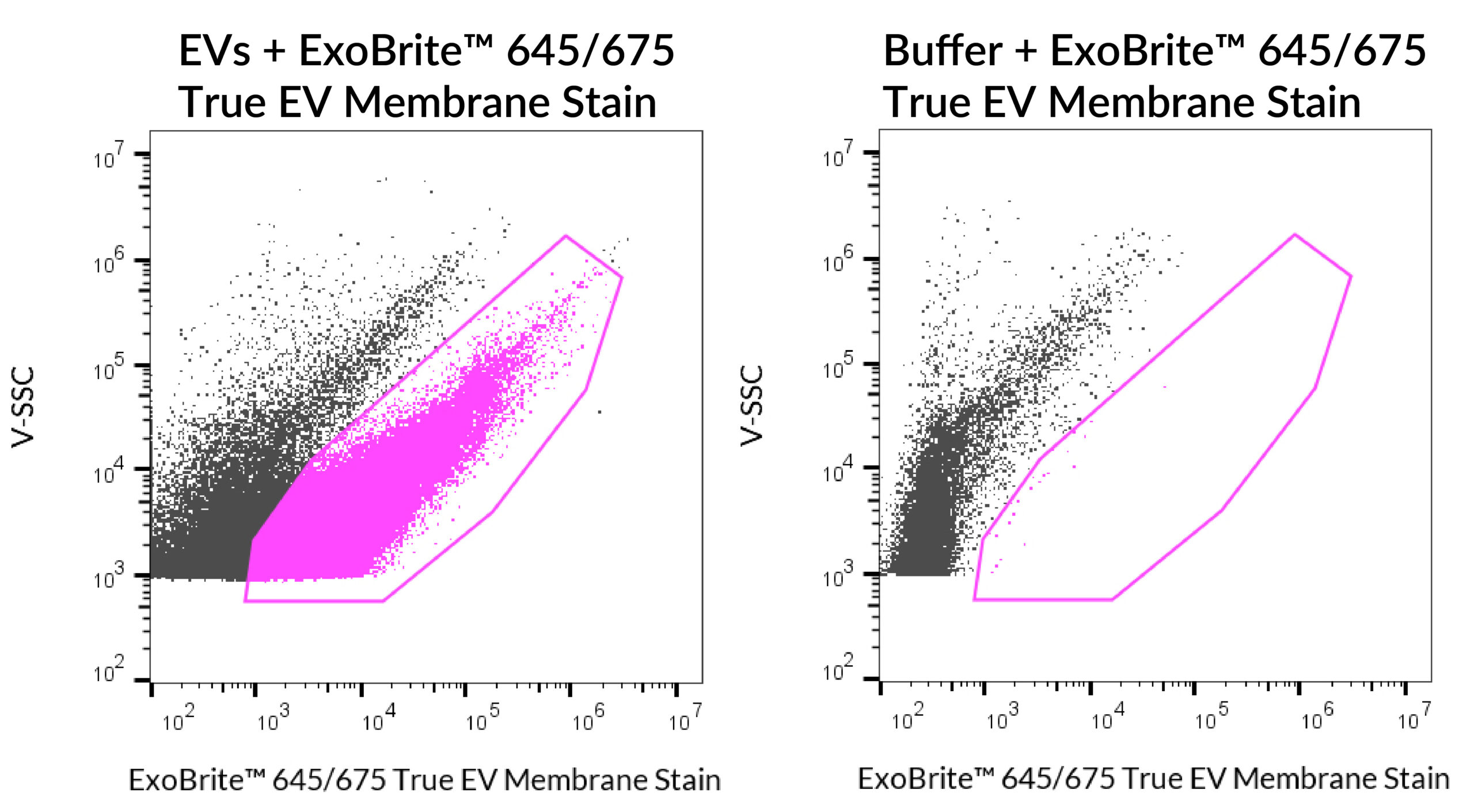
ExoBrite™ 645/675 True EV Membrane Stain was used to stain SEC EVs isolated from MCF-7 cells (left panel). The samples were run on a CytoFLEX LX flow cytometer in the R660 (APC) channel. The gated population shows that stained EVs are easily distinguished from background, compared to the dye in buffer alone (right panel).
Full Citation
Whitney, L. S., College, E. R., Peña-Ekker, J., Cannon, C. A., Mason, K. M., Wilson, J. N., & Pullan, J. E., PhD. (2026). Isolation and characterization of bovine-milk derived extracellular vesicles using a modified aqueous two-phase system. BioTechniques, 78(1–12), 55–64. https://doi.org/10.1080/07366205.2026.2633107