New Products
 Earth-Friendly Products
 Biotium Choice Antibodies
 Special Offers
 Quick Order Tool


Content #1
Content #1
Content #1
Alzheimer’s disease (AD) is characterized by the accumulation of abnormal tau protein structures, with increasing evidence suggesting that small, soluble tau oligomers may be among the earliest and most toxic forms of tau pathology. More research is being conducted specifically on the effects of tau oligomerization on the nuclear lamina, which maintains nuclear structure and organizes chromatin. Understanding how these toxic aggregates disrupt the lamina’s function may provide new insight into how tau pathology alters gene regulation and other essential cellular processes.
In a 2026 Acta Neuropathologica publication, Shuo Yuan et al. investigated how tau oligomerization affects nuclear structure and function in Alzheimer’s disease using a previously-developed OptoTau system. This system uses mCherry linked with the protein Cry2Olig which dimerizes under 488λ blue light exposure, thus allowing light-inducible oligomerization and subsequent fluorescence imaging of tau oligomers. Induced pluripotent stem cell (iPSC)-derived neurons were transduced with OptoTau or mCherry control and subjected to light-induced activation. Biotium's low-toxicity NucSpot® Live 488 Nuclear Stain was used to label the cell nuclei during time-lapse tracking of nuclear morphology and mCherry-tau oligomer localization.
The researchers identified significant nuclear deformation compared with mCherry-only controls, particularly that oligomeric tau directly interacts with nuclear lamina proteins and causes the nuclear membrane to fold inward and chromatin to become decompacted. They showed that nuclear lamina disruption is an early indicator of AD pathology and coincides with the emergence of pathological tau, with the damage becoming progressively more severe as tau pathology advances.
These findings are promising for a better understanding of how early tau pathology may trigger nuclear dysfunction in Alzheimer’s disease, and they suggest that protecting nuclear integrity could represent a potential therapeutic strategy. By enabling researchers to visualize nuclei in living neurons while tau aggregation is actively occurring, Biotium’s NucSpot® Live Cell Nuclear Stains can help advance research into the spatial and temporal relationship between protein aggregation, nuclear architecture, and neuronal dysfunction.
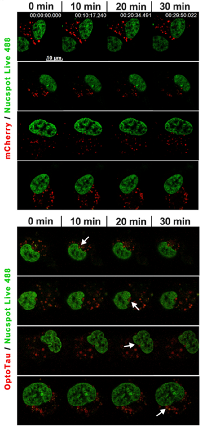
Time-lapse images of mCherry control vs. OptoTau neurons over 0, 10, 20, and 30 minutes of blue light with nuclei marked with NucSpot® Live 488. OptoTau granules progressively cluster at and deform the nucleus; mCherry granules move randomly and don't localize. Figure adapted from Shuo Yuan et al. 2026. Reproduced under CC BY 4.0.
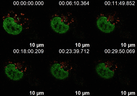
Supplemental video-2: Live cell imaging of 30 minutes of blue light activation shows that OptoTau granules quickly aggregated toward the nuclear envelope, leading to nuclear deformation. Reproduced under CC BY 4.0.
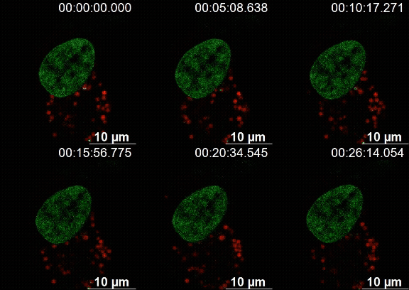
Live cell imaging of 30 minutes of blue light activation shows that mCherry particles move about randomly within the cell. Reproduced under CC BY 4.0.
Learn more about Biotium’s fluorescent cellular stains with sensitive visualization of cellular structures for neuroscience research and other complex biological processes.
Full Citation
Yuan, S., Essepian, N., Roberts, R. et al. Tau oligomerization induces nuclear lamina invagination and chromatin remodeling in Alzheimer’s disease. Acta Neuropathol 151, 43 (2026). https://doi.org/10.1007/s00401-026-03018-1