New Products
 Earth-Friendly Products
 Biotium Choice Antibodies
 Special Offers
 Quick Order Tool







Content #1
Content #1
Content #1
Validated CD9 antibody for detection of EVs, formulated to minimize aggregation, and conjugated to high-performance CF® Dyes for STORM.
ExoBrite™ STORM CD9 Antibody is designed for detecting the EV marker CD9 using stochastic optical reconstruction microscopy (STORM). Conjugated to optimized CF® Dyes for superior STORM performance, the antibody and its buffer are also specifically formulated for reliable detection of isolated EVs.
The most common proteins used as EV markers are CD9, CD63, and CD81, members of the tetraspanin family. These tetraspanins are broadly expressed on many cell types and can therefore be detected on many types of EVs, but their expression levels vary depending on the cell type of origin.
Other commercially available antibodies for tetraspanin proteins CD9, CD63, and CD81 are generally not validated for isolated EVs or super-resolution imaging, and may require tedious optimization for your EV prep and staining protocol.
ExoBrite™ STORM Antibodies are powered by our industry-leading CF® Dyes for STORM. Available in CF®498, CF®568, CF®583R, and CF®647Plus, these dyes offer exceptional photochemical switching properties—delivering superior signal quality and image reconstruction for unmatched performance in STORM.
The labeled antibody was carefully selected and validated for robust detection of isolated EVs. In addition, the antibodies are provided in a proprietary buffer formulation for reduced antibody aggregation and brighter EV staining for optimal accuracy and signal-to-noise. ExoBrite™ STORM Antibodies are compatible with co-staining with other fluorescently labeled antibodies or for bead-bound EV staining protocols.
Cholera toxin subunit B (CTB) conjugated to high-performance CF® Dyes for STORM are also available, as well as tetraspanin antibodies optimized for detection of EVs by flow cytometry and western blotting.
Learn more about our CF® Dyes for super-resolution imaging, as well as our other ExoBrite™ stains, antibodies, and other reagents developed specifically for EV detection and analysis.
| Antibody | Ex/Em | Size | Conc. | Detection Channel | Catalog No. |
|---|---|---|---|---|---|
| ExoBrite™ STORM CF®498 CD9 Antibody | 498/519 nm | 500 uL | 100 ug/mL | FITC | P003-498ST-500 |
| ExoBrite™ STORM CF®568 CD9 Antibody | 562/584 nm | 500 uL | 100 ug/mL | Cy®3 | P003-568ST-500 |
| ExoBrite™ STORM CF®583R CD9 Antibody | 585/609 nm | 500 uL | 100 ug/mL | Texas Red® | P003-583RST-500 |
| ExoBrite™ STORM CF®647Plus CD9 Antibody | 652/668 nm | 500 uL | 100 ug/mL | Cy®5 | P003-647PST-500 |
| ExoBrite™ STORM CF®498 CD63 Antibody | 498/519 nm | 500 uL | 100 ug/mL | FITC | P004-498ST-500 |
| ExoBrite™ STORM CF®568 CD63 Antibody | 562/584 nm | 500 uL | 100 ug/mL | Cy®3 | P004-568ST-500 |
| ExoBrite™ STORM CF®583R CD63 Antibody | 585/609 nm | 500 uL | 100 ug/mL | Texas Red® | P004-583RST-500 |
| ExoBrite™ STORM CF®647Plus CD63 Antibody | 652/668 nm | 500 uL | 100 ug/mL | Cy®5 | P004-647PST-500 |
| ExoBrite™ STORM CF®498 CD81 Antibody | 498/519 nm | 500 uL | 100 ug/mL | FITC | P005-498ST-500 |
| ExoBrite™ STORM CF®568 CD81 Antibody | 562/584 nm | 500 uL | 100 ug/mL | Cy®3 | P005-568ST-500 |
| ExoBrite™ STORM CF®583R CD81 Antibody | 585/609 nm | 500 uL | 100 ug/mL | Texas Red® | P005-583RST-500 |
| ExoBrite™ STORM CF®647Plus CD81 Antibody | 652/668 nm | 500 uL | 100 ug/mL | Cy®5 | P005-647PST-500 |
| ExoBrite™ STORM CF®568 CD9 (Mouse) Antibody | 562/584 nm | 500 uL | 100 ug/mL | Cy®3 | P018-568ST-500 |
| ExoBrite™ STORM CF®647Plus CD9 (Mouse) Antibody | 652/668 nm | 500 uL | 100 ug/mL | Cy®5 | P018-647PST-500 |
| ExoBrite™ STORM CF®568 CD63 (Mouse) Antibody | 562/584 nm | 500 uL | 100 ug/mL | Cy®3 | P022-568ST-500 |
| ExoBrite™ STORM CF®647Plus CD63 (Mouse) Antibody | 652/668 nm | 500 uL | 100 ug/mL | Cy®5 | P022-647PST-500 |
| ExoBrite™ STORM CF®568 CD81 (Mouse/Rat) Antibody | 562/584 nm | 500 uL | 100 ug/mL | Cy®3 | P019-568ST-500 |
| ExoBrite™ STORM CF®647Plus CD81 (Mouse/Rat) Antibody | 652/668 nm | 500 uL | 100 ug/mL | Cy®5 | P019-647PST-500 |
Extracellular vesicles (EVs) are nanoscale, membrane-bound particles released by cells that play important roles in intercellular communication by transporting proteins, lipids, and nucleic acids. Bovine milk is an abundant and accessible source of extracellular vesicles with potential applications in therapeutic delivery, diagnostics, and biomedical research. However, efficiently isolating these vesicles from milk remains challenging due to the complexity of the biological matrix and limitations of conventional isolation methods. Conventional ultracentrifugation or size exclusion chromatography approaches can require expensive, specialized equipment and may produce lower yields or affect vesicle integrity. Developing simpler, scalable, and cost-effective isolation methods could make EV research more accessible to laboratories with limited resources.
In a 2026 BioTechniques publication, Whitney, L. et al. investigated a modified aqueous two-phase system (ATPS) for isolating and characterizing EVs from raw bovine milk. Biotium’s ExoBrite™ 645/675 True EV Membrane Stain was used to fluorescently label the isolated EV membranes for flow cytometry analysis. The researchers combined the membrane stain with antibodies against EV-associated proteins CD63 and TSG101 to confirm the presence and identity of the isolated vesicles. Flow cytometry detected substantial populations of CD63- and TSG101-positive EVs above background, supporting successful EV recovery using the ATPS approach. Additional characterization found that the isolated particles exhibited typical EV morphology and size, as well as protein, lipid, and nucleic acid profiles consistent with extracellular vesicles.
Together, these findings demonstrated that the modified ATPS method can recover intact, biologically relevant EVs from raw bovine milk while requiring less specialized equipment than conventional isolation approaches. These findings are promising toward expanding access to extracellular vesicle research and unlocking the therapeutic and biomedical potential of milk-derived EVs.
By providing reliable fluorescent tools for identifying and characterizing EV populations, Biotium’s ExoBrite™ True EV Membrane Stains can help researchers visualize and distinguish extracellular vesicles during flow cytometry and other analytical workflows, supporting research into EV biology, isolation methods, and potential applications in drug delivery and diagnostics.
Learn more about Biotium’s wide range of EV stains, including ExoBrite™ WGA, CTB, and Annexin V conjugates for bright and sensitive EV surface staining. Biotium also offers ExoBrite™ Flow Antibody Conjugates and ExoBrite™ Antibody Cocktails for optimized detection of EV Markers by flow and fNTA.
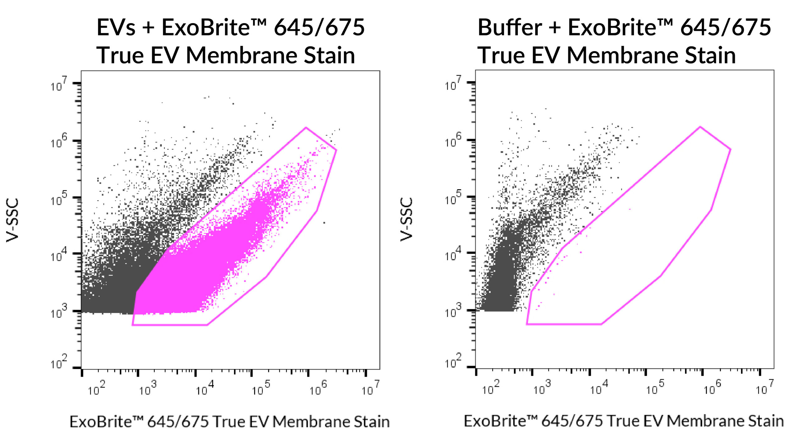
ExoBrite™ 645/675 True EV Membrane Stain was used to stain SEC EVs isolated from MCF-7 cells (left panel). The samples were run on a CytoFLEX LX flow cytometer in the R660 (APC) channel. The gated population shows that stained EVs are easily distinguished from background, compared to the dye in buffer alone (right panel).
Full Citation
Whitney, L. S., College, E. R., Peña-Ekker, J., Cannon, C. A., Mason, K. M., Wilson, J. N., & Pullan, J. E., PhD. (2026). Isolation and characterization of bovine-milk derived extracellular vesicles using a modified aqueous two-phase system. BioTechniques, 78(1–12), 55–64. https://doi.org/10.1080/07366205.2026.2633107
Extracellular vesicles (EVs) derived from mesenchymal stem cells (MSCs) are emerging as powerful, cell-free immunomodulatory therapies for inflammatory diseases such as COVID-19. However, because the mechanism is poorly understood, optimizing EV-based therapies remains challenging.
In a 2025 Springer Nature study, Infante et al. investigated how COVID-19 patient serum reshapes the transcriptome and paracrine activity of Wharton’s jelly–derived MSC stem cells (WJ-MSCs). WJ-MCSs exposed to serum from hospitalized COVID patients showed downregulation of NEAT1 and MALAT1, two pro-inflammatory two long noncoding RNAs (lncRNAs). Furthermore, the researchers found that EVs derived from the treated cells had enhanced immunosuppressive activity when administered to T-cells.
The researchers isolated EVs from WJ-MSC cells after NEAT1 and/or MALAT1 knockdown, and tested whether there was an effect on T-cell proliferation. A Western blot of EVs derived from control and lncRNA-knockdown MSCs were probed with ExoBrite™ 680/700 CD81 Western Antibody. ExoBrite™ 770/800 Calnexin Western Antibody was also used as an endoplasmic reticulum marker to assess cellular contamination.
EV enriched samples in control, NEAT1 knockdown, MALAT1 knockdown, and NEAT1/MALAT1-double knockdown were confirmed by bright CD81 detection and the absence of Calnexin. They found that the MALAT1 knockdown EVs were found to have an inhibitory effect on T-cell proliferation. These results illustrate the importance of EV characterization using tools like Biotium’s ExoBrite™ antibodies in translational EV research.
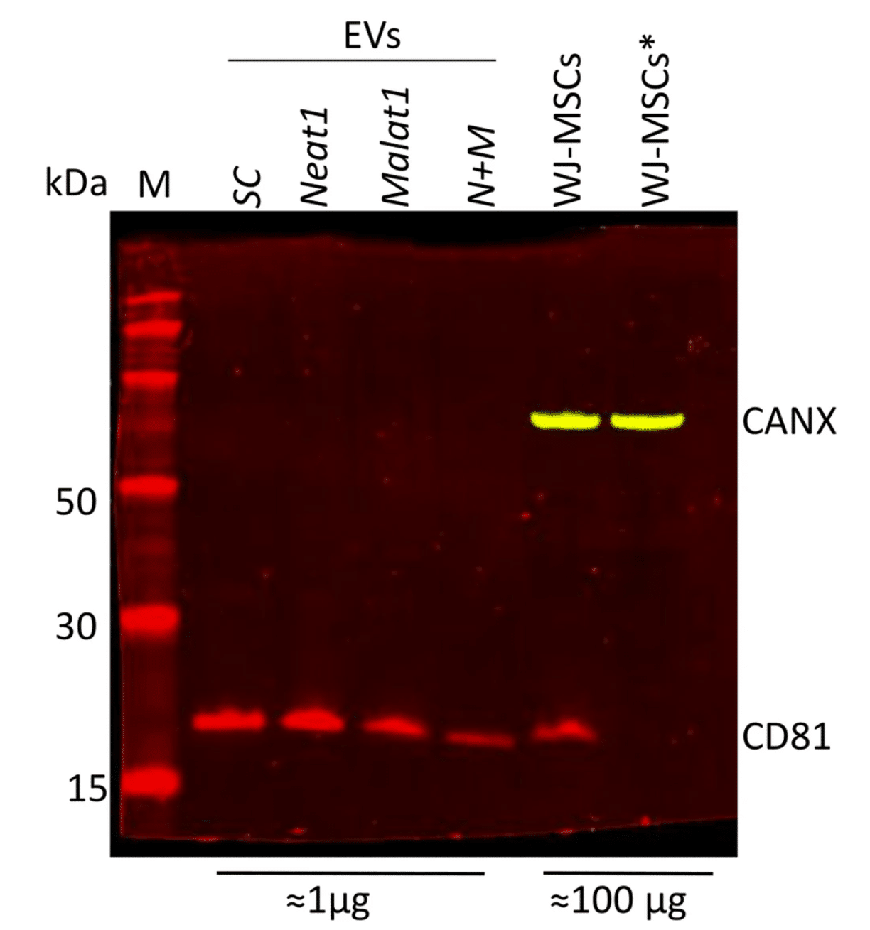
Isolation and characterization of EVs from various lncRNA knock-down WJ-MSCs. Western blot analysis using ExoBrite™ 680/700 CD81 and ExoBrite™ 770/800 Calnexin in EV and MSC lysates. Asterisk (*) indicates reduced conditions used in the MSCs lysate. Modified from Infante et. al. Reproduced under CC BY 4.0.
Learn more about Biotium’s many stains and antibodies for EV research, including ExoBrite™ CD9/CD63/CD81 Antibody Cocktails for flexible and bright multiplexing detection by flow cytometry. Biotium also offers ExoBrite™ stains for pan-EV labeling, optimized fluorescent conjugates of CTB, WGA, and Annexin V for EV detection, ExoBrite™ antibodies for STORM imaging, and more.
Full Citation:
Infante, A., Cabodevilla, L., Gener, B. et al. Modulation of NEAT1 and MALAT1 expression in WJ-MSCs by Covid-19 serum: a foundation for EVs-mediated therapy. Respir Res 26, 313 (2025). https://doi.org/10.1186/s12931-025-03394-4
While early studies of EVs attempted to use first-generation membrane dyes like DiI or PKH to stain EVs, more recently this class of dyes has been found to be largely unsuitable for EV staining due to their high degree of aggregation. Dye aggregation not only generates nonspecific particles that are indistinguishable from EVs in flow cytometry, but also results in poor EV labeling efficiency. Biotium developed the ExoBrite™ True EV Membrane Stains in response to our customers difficulties with using traditional membrane dyes to stain EVs. See our Literature Digest for more information.