 New Products
 Earth-Friendly Products
 Biotium Choice Antibodies
 Special Offers


Content #1
Content #1
Content #1
Unique kits designed for rapid, simple, and gentle covalent labeling of glycoproteins on the surface of live cells with fluorescent dyes or biotin.
GlycoLiner™ Kits are designed for labeling glycoproteins on the surface of live cells with fluorescent dyes or biotin. The reaction is rapid, simple, and gentle, and produces highly selective, covalent labeling of the cell surface that is compatible with subsequent fixation, permeabilization, and immunofluorescence staining or cell lysis.
GlycoLiner™ Kits were designed as a superior alternative for labeling cell surfaces in live cultures. Unlike other covalent cell surface labels which can have significant cytoplasmic background, GlycoLiner™ staining of dead cell cytoplasm is less intense, providing easier imaging of cell surfaces.
The labeling procedure uses aminooxy chemistry to label live cells after a mild oxidation treatment to convert glycoprotein sugars into carbonyl groups (aldehydes and ketones). The reactive GlycoLiner™ Dye or Biotin then reacts with the carbonyl groups to form a stable covalent oxime bond. GlycoLiner™ technology uses novel self-catalyzing aminooxy groups that are highly reactive at neutral pH without the need to add a catalyst. This is in contrast to glycoprotein labeling using conventional aminooxy labels, which is not only slow but also requires the use of high concentrations of reactive dye and catalyst in acidic buffer. The GlycoLiner™ labeling procedure is complete in about 20 minutes, and can be performed at room temperature or 4°C.
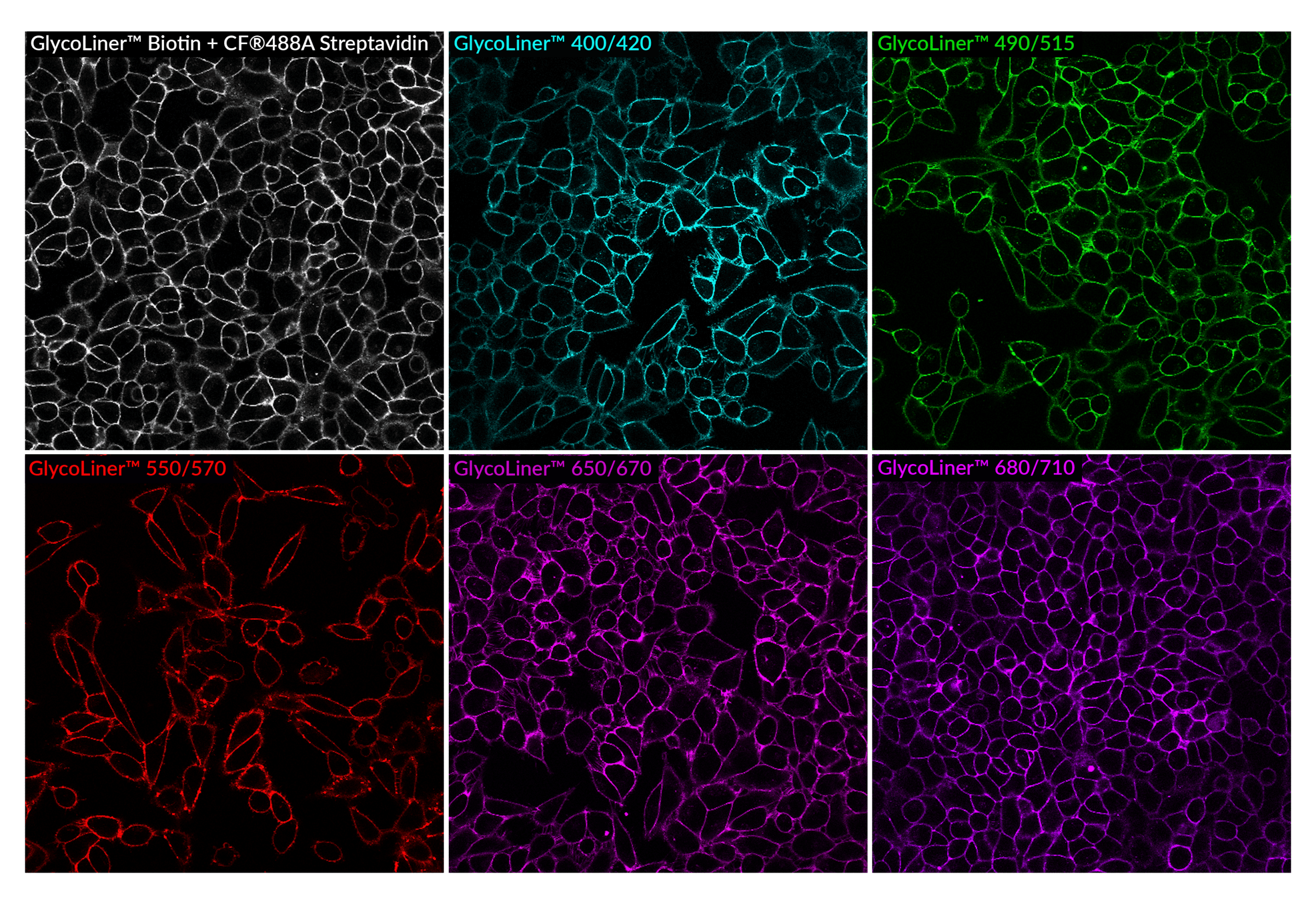
Live HeLa cells labeled with GlycoLiner™ Reactive Biotin detected with CF®488A streptavidin, or GlycoLiner™ Reactive Dyes, imaged by confocal microscopy using a 40X objective.
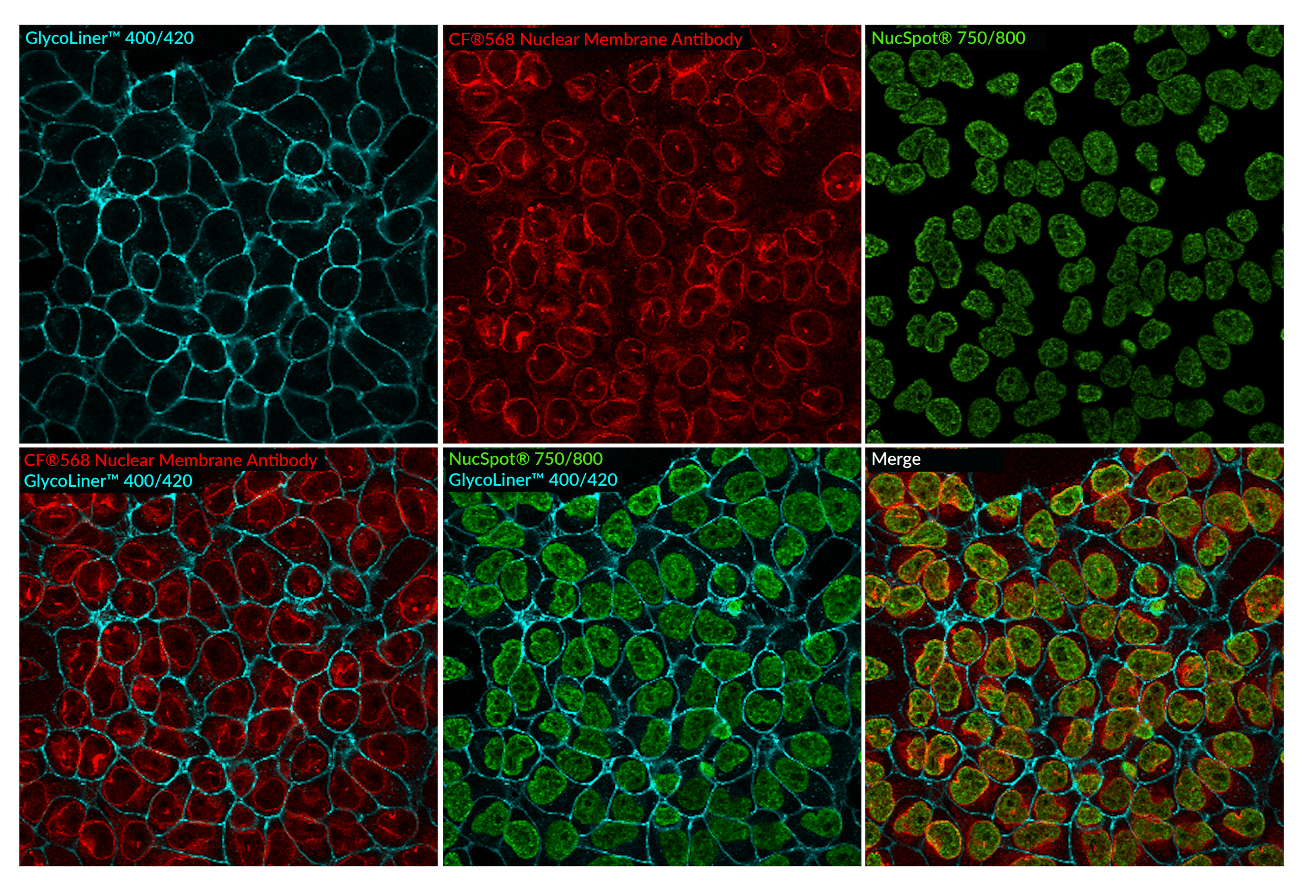
HeLa cells stained with GlycoLiner™ 400/420 (blue), then fixed with PFA, blocked and permeablized, and stained with CF®568 Recombinant Monoclonal Mouse anti-Nuclear Membrane Antibody (2406.NM) (red) and NucSpot® 750/800 Nuclear Stain (green). Imaged on an Evident FV4000 confocal system with 405 nm, 568 nm, and 730 nm laser lines.
GlycoLiner™ Kits are part of Biotium's family of unique and innovative cell membrane stains designed for specific applications. For staining cells already fixed with formaldehyde, we recommend our CytoLiner™ Fixed Cell Membrane Stains. For workflows requiring staining live cells just before fixation, Biotium also offers CellBrite® Fix Membrane Stains and MemBrite® Fix Cell Surface Staining Kits. CF® Dye conjugated lectins are available, including Concanavalin A (Con A) and Wheat Germ Agglutinin (WGA), for visualizing cell surfaces in live or fixed cells. For longer term monitoring of cell surfaces in live cells, we recommend CellBrite® Steady Dyes. To find the right stain for your application, see our Membrane & Cell Surface Stains Comparison, or download our Membrane & Surface Stains Brochure.
| Dye | Abs/Em (nm) | Detection channel | Size | Catalog No. |
|---|---|---|---|---|
| GlycoLiner™ 400/420 | 400/419 | DAPI, Alexa Fluor® 405 | 100 labelings | 30144-T |
| 500 labelings | 30144 | |||
| GlycoLiner™ 490/515 | 492/516 | FITC, Alexa Fluor® 488 | 100 labelings | 30145-T |
| 500 labelings | 30145 | |||
| GlycoLiner™ 550/570 | 550/570 | Cy®3, PE | 100 labelings | 30151-T |
| 500 labelings | 30151 | |||
| GlycoLiner™ 650/670 | 652/673 | Alexa Fluor® 647 | 100 labelings | 30146-T |
| 500 labelings | 30146 | |||
| GlycoLiner™ 680/710 | 680/706 | Alexa Fluor® 680 | 100 labelings | 30147-T |
| 500 labelings | 30147 | |||
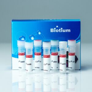
| GlycoLiner™ Biotin | N/A | N/A | 100 labelings | 30143-T |
| 500 labelings | 30143 |
ALEXA FLUOR is a registered trademark of Thermo Fisher Scientific; CY DYE is a registered trademark of Cytiva.
Advances in gene therapy increasingly depend on understanding how viral vectors behave within complex, multilayered human tissues. While retinal organoids serve as a powerful model for studying AAV efficacy, their dense, light-scattering architecture has historically limited the ability to visualize and quantify transduction at single-cell resolution. Conventional nuclear stains suffer from rapid photobleaching, cytotoxicity, and shallow imaging depth which hinder repeated live imaging and prevent accurate 3D cell segmentation throughout the organoid. Conventional membrane dyes also pose challenges for staining organoids due to poor penetration, uneven labeling, and rapid internalization by endocytosis.
In a 2025 Small Methods publication, Rogler et. al. developed a longitudinal imaging and deep-learning pipeline to map single-cell AAV transduction dynamics in intact human retinal organoids. This approach required robust and photostable live-cell stains compatible with deep (>100 µm) confocal imaging and repeated imaging over many days. To meet this need, the authors selected Biotium’s far-red NucSpot® Live 650 Nuclear Stain, which provides bright, uniform labeling with minimal phototoxicity and exceptional light penetration compared to blue- or green-excitable DNA dyes. CellBrite® Steady 550, a unique stain for long-term labeling of membranes in live cells, was also used for manual quantification of transduced cells to gauge the performance of their deep-learning method.
Using NucSpot® Live 650, the team captured high-contrast 3D nuclear signals across entire organoids and enabled the use of Cellpose, a deep-learning segmentation algorithm. Paired with GFP-expressing AAV reporters, this allowed precise quantification of transduced cells, as well as quantification of how transduction patterns evolve over time and spatial depth.
The end result revealed heterogeneous AAV penetration profiles, cell-type-specific susceptibility, and spatial gradients of transduction that would have been obscured using conventional methods. Biotium’s NucSpot® Live 650 Nuclear Stain and CellBrite® Steady 550 Membrane Stain enabled high-fidelity, longitudinal imaging in thick living tissues, making quantitative AAV mapping in 3D retinal models possible.
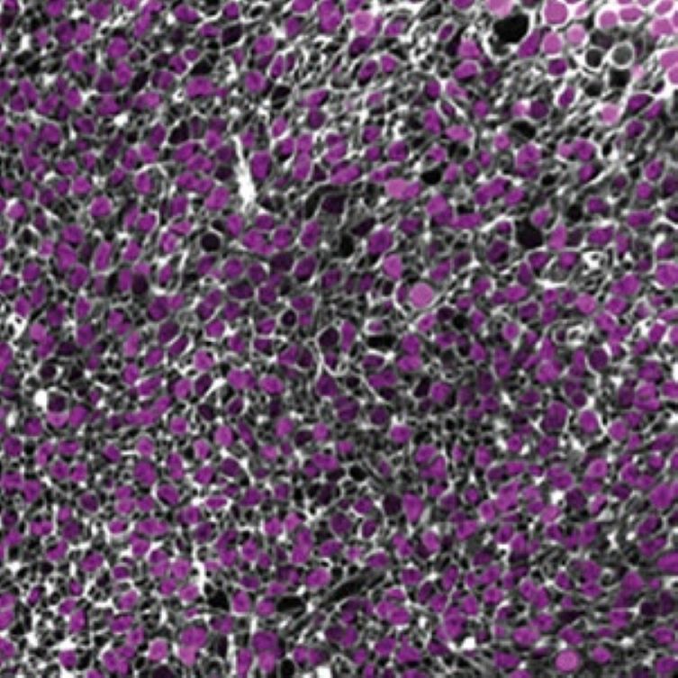
Confocal image of the center plane of the 3D stack of a 264 days old human retinal organoid without virus, stained with NucSpot Live 650 (magenta) and CellBrite Steady 550 (white). Credit: Rogler et al., Small Methods (2025). Reproduced under CC BY 4.0.
Biotium offers an extensive portfolio of bright and specific nuclear and membrane stains, with color options in the near-infrared for deep imaging. View our full selection of cell stains compatible with organoids and other 3D cultures.
Full Citation:
Rogler, T. S., Salbaum, K. A., Brinkop, A. T., Sonntag, S. M., James, R., Shelton, E. R., Thielen, A., Rose, R., Babutzka, S., Klopstock, T., Michalakis, S., & Serwane, F. (2025). 3D quantification of viral transduction efficiency in living human retinal organoids. Small Methods, 2025 Jun 12, e2401050. https://doi.org/10.1002/smtd.202401050
While CellBrite® Cytoplasmic Membrane Dyes can stain formaldehyde-fixed cells, they generally do not give good results in cryosections, possibly because the cell membrane integrity is disrupted, exposing other membrane structures to the dyes. Some customers have reported success using these dyes with vibratome sections.
CellBrite® Cytoplasmic Membrane Dyes are not suitable for membrane staining in FFPE samples as membrane lipids are extracted during the dewaxing and rehydration process. Similarly, acetone or methanol fixation of cryosections will extract lipids, leading to poor staining.
CellBrite® Fix, MemBrite® Fix, and CellBrite® Steady are recommended for use on live cells only. In fixed cells or sections they will label intracellular structures.
In some tissue types, lectins such as CF® Dye WGA Conjugates, CF® Dye Concanavalin A Conjugates, or CF® Dye PNA Conjugates may be useful for staining cell boundaries in FFPE or frozen sections. However, the staining pattern of lectins is highly dependent on cell and tissue type, so we recommend consulting the literature before trying these stains for your tissue of interest.
Alternatively, immunostaining using cell surface-specific antibodies could be done.
So far we have not found a universal plasma membrane stain for tissue sections. This is an application of interest to us and our customers, so we are working to find new solutions.
CellBrite® Cytoplasmic Membrane Dyes are too prone to aggregation to efficiently stain EVs. Some of the CellBrite® Fix, MemBrite® Fix, and CellBrite® Steady dye options have been reported for this application, however we do not recommend them. For optimal staining of exosome membranes we recommend our ExoBrite™ True EV Membrane Stains, which are novel lipophilic membrane dyes specifically designed and optimized for efficient staining of EV membranes with minimal dye aggregation. See our Extracellular Vesicle Research page for more information about our complete line of EV stains and antibodies.
