 New Products
 Earth-Friendly Products
 Biotium Choice Antibodies
 Special Offers


Content #1
Content #1
Content #1
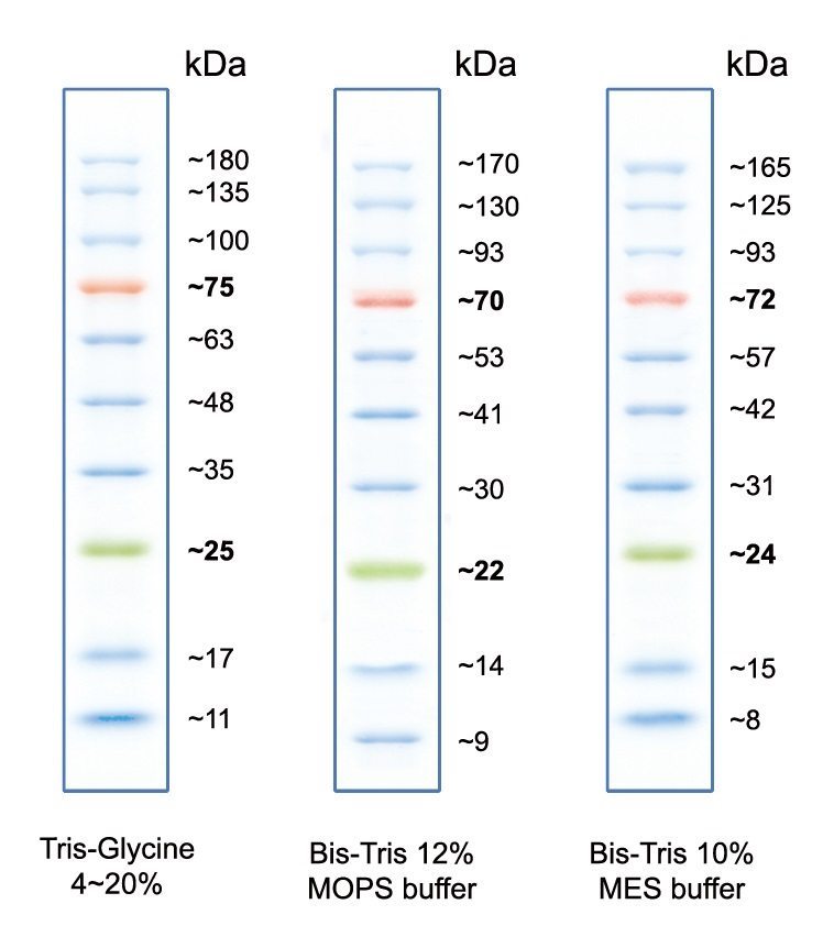
A visible three-color protein marker for SDS-PAGE or western blotting, with 10 bands ranging from 10 to 180 kDa.
Peacock™ Prestained Protein Marker is a three-color protein ladder that allows you to visually monitor protein separation during SDS-PAGE or protein transfer to membranes for western blotting. We also offer Peacock™ Plus Prestained Protein Marker, a three color marker with 12 bands ranging from 8 kDa to 245 kDa.
The Peacock™ Prestained Marker contains a total of 10 visible bands. This includes 8 blue bands ranging from 10 kDa to 180 kDa, plus a red band at 75 kDa and green band at 25 kDa for easy band identification. Peacock™ Prestained Protein Marker is ready to load with no heating or other preparation needed. Recommended loading is 3-5 uL per well for mini-gels.
Also visit our Protein Detection, Quantitation, & Analysis technology page to learn about our safe and sensitive protein gel stains and western blotting normalization reagents.
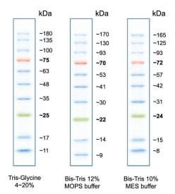
Peacock™ Prestained Protein Marker has 10 total protein bands (8 blue, 1 red, 1 green), spanning 10-180 kDa. Shown are the predicted banding patterns on different types of PAGE gels.

Comparison of Peacock™ Prestained Protein Marker visible and fluorescent bands on a 10% MOPS PAGE gel. The blue and green marker bands, but not the orange marker band, can be detected using a near-IR fluorescence image. Fluorescence was imaged in the 700 channel on a LICORbio Odyssey® M imaging system. Approximate band molecular weights are shown next to each marker.
Even though AccuOrange™ buffer does contain SDS, which is required for the dye to bind proteins, the assay is very sensitive to small changes in SDS concentration, and also cannot tolerate non-ionic detergents that form mixed micelles with SDS, like Triton®. Therefore we don't recommend using the kit for cell lysates or other samples with significant amounts of detergents.
Gels stained with One-Step Blue® can be dried just like gels stained with Coomassie. The stain will not interfere with the detection of radiolabeled proteins.
The AccuOrange™ assay is a fluorescent dye-based assay. The dye binds to proteins primarily through hydrophobic interactions. Proteins denature upon heating; the dye binds to the exposed hydrophobic pockets of the protein after cooling. The free AccuOrange™ dye is fluorogenic due to non-radioactive decay but becomes highly fluorescent due to the rigid conformation inside the pocket.
The AccuOrange™ assay more sensitive than traditional protein quantitation assays such as BCA, Bradford and Lowry, and shows superior linearity and reproducibility than the NanoOrange® protein quantitation assay (Thermo Fisher Sci.), but has low tolerance for detergents like SDS and Triton® X-100.
