 New Products
 Earth-Friendly Products
 Biotium Choice Antibodies
 Special Offers


 Powered by Bioz


Content #1
Content #1
Content #1
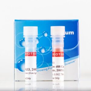
Fixable cytoplasmic stains for monitoring cell division by flow cytometry. The dyes can also be used to track cell populations in co-culture.
ViaFluor® SE Cell Proliferation Kits use amine-reactive dyes to covalently label cells throughout the cell cytoplasm and intracellular compartments for fixable fluorescent staining. Cell proliferation dyes are commonly used to monitor cell division by flow cytometry. The dyes also can be used to stably label cells to image cell morphology, or to track cell populations in mixed co-culture experiments.
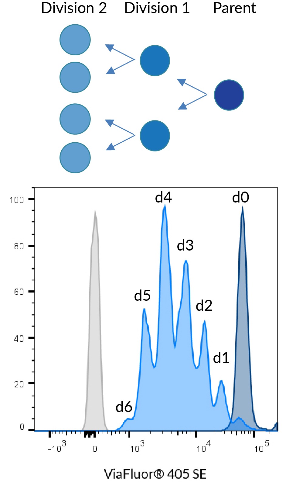
ViaFluor® SE dyes are membrane-permeant compounds that are initially non-fluorescent esters, but are converted to fluorescent dyes by intracellular esterases and will covalently react with amine groups on intracellular proteins at the same time, forming fluorescent conjugates that are retained in the cell. Immediately after staining, a single bright fluorescent population will be detected by flow cytometry. With each cell division, daughter cells inherit roughly half of the fluorescent label, allowing the number of cell divisions that occur after labeling to be detected by the appearance of successively dimmer fluorescent peaks on a flow cytometry histogram compared to cells analyzed immediately after staining. Thus, cell proliferation dyes can be used to track multiple cell divisions of cells grown in culture or injected in vivo after labeling with the ViaFluor® SE dye.
The number of assays that can be performed per kit depends on the dye concentration used (see the product protocol for more information). When used at 1 uM to label 106 cells in one mL, each dye vial can be used for 90-100 labelings.
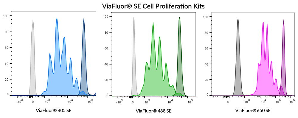
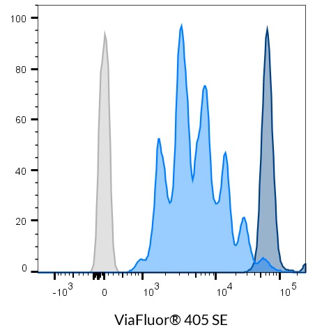
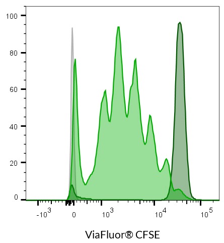
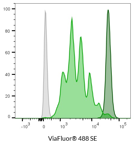
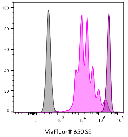
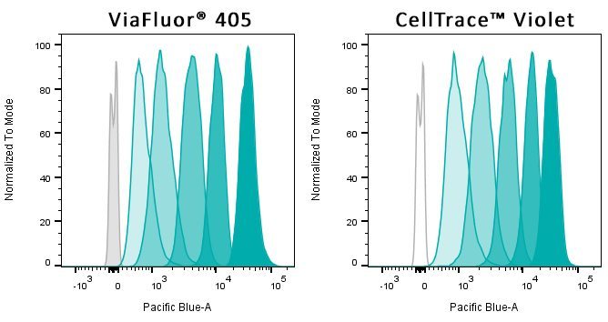
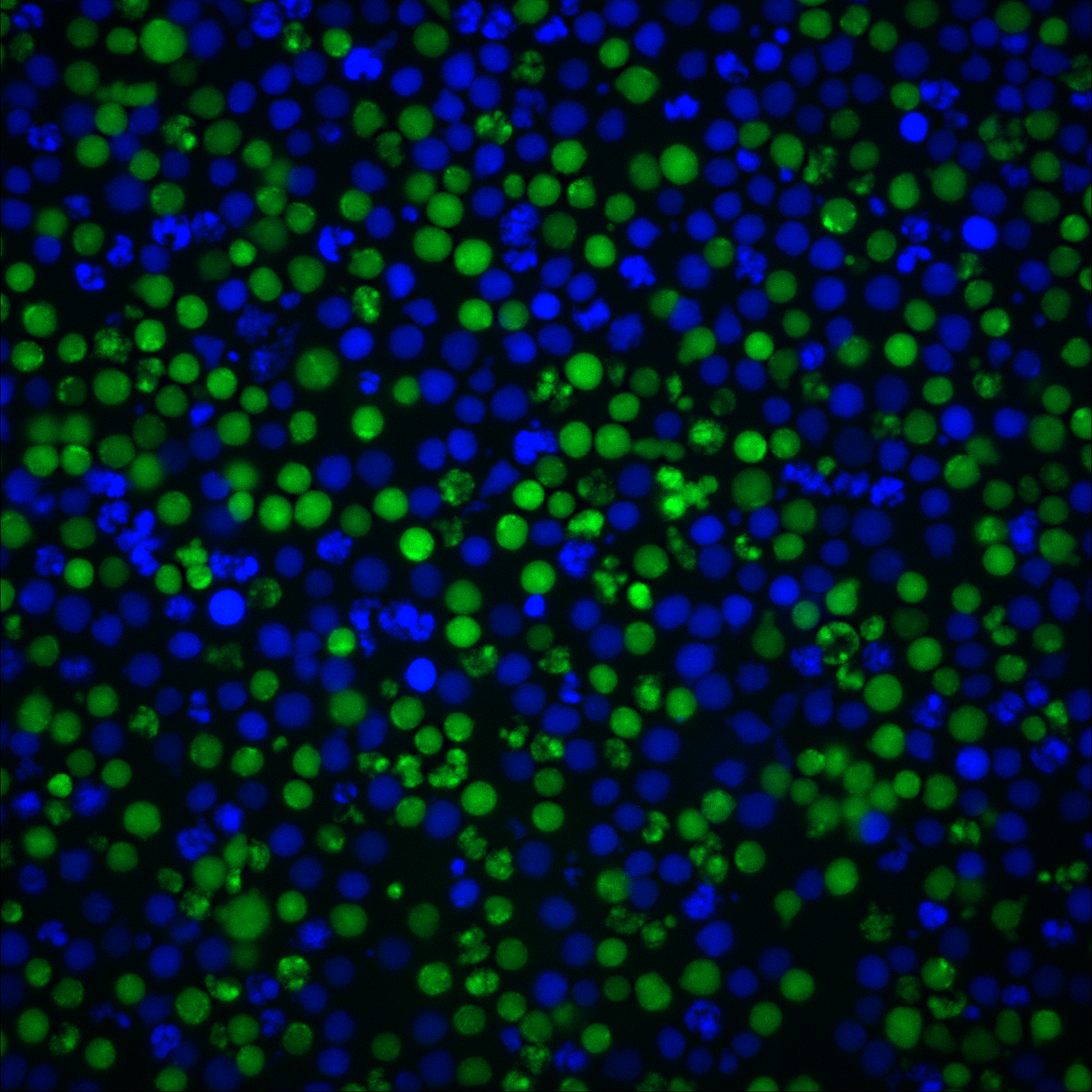
Human PBMCs were stained with ViaFluor® 405 (left), ViaFluor® 488 (center), or ViaFluor® 650 (right). Cells were stimulated with Dynabeads® Human T-Activator CD3/CD28 beads and 100 ng/mL IL-2. Cells were analyzed 4 days post-induction. CD3+ T-cells are shown. Unstimulated cells (dark peaks) and unstained cells (gray) are shown for comparison.
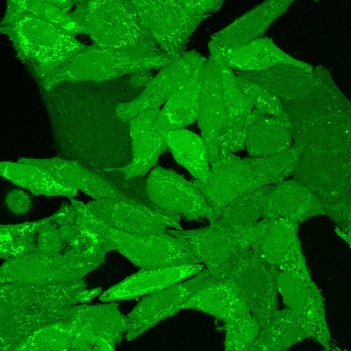
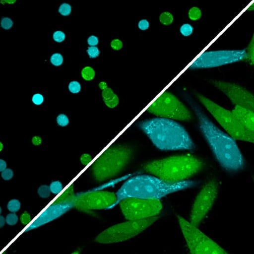
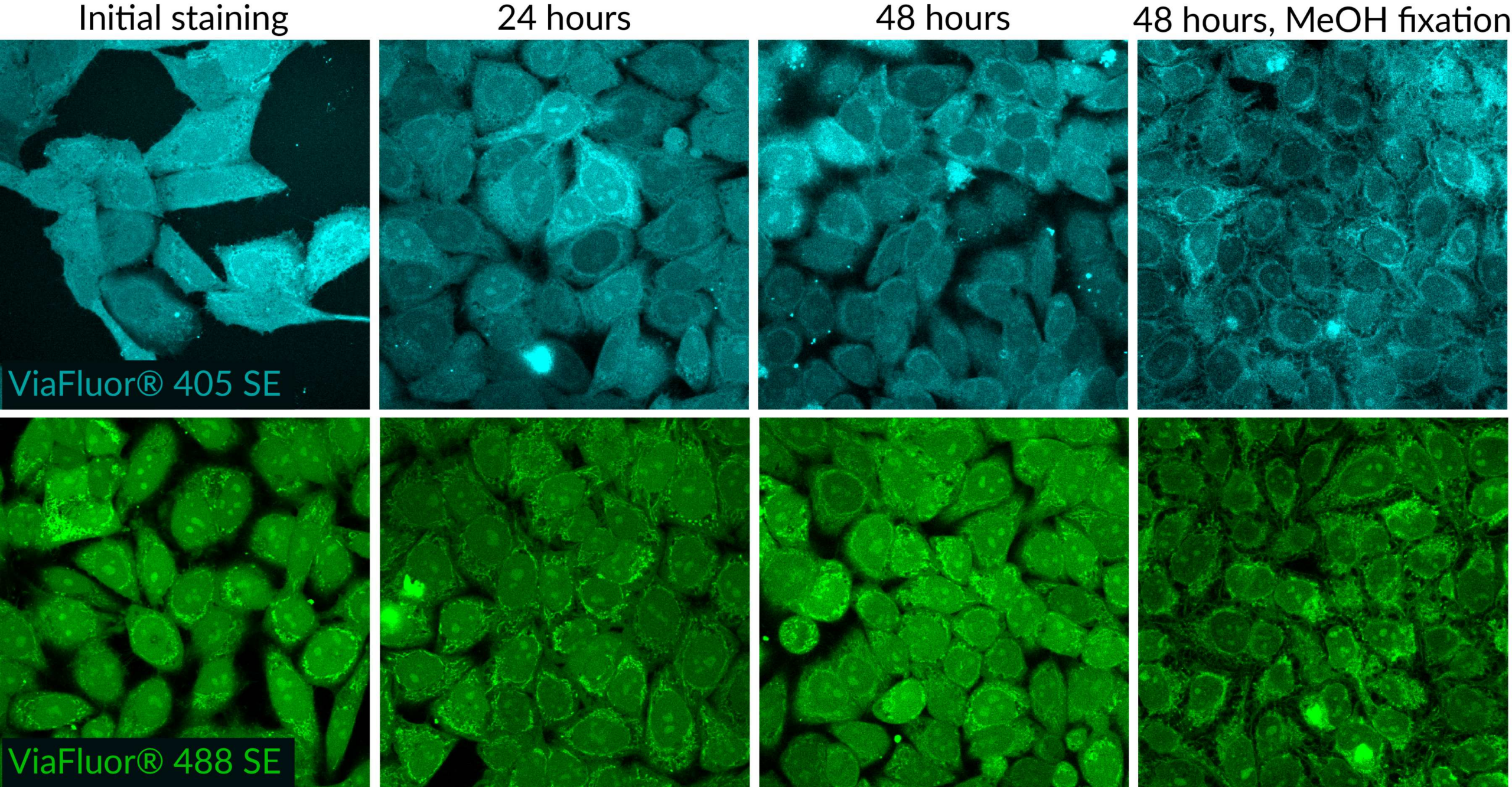
ViaFluor® SE dyes also can be used for imaging cell morphology or identifying cells in co-culture by microscopy. Because the staining is non-toxic and well-retained, it can be used for imaging live cells over time. See our Tech Tip: Using ViaFluor® SE Stains for Cell Tracing and Co-Culture.
All three ViaFluor® SE dyes can stain gram-positive bacteria, but not gram-negative bacteria. ViaFluor® CFSE stains the cytoplasm in yeast, but ViaFluor® 405 & ViaFluor® 488 stain the yeast cell periphery. See our Cellular Stains Table for more information on how our dyes stain various organisms.
Cell Division | Catalog No. | Ex/Em (nm) | Flow detection | Features |
|---|---|---|---|---|
| ViaFluor® 405 SE Cell Proliferation Kit | 30068 | 387/446 | Pacific Blue® | • ViaFluor® 405 SE replaces CellTrace™ Violet |
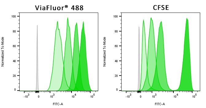
| ViaFluor® 488 SE Cell Proliferation Kit | 30086 | 495/524 | FITC | • ViaFluor® 488 SE is a unique, improved green dye to replace CFSE |
| ViaFluor® 650 SE Cell Proliferation Kit | 30139 | 653/682 | APC | • ViaFluor® 650 SE is an improved and cost-effective alternative to CellTrace™ Far Red |
| ViaFluor® CFSE Cell Proliferation Kit | 30050 | 495/519 | FITC | • Classic cell division tracing dye, see our ViaFluor® 488 SE for an improved alternative |
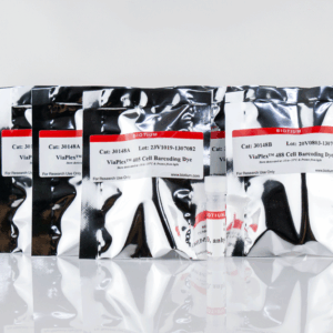
Biotium also offers the the ViaPlex™ 2-Color Cell Barcoding Kits for analyzing up to 15 samples in a single tube to save time and reagents, as well as Live-or-Dye™ Fixable Viability Staining Kits for robust dead cell detection in 18 colors spanning blue to near-IR. Learn more about our products for flow cytometry.
It has been reported in publications that concentrations of serum above 10% in the assay may affect the results.
See the following publications for more information
Our ViaFluor® SE Cell Proliferation assay is a dye dilution assay for cell division, like CFSE and CellTrace™ Violet from Thermo. This type of assay is commonly used to measure lymphocyte proliferative responses in culture and in vivo (if the labeled cells are injected back into mice). It requires flow cytometry to analyze and allows you to count how many cell divisions have occurred in the labeled cells.
For more information and a typical procedure for using fluorescent ViaFluor® SE Dyes with PMBCs, which can easily be adapted for use with other cell types, please see our Tech Tip: Measuring Cell Division in PMBCs by Flow Cytometry
If flow cytometry is not an option, we offer absorbance-based and fluorescence-based microplate assays for quantitating cell numbers. These measure mitochondrial activity (resazurin/MTT/XTT) or intracellular esterase activity (calcein AM) as a readout of viable cell numbers. Please visit the Cell Viability and Apoptosis technology page for more information.
The ATP-Glo™ assay is a luminescence assay for cellular ATP levels, which are proportional to the number of live cells. This assay requires a luminometer.
CellTrace is a trademark of Thermo Fisher Scientific.
Our Resazurin Cell Viability Assay (Cat. No. 30025) has red fluorescence (Ex/Em 530-560/590 nm), and is specifically designed for microplate reader. It is an economical, easy-to-use, and homogeneous (no-wash) assay for quantifying live cells. It is similar to alamarBlue®, PrestoBlue®, and CellTiter-Blue®.
The Calcein AM Cell Viability Assay (Cat. No. 30026) has green fluorescence (Ex/Em 485/530 nm), and also works well for microplate reader. This assay requires culture medium to be removed from cells before adding the viability dye in buffer. We also offer the Viability/Cytotoxicity Assay for Animal Live & Dead Cells, which combines calcein-AM with the fluorescent dead cell stain EthD-III, and is compatible with microplate reader.
To date, we have not identified a fluorescent cellular stain that will detect bacteria but not mammalian cells with high specificity, or vice versa. While some mammalian cell stains show weak staining of bacteria, they usually do show some signal, and will frequently stain dead bacteria more intensely than live bacteria.
We offer a selection of antibodies for specific bacterial antigens, which potentially have applications for differential staining of bacteria vs. mammalian cells, but we have not validated them in co-culture models.
Also see our Viability PCR Technology Page to learn about how PMA dye can be used for highly specific detection of microbial cell viability in complex samples.
CellBrite® and MemBrite® Stains were originally developed for staining mammalian cells in culture, but some of the stains also have been validated for other organisms and applications. For dyes to stain yeast or bacteria membranes, see Cellular Stains in Different Organisms. For information on staining other organisms or cell types, please see our Tech Tip: Researching Applications for Membrane Dyes.
The CellBrite® Cytoplasmic Membrane Dyes do not stain bacteria. The reactive CellBrite® Fix dyes stain both gram-positive and gram-negative bacteria, while the MemBrite® Fix dyes stain only gram-positive bacteria. However we have not tested these dyes for cell division tracking in bacteria.
There is literature describing the use of CFSE to track bacterial cell division, the ViaFluor® SE cell proliferation dyes are likely to work in a similar manner, but we have not tested this.
See our Cellular Stains Table for a comprehensive list of cellular stains with their ability to stain various cell types.
