 New Products
 Earth-Friendly Products
 Biotium Choice Antibodies
 Special Offers


 Powered by Bioz


Content #1
Content #1
Content #1
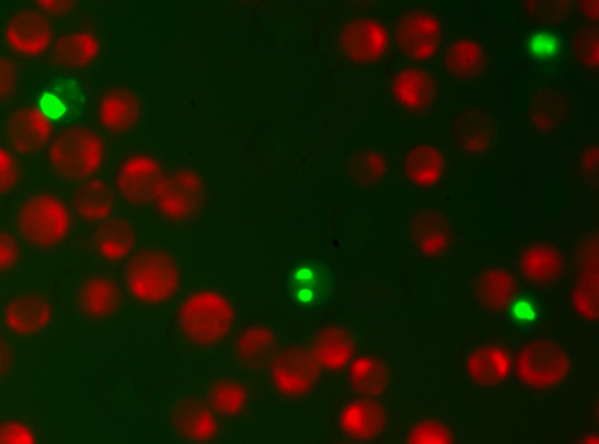
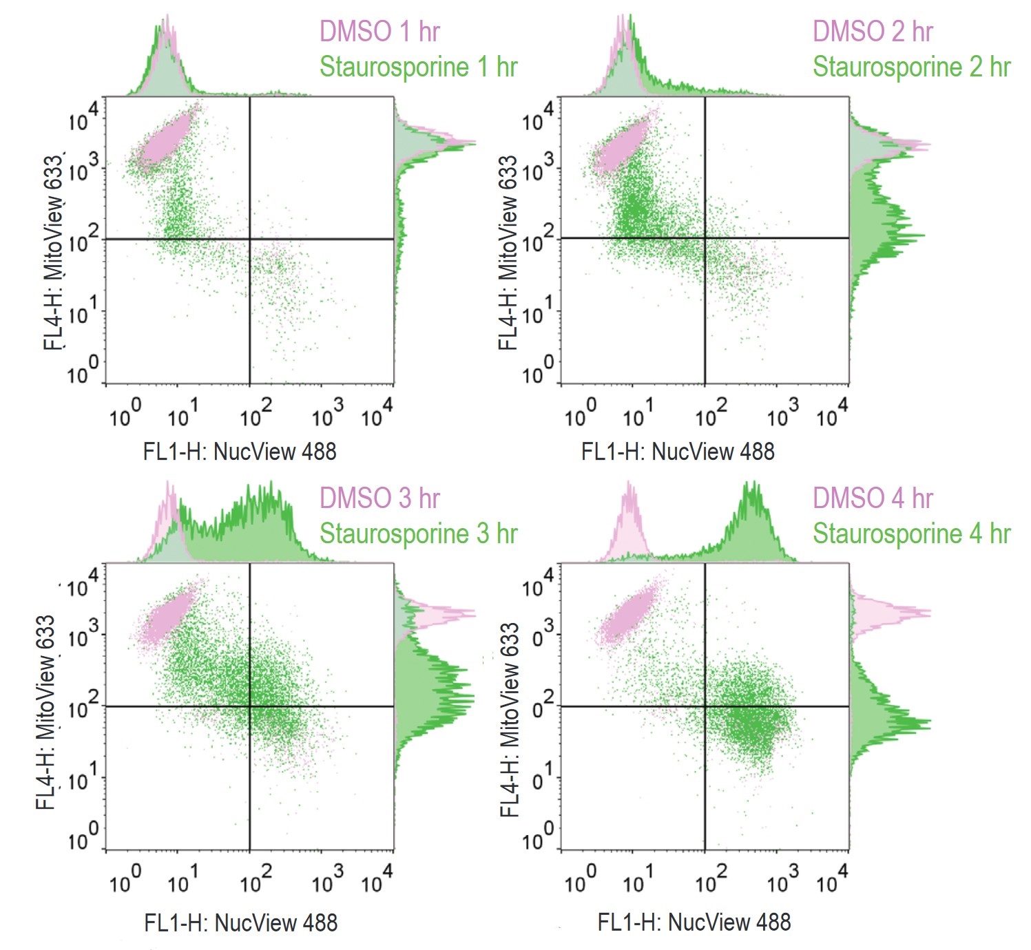
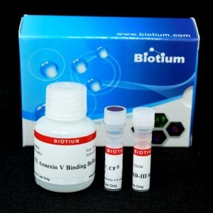
No wash, homogeneous assay for profiling apoptotic cells based on caspase-3 activity and changes in the mitochondrial membrane potential using fluorescence microscopy, real-time imaging, or flow cytometry.

This kit contains NucView® 488 Caspase-3 Substrate to detect apoptotic cells and MitoView™ 633 mitochondrial membrane potential dye.
NucView® 488 Caspase-3 Substrate is able to detect both intracellular caspase-3 and at the same time stain the cell nucleus, which is known to undergo morphological changes during apoptosis. MitoView™ 633 is a far-red fluorescent mitochondrial dye. The staining is dependent upon the mitochondrial membrane potential; thus, apoptotic cells exhibit a much lower MitoView™ 633 dye staining compared to healthy cells. Furthermore, the spectral separation of these two dyes minimizes fluorescence overlap. See a video of NucView® & MitoView™ in real-time imaging.
Note: The optimal detection settings for MitoView™ 633 are the same as for Cy®5 and other far-red dyes. However, the dye also has visible red fluorescence and can be imaged using Cy®3 settings as well. As a consequence, the dye cannot be used for two-color imaging with other red probes.
To learn about the advantages of monitoring apoptosis using NucView® caspase-3 substrates, visit the NucView® Technology Page.
Download list of curated NucView® references and validated cell lines.
Download list of curated NucView® references and validated cell lines.
Bioscience kits
The guaranteed shelf life from date of receipt for bioscience kits is listed on the product information sheet. Some kits have an expiration date printed on the kit box label, this is the guaranteed shelf life date calculated from the day that the product shipped from our facility. Kits often are functional for significantly longer than the guaranteed shelf life. If you have an older kit in storage that you wish to use, we recommend performing a small scale positive control experiment to confirm that the kit still works for your application before processing a large number of samples or precious samples.
Antibodies and other conjugates
The guaranteed shelf life from date of receipt for antibodies and conjugates is listed on the datasheet sheet which can be downloaded on the product page. Antibodies and other conjugates often are functional for significantly longer than the guaranteed shelf life. If you have an older conjugate in storage that you wish to use, we recommend performing a small scale positive control experiment to confirm that the product still works for your application before processing a large number of samples or precious samples.
For lyophilized antibodies, we recommend reconstituting the antibody with glycerol and antimicrobial preservative like sodium azide for the longest shelf life (note that sodium azide is not compatible with HRP-conjugates).
Chemicals, dyes, and gel stains
Biotium guarantees the stability of chemicals, dyes, and gel stains for at least a year from the date you receive the product. However, the majority of these products are highly stable for many years, as long as they are stored as recommended. Storage conditions can be found on the product information sheet or product safety and data sheet, material safety data sheet, and on the product label. Fluorescent compounds should be protected from light for long term storage.
If you have a Biotium compound that has been in storage for longer than one year that you wish to use, we recommend performing a small scale positive control experiment to confirm that the compound still works for your application before processing a large number of samples or precious samples.
Expiration date based on date of manufacture (DOM)
If your institution requires you to document expiration date based on date of manufacture for reagents, please contact [email protected] for assistance.
Chemical products with special stability considerations:
Esters
Ester compounds include the following:
Ester dyes are stable in solid form as long as they are protected from light and moisture. Esters are not stable in aqueous solution. Concentrated stock solutions should be prepared in anhydrous DMSO (see Biotium catalog no. 90082). Stock solutions in anhydrous DMSO can be stored desiccated at -20°C for one month or longer. Esters should be diluted in aqueous solution immediately before use. Succinimidyl esters (SE) should be dissolved in a solution that is free of amine-containing compounds like Tris, glycine, or protein, which will react with the SE functional group. AM esters and diacetate compounds should be dissolved in a solution that is free of serum, because serum could contain esterases that would hydrolyze the compound.
A note on CF® Dye succinimidyl ester stability
Succinimidyl esters (SE) are generally susceptible to hydrolysis, which can result in lower labeling efficiency. Many commercially available fluorescent dyes used for life science research are heavily sulfonated dyes which makes them particularly hygroscopic, worsening the hydrolysis problem. In addition, for several commercially available SE reactive dyes, the SE group is derived from an aromatic carboxylic acid, while the SE group in all of Biotium’s CF® Dyes is prepared from an aliphatic carboxylic acid. This structural difference reduces the susceptibility of CF® Dye SE reactive groups to hydrolysis, resulting in relatively stable reactive dyes with consistently higher labeling efficiency compared to other SE derivatives of other fluorescent dyes.
Maleimides, MTS and thiosulfate dyes
Like the succinimidyl ester dyes, these dyes are also susceptible to hydrolysis, although generally to a much lower degree. Thus, for long term storage, anhydrous DMSO is recommended for making stock solutions.
Other reactive dyes
Amines, aminooxy (also known as oxylamine), hydrazide, azide, alkyne, BCN, and tyramide reactive dyes, as well as dye free acids, are generally stable in aqueous solution when stored at -20°C for 6-12 months or longer, as long as no compounds are present that may react with the dye’s functional group. See the product information sheets for specific reactive dyes more information.
Coelenterazines and D-luciferin
Coelenterazines are stable in solid form when stored as recommended; they are not stable in aqueous solution. Concentrated coelenterazine stock solutions (typically 1-100 mg/mL) should be prepared in ethanol or methanol; do not use DMSO or DMF to dissolve coelenterazines, because these solvents will oxidize the compounds. Ethanol or methanol stocks of coelenterazine can be stored at -20°C or below for six months or longer; alcohol stocks may evaporate during storage, so use tightly sealing screw cap vials and wrap the vials with Parafilm for long term storage. Propylene glycol also can be used as a solvent to minimize evaporation. If the solvent evaporates, the coelenterazine will still be present in the vial, so note the volume in the vial prior to storage so that you can adjust the solvent volume to correct for evaporation if needed. Prepare working solutions in aqueous buffers immediately before use. Coelenterazines are stable for up to five hours in aqueous solution.
Aquaphile™ coelenterazines are water soluble formulations of coelenterazines. They are stable in solid form when stored as recommended. Aquaphile™ coelenterazines should be dissolved in aqueous solution immediately before use. They are stable for up to five hours in aqueous solution.
Note that coelenterazines are predominantly yellow solids, but may contain dark red or brown flecks. This does not affect product stability or performance. If your coelenterazine is uniformly brown, then it is oxidized and needs to be replaced.
D-luciferin is stable in solid form and as a concentrated stock solution when stored as recommended; it is not stable at dilute working concentrations in aqueous solution. Prepare concentrated D-luciferin stock solutions (typically 1-100 mg/mL) in water, and store in aliquots at -20°C or below for six months or longer. Prepare working solutions immediately before use.
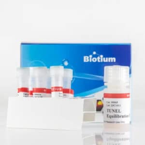
The NucView® caspase-3 substrates are activity-dependent i.e. require active caspase-3 enzyme. In dead, fixed and preserved cells or tissue sections, there are no active caspases and hence these substrates cannot be used. We also offer TUNEL assay kits that are suitable for apoptosis detection in fixed cells and tissues. The kits employ dUTPs conjugated to our exceptionally bright and photostable CF® dyes for single-step fluorescent TUNEL labeling of DNA strand breaks, a hallmark of apoptotic cells, and are suitable for analysis by fluorescence microscopy or flow cytometry.
The NucView® 488 Assay Kit for Live Cells (30029) contains NucView® 488 substrate at 0.2 mM in DMSO and caspase-3 inhibitor Ac-DEVD-CHO. The final DMSO concentration in the kit assay can be fairly high, which is undesirable for sensitive cell types. Because of this, we offer NucView® 488 Caspase-3 Substrate at 1 mM in DMSO (10402) and at 1 mM in PBS (10403), so customers can control the amount of DMSO in their assay. Note that in non-DMSO sensitive cell types, adding DMSO during the substrate incubation can enhance NucView® staining.
We also offer blue fluorogenic NucView® 405 in DMSO (10405) and PBS (10407), and orange fluorogenic NucView® 530 in DMSO (10406) and PBS (10408).
