Alveoli, the gas-exchange sites of the lungs, play a central role in lung function and disease. Damaged or insufficient alveoli are primary underlying factors in infant and chronic adult lung diseases. Effective treatments for these conditions are almost completely lacking. A deeper understanding of the process of alveolar development (alveologenesis) is therefore critical not only to understanding lung homeostasis and dysregulation leading to disease, but also for the development of therapies to repair and regenerate lung tissue.
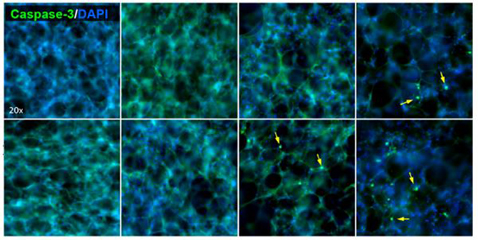
In a recent Nature Communications article, Akram et al. establish a method to visualize live alveologenesis in real-time using precision-cut lung slices (PCLS). In contrast to alveolar co-cultures or organoids, PCLS retain cell type ratios and cell–cell and cell–matrix interactions as seen in vivo. Cell viability in PCLS (300 μm thick) prepared from postnatal mice was determined by metabolic activity measurements, Live/Dead stains, and NucView® 488 caspase assays. Long-term, time-lapse imaging using a combination of live cell stains and conjugated antibodies reveals that the alveolar epithelial cells are highly mobile and show alveologenesis-specific cell behaviors: migration, septation, clustering, hollowing, and extension. Treatment with cytoskeleton inhibitors blebbistatin and cytochalasin D demonstrates that cell migration is a key driver of alveologenesis. This study reveals novel information about alveologenesis and lung biology, and provides an ex vivo system for genetic and pharmacological manipulation.
Full citation
Akram KM, Yates LL, Mongey R, Rothery S, Gaboriau DCA, Sanderson J, Hind M, Griffiths M, Dean CH. Live imaging of alveologenesis in precision-cut lung slices reveals dynamic epithelial cell behaviour. Nat Commun. 2019 Mar 12;10(1):1178. doi: 10.1038/s41467-019-09067-3.