As genetics and gene therapy techniques become more widely implemented in the treatment of genetic disorders and cancer, developing safe and effective gene delivery methods remains a challenge. Two of the most prevalent gene delivery vehicles are viral agents and synthetic chemical vectors. Despite viral agents being highly effective, they have many drawbacks, including limited cargo capacity, safety issues, and difficult production and quality control. Synthetic chemical carriers are superior to viral agents because they minimize these drawbacks. Metal complexes are emerging as a leading synthetic gene delivery vector option, but few have been tested for this application yet.
In a recent publication in Nucleic Acids Research, J. Malina et al. evaluated three enantiomeric pairs of Fe(II) metallohelices as nonviral DNA vectors for gene delivery. Using GelRed® in a gel retardation assay, they evaluated the DNA condensing ability of the Fe metallohelices. Results show all three metallohelices effectively reduced the amount of free DNA in the gel.
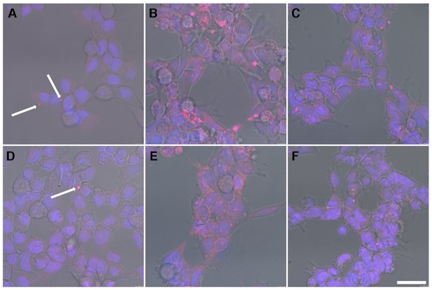
The authors then measured the efficiency of cellular uptake by treating HEK293 cells with a mixture of metallohelices and plasmid DNA prestained with GelRed®. Multitudinous red fluorescent spots were observed during confocal microscopy, confirming the successful cellular uptake of the vectors. Lastly, the authors conducted transfection studies on HEK293 cells using a GFP reporter gene to further evaluate the metallohelix gene delivery vectors. Fluorescence microscopy analysis demonstrated 2 out of the 3 enantiomer pairs yielded substantial numbers of GFP-positive cells, confirming successful transfection.
These findings indicate that Fe(II) metallohelices are suitable as DNA transfection agents for efficient gene delivery. Further studies on the design of these systems may lead to improved delivery of gene therapy treatments.
Learn more about Biotium’s highly sensitive and cell-impermeant GelRed® and GelGreen® Nucleic Acid Gel Stains.
Full Citation:
Malina, J., Kostrhunova, H., Novohradsky, V., Scott, P., & Brabec, V. (2022). Metallohelix vectors for efficient gene delivery via cationic DNA nanoparticles. Nucleic Acids Research, 50(2), 674–683. https://doi.org/10.1093/NAR/GKAB1277