Tyramide Signal Amplification (TSA) uses peroxidase (POD) catalysis to produce high-intensity labeling of biomolecular targets and enables the detection of low-abundance targets in immunocytochemistry (ICC), immunohistochemistry (IHC), and in situ hybridization (FISH) applications. Specifically, POD reacts with hydrogen peroxide (H2O2) to catalyze production of reactive tyramide radical intermediates, which covalently bind to electron-rich molecules such as tyrosine, phenylalanine, and tryptophan amino acids near the site of catalysis. The ability of POD to catalyze the deposition of many fluorochromes at the labeling site results in signal amplification and a significant increase in sensitivity compared to traditional immunofluorescence (IF). Multiplex TSA-based imaging has emerged as a central tool in spatial biology studies, with several comprehensive kits commercially available along with various stand-alone TSA components for customized and cost-effective panel design. While multiplex TSA staining has become an indispensable tool, further refinement could make the technique more sensitive and accessible.
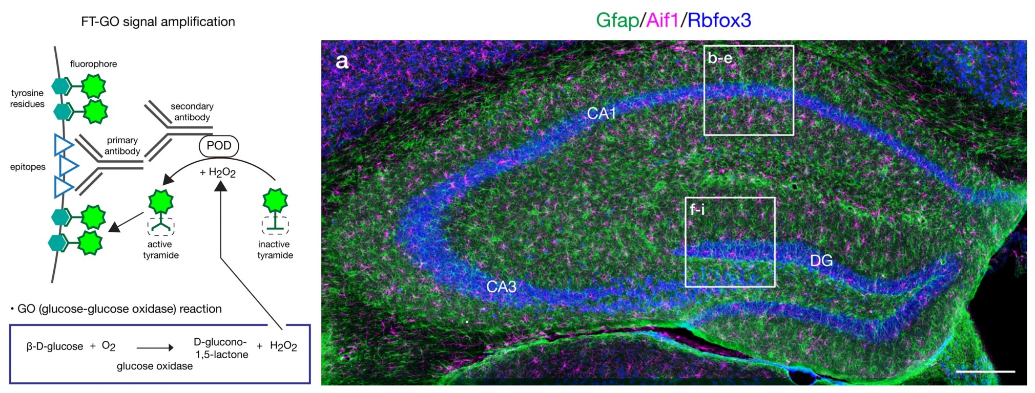
In a recent Scientific Reports article, Yamauchi et al. eloquently showed the utility of a novel multiplex TSA method using a custom panel of Biotium’s CF® Dye Tyramides. A limitation of standard TSA is that H2O2 is relatively unstable, which limits the staining time in the reaction. To overcome this, Yamauchi et al. recently developed biotinyl tyramine-glucose oxidase (BT-GO) and have now pushed BT-GO into the realm of multiplex IF with the novel fluorochromized tyramide (FT)-glucose oxidase (FT-GO) method. Distinct from other TSA systems, where H2O2 is added directly to the staining reaction, BT-GO and FT-GO rely on H2O2 produced during glucose oxidation by glucose oxidase (GO) to deposit tyramide stains onto their targets at a more sustained rate for longer periods (Figure 1, Left). Following preincubation in a mixture containing 10 µM FT and 3 µg/mL GO, the reaction was initiated by the addition of 20 µg/mL β-D-glucose, followed by a 30-minute incubation. The FTs used were Biotium CF®488A, CF®568, and CF®647 Dye Tyramides, often in conjunction with Biotium secondary antibodies for IF. Antibody-POD conjugates were inactivated between each round of FT deposition with 2% NaN3. Using combined FT-GO and IF in diverse brain tissue samples, the team achieved bright quadruple staining with low background signals. Along with distinguishing neocortical interneuron subtypes and increased detection of monoaminergic projection systems in mouse (Figure 1, Right) and marmoset brains, FT-GO also capably detected a viral tracer. Given its simplicity, sensitivity, and high signal-to-noise ratio, FT-GO offers a versatile alternative protocol.
Learn more about Biotium’s tyramide signal amplification products available in a wide selection of bright and photostable CF® Dyes, or find tips and protocols for their use in multiplex assays. Biotium also offers secondary antibodies conjugated to fluorophores or enzymes and a broad assortment of reagents for immunofluorescence microscopy.
Full Citation
Yamauchi, K., Okamoto, S., Ishida, Y., et al. Fluorochromized tyramide-glucose oxidase as a multiplex fluorescent tyramide signal amplification system for histochemical analysis. Scientific Reports, 12(1), 1-12 (2022). https://doi.org/10.1038/s41598-022-19085-9