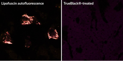
Efforts to develop diagnostics for early detection of tumor metastasis focus on accurate detection of individual tumor cells in blood and bone marrow. Immunofluorescence staining offers important advantages in the detection circulating tumor cells (CTCs) in blood and disseminated tumor cells (DTCs) in bone marrow, allowing multiplex target detection and cell morphology analysis of extremely rare cells. However, non-specific fluorescence background in blood and tissues hinders the specific detection of tumor cells, especially in bone marrow, which contains multiple autofluorescent cell types and tissue components. In a recent publication, Axelrod and associates from Johns Hopkins University identified tissue autofluorescence and non-specific binding of fluorescent secondary antibodies as the main sources of background in human bone marrow. By using TrueBlack® Lipofuscin Autofluorescence Quencher to reduce autofluorescence, together with Image-iT® FX background suppressor to reduce non-specific binding of fluorescent secondary antibodies, they were able to enhance the specificity of staining enough to detect rare model DTCs in human bone marrow using automated imaging software.
Learn more about Biotium’s TrueBlack® Lipofuscin Autofluorescence Quencher and TrueBlack® IF Background Suppressor System for blocking non-specific secondary antibody binding.
Full Citation
Axelrod, H. D., Pienta, K. J., & Valkenburg, K. C. (2018). Optimization of Immunofluorescent Detection of Bone Marrow Disseminated Tumor Cells. Biological Procedures Online, 20(1), 13. https://doi.org/10.1186/s12575-018-0078-5