 New Products
 Earth-Friendly Products
 Biotium Choice Antibodies
 Special Offers


Content #1
Content #1
Content #1
Biotium’s next-generation tyramide dyes with the widest selection of bright, photostable, and chemically stable dyes for spatial biology.
Test how it performs with a free 20 uL sample of any TyraMax™ Amplification Dye, limited time only!

TyraMax™ Dyes are Biotium’s next generation of tyramide amplification dyes for spatial biology. The dyes have been designed to yield a brighter signal compared to our original CF® Dye tyramides, and have advantages in brightness, photostability, and working solution stability compared to other TSA dyes.
Power Your Multiplex Panels with TyraMax™
Tyramide signal amplification (TSA), also known as Catalyzed Reporter Deposition (CARD), is a highly sensitive technique for detecting low-abundance targets in fluorescent immunocytochemistry (ICC), immunohistochemistry (IHC), and in situ hybridization (FISH). The superior sensitivity of TSA and its compatibility with multiplexing by cyclic immunofluorescence (CycIF) has made it indispensable for spatial biology applications.
TyraMax™ Dyes were developed as high-performance TSA dyes, offering brighter, more photostable signals than Aluora® and Opal® reagents. They also remain stable in amplification buffer for up to 24 hours, facilitating automated staining workflows. For optimal results, Biotium recommends using Tyramide Amplification Buffer Plus (Cat. No. 22029) with TyraMax™ Amplification Dyes.
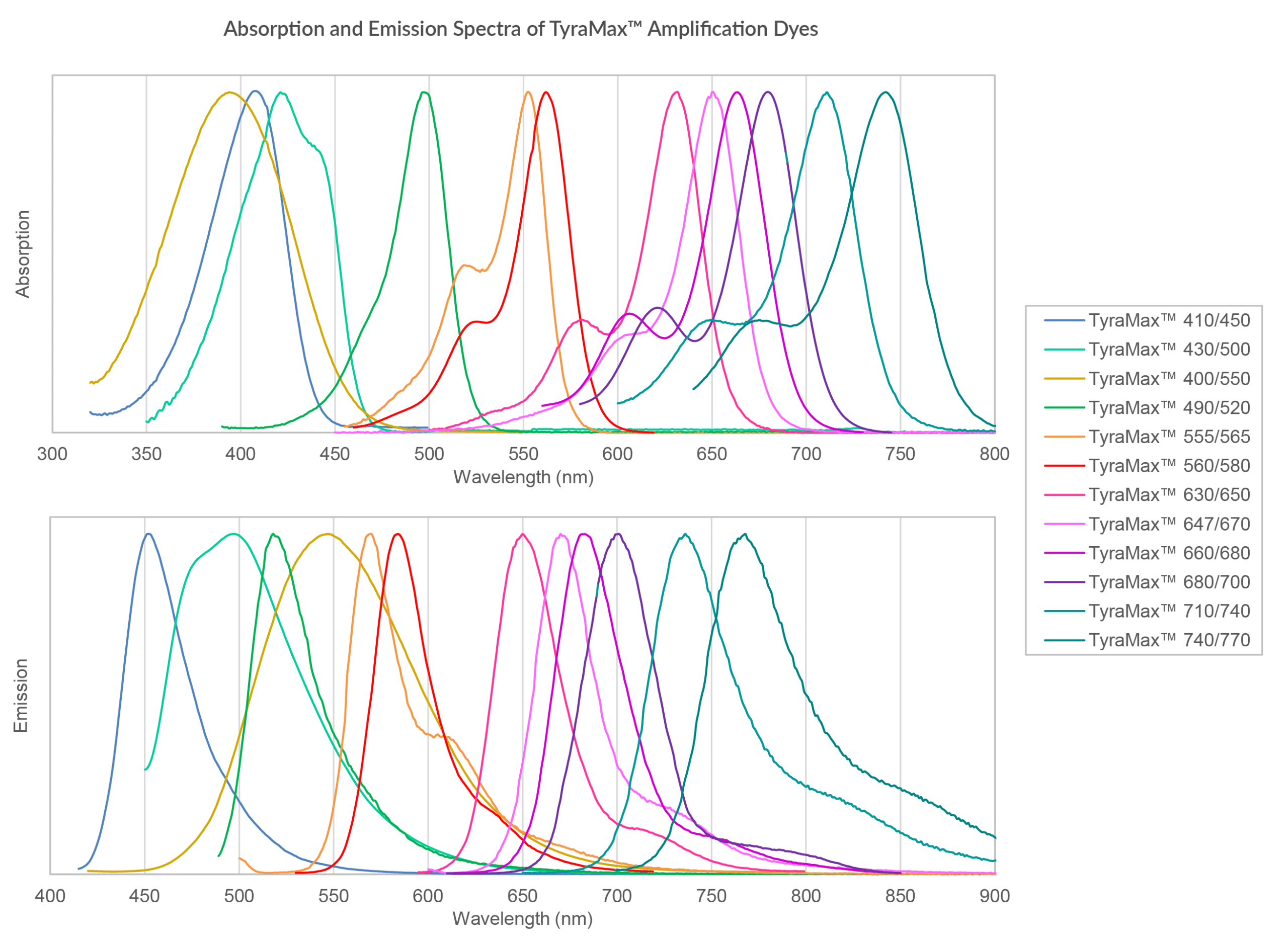
Widest Selection of Colors for Unrivaled Panel Flexibility
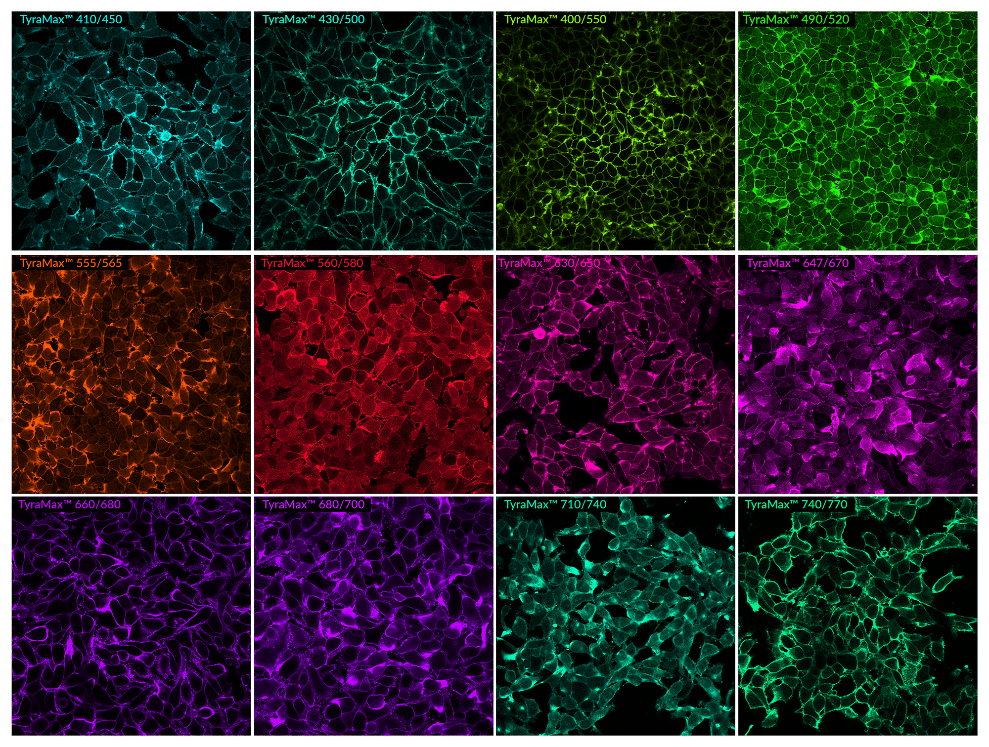
PFA-fixed HeLa cells stained with WGA-HRP detected with TyraMax™ Amplification Dyes in Biotium's Tyramide Amplification Buffer Plus. Imaged using a 40X oil objective on Evident FV4000 confocal system.
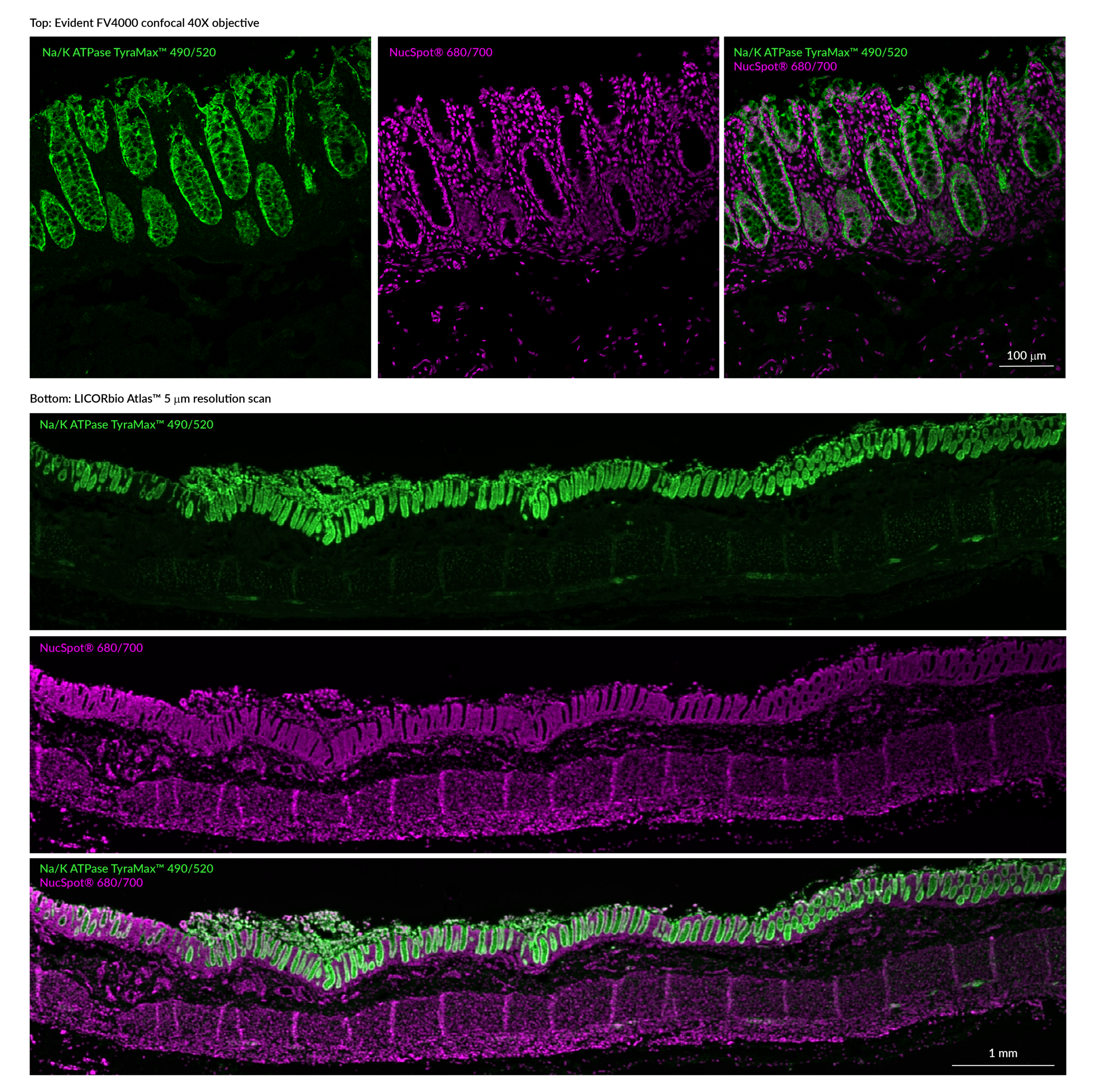
Ideal for Multiplex Imaging and CycIF Workflows
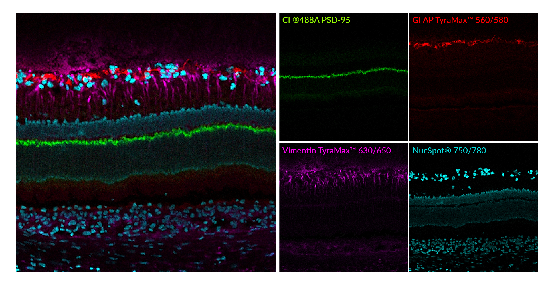
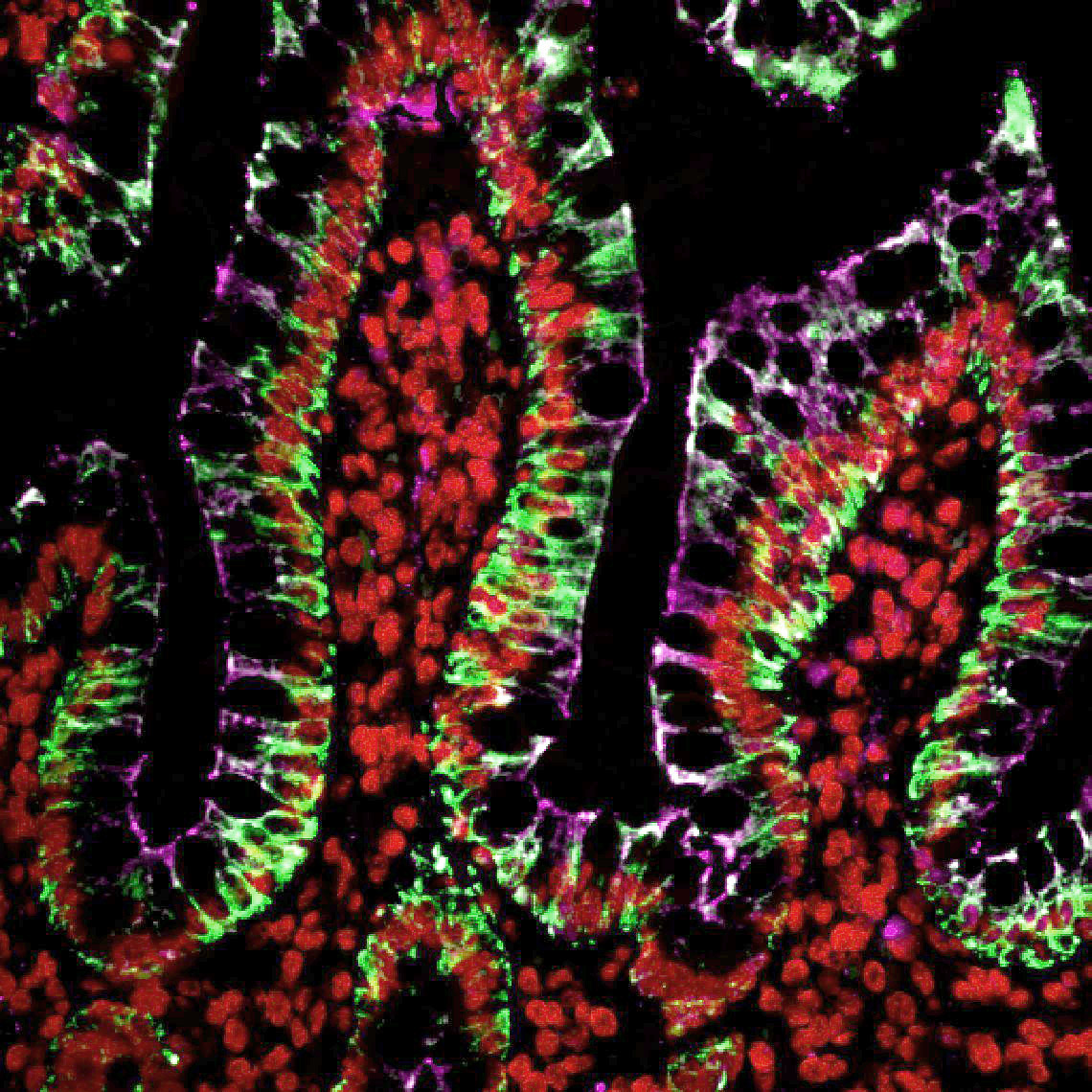
PFA-fixed rat eye cryosection stained with Vimentin Recombinant Monoclonal Mouse Antibody (rV9) HRP conjugate with TyraMax™ 630/650 (magenta), GFAP Recombinant Monoclonal Mouse Antibody (rGA5) HRP conjugate with TyraMax™ 560/580 (red), and CF®488A PSD95 Recombinant Monoclonal Mouse Antibody (rK28/43) (green). Nuclei are stained with NucSpot® 750/780 (cyan).
Superior Brightness Compared to Other TSA Reagents
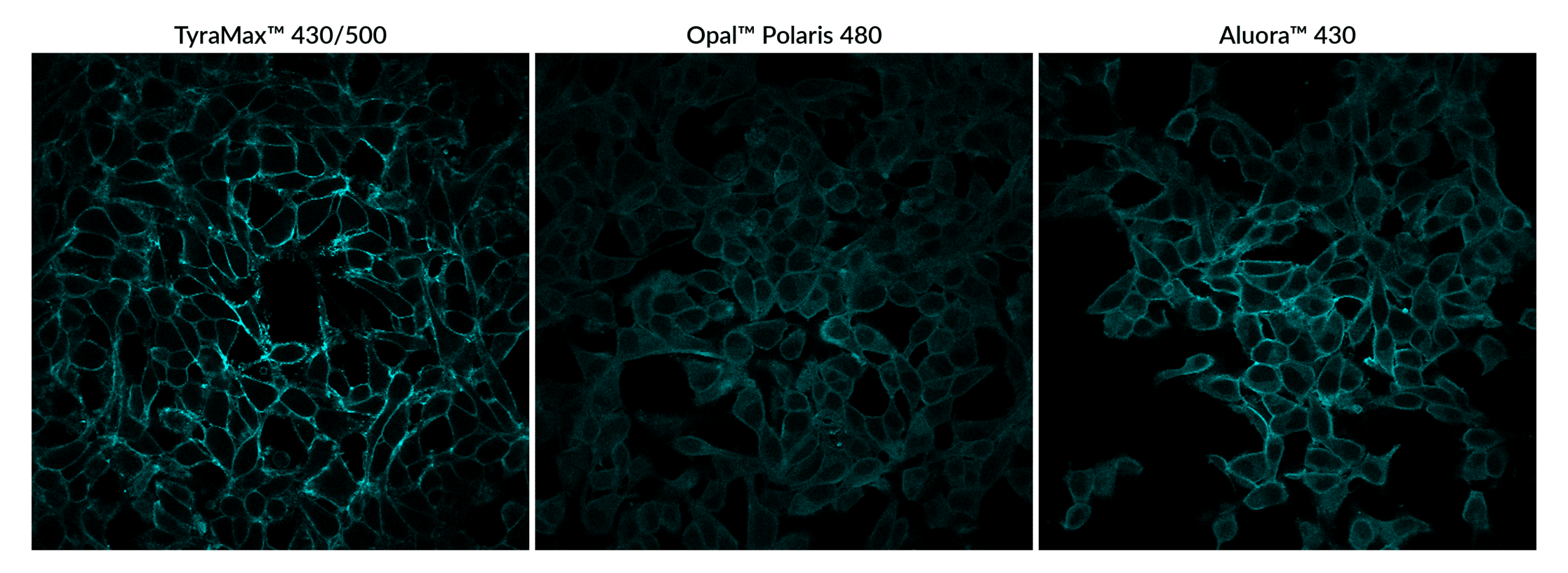
TyraMax™ 430/500 has excellent brightness compared to other commerically available TSA dyes. PFA-fixed HeLa cells stained with WGA-HRP and detected with TyraMax™ 430/500 Amplification Dye, Opal® Polaris 480 Reagent (Ex/Em 450/500 nm), or Aluora® 430 Spatial dye in Biotium's Tyramide Amplification Buffer Plus. Imaged using a 40X oil objective on Evident FV4000 confocal system with 405 nm excitation in the Alexa Fluor® 430 detection channel.
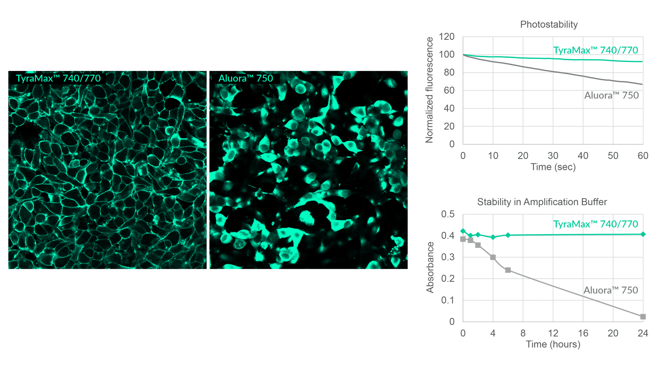
Signal Stability You Can Count On
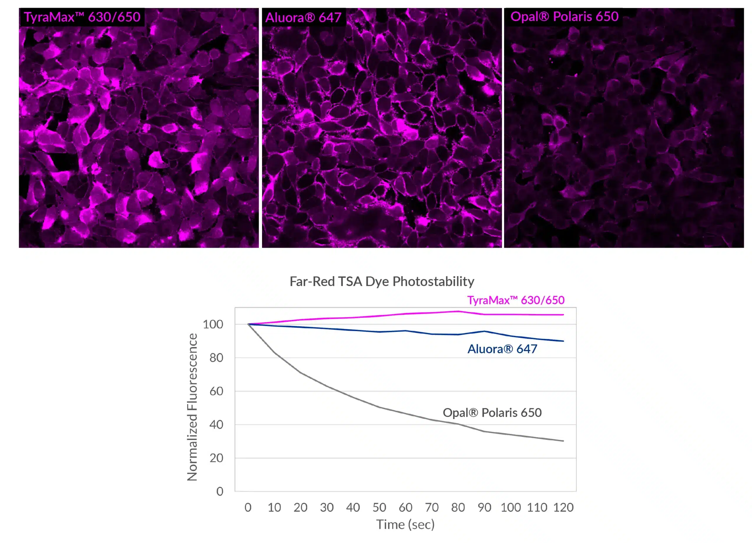
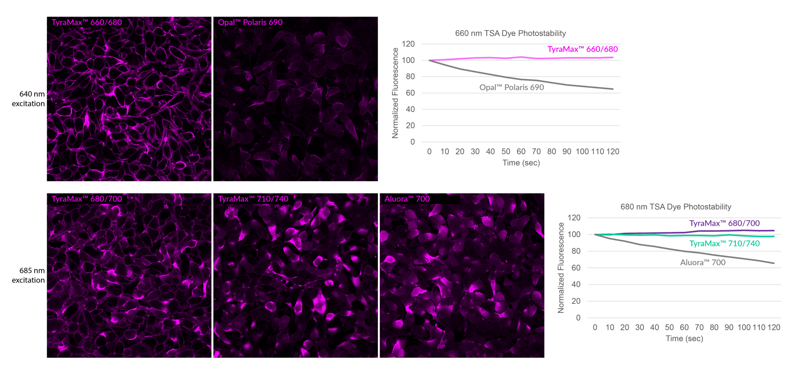
TyraMax™ 630/650 has excellent brightness and photostability compared to other commercially available TSA dyes. PFA-fixed HeLa cells stained with WGA-HRP and detected with TyraMax™630/650 Amplification Dye, Opal™ Polaris 650 Reagent, or Aluora™ 647 Spatial dye. Imaged using a 40X oil objective on Evident FV4000 confocal system with 640 nm excitation in the Alexa Fluor® 647 detection channel. Photostability measurements were done on an Olympus IX71 epifluorescence microscope using a Cy®5 filter cube. Stained cells were imaged every 10 seconds for 120 seconds, and mean fluorescence was normalized to the first image taken.
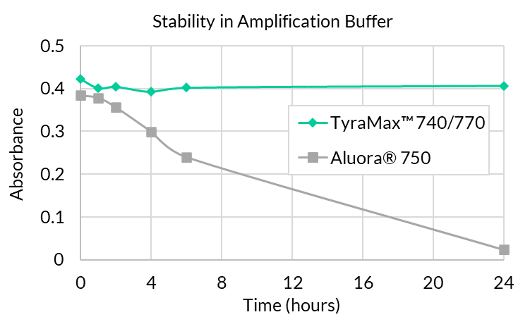
TyraMax™ 740/770 is stable in working solution for at least 24 hours. OD-matched solutions of TyraMax™ 740/770 and Aluora® 750 dyes were prepared in Tyramide Amplification Buffer Plus with 0.0015% hydrogen peroxide. Absorbance was measured on an Agilent Cary 60 spectrophotometer, and then the solutions were left at room temperature, protected from light, and absorbance was measured at various time points up to 24 hours. TyraMax™ 740/770 was stable in the presence of hydrogen peroxide over 24 hours, while Aluora® 750 completely degrades overnight.
TyraMax™ Amplification Dyes are available with a wide choice of dye options for multiplexing by conventional or spectral imaging. TyraMax™ Dyes are offered as standalone dye solutions, as 3-color or 5-color dye sets plus DAPI counterstain, and in a sampler for custom panel optimization. See the tables below for a full list of TyraMax™ Dyes, kits, and recommended channels.
| Dye | Abs/Em (nm) | Laser Line | Detection channel | Dye Features | Size | Catalog No. |
|---|---|---|---|---|---|---|
| TyraMax™ 410/450 | 408/452 | 405 nm | DAPI/Alexa Fluor® 405 | Unique tyramide color, spectrally similar to Alexa Fluor® 405 | 20 uL | 96134-20UL |
| 100 uL | 96134-100UL | |||||
| TyraMax™ 430/500 | 421/497 | 405 nm | FITC | Brighter than Aluora® 430 and Opal® 480 | 20 uL | 96135-20UL |
| 100 uL | 96135-100UL | |||||
| TyraMax™ 400/550 | 394/547 | 405 nm | FITC | Unique tyramide color, spectrally similar to Pacific Orange® | 20 uL | 96136-20UL |
| 100 uL | 96136-100UL | |||||
| TyraMax™ 490/520 | 497/518 | 488 nm | FITC | Brighter than Opal® 520, replacement for Aluora® 488 | 20 uL | 96137-20UL |
| 100 uL | 96137-100UL | |||||
| TyraMax™ 555/565 | 552/569 | 555 nm or 561 nm | TRITC | Brighter than Opal® 570, replacement for Aluora® 555 | 20 uL | 96138-20UL |
| 100 uL | 96138-100UL | |||||
| TyraMax™ 560/580 | 562/584 | 555 nm or 561 nm | TRITC | Alternative to Aluora® 555, Opal® 570 with superior photostability | 20 uL | 96139-20UL |
| 100 uL | 96139-100UL | |||||
| TyraMax™ 630/650 | 631/650 | 633 nm or 640 nm | Cy®5 | Bright and photostable alternative to Aluora® 647, Opal® 650 | 20 uL | 96140-20UL |
| 100 uL | 96140-100UL | |||||
| TyraMax™ 647/670 | 650/670 | 633 nm or 640 nm | Cy®5 | Brighter than Opal® 650, replacement for Aluora® 647 | 20 uL | 96141-20UL |
| 100 uL | 96141-100UL | |||||
| TyraMax™ 660/680 | 663/683 | 633 nm or 640 nm | Alexa Fluor® 680 | Unique tyramide color, spectrally similar to Alexa Fluor® 660. Brighter and more photostable Opal® 690 when excited at 640 nm | 20 uL | 96142-20UL |
| 100 uL | 96142-100UL | |||||
| TyraMax™ 680/700 | 680/701 | 685 nm (detectable with 640 nm excitation) | Alexa Fluor® 680 | Brighter and more photostable than Opal® 690 when excited at 685 nm | 20 uL | 96143-20UL |
| 100 uL | 96143-100UL | |||||
| TyraMax™ 710/740 | 711/736 | 685 nm | Alexa Fluor® 700 | Brighter and more photostable than Aluora® 700 | 20 uL | 96144-20UL |
| 100 uL | 96144-100UL | |||||
| TyraMax™ 740/770 | 742/768 | 730 nm | Alexa Fluor® 750 | Single-step detection, unlike Opal® 780. Stable in amplification buffer for up to 24 hours, unlike Aluora® 750 | 20 uL | 96145-20UL |
| 100 uL | 96145-100UL |
| Component # | Component Name | Size |
|---|---|---|
| 96137-100UL | TyraMax™ 490/520 Amplification Dye, 100X, 100 uL | For 100 samples using 100 uL staining volume |
| 96139-100UL | TyraMax™ 560/580 Amplification Dye, 100X, 100 uL | |
| 96140-100UL | TyraMax™ 630/650 Amplification Dye, 100X, 100 uL | |
| 99897-50UL | DAPI, 1000X, 50 uL |
| Component # | Component Name | Size |
|---|---|---|
| 96137-100UL | TyraMax™ 490/520 Amplification Dye, 100X, 100 uL | For 100 samples using 100 uL staining volume |
| 96139-100UL | TyraMax™ 560/580 Amplification Dye, 100X, 100 uL | |
| 96140-100UL | TyraMax™ 630/650 Amplification Dye, 100X, 100 uL | |
| 96143-100UL | TyraMax™ 680/700 Amplification Dye, 100X, 100 uL | |
| 96145-100UL | TyraMax™ 740/770 Amplification Dye, 100X, 100 uL | |
| 99897-50UL | DAPI, 1000X, 50 uL |
| Component # | Component Name | Size |
|---|---|---|
| 96134-20UL | TyraMax™ 410/450 | For 20 samples per vial using 100 uL staining volume |
| 96135-20UL | TyraMax™ 430/500 | |
| 96136-20UL | TyraMax™ 400/550 | |
| 96137-20UL | TyraMax™ 490/520 | |
| 96138-20UL | TyraMax™ 555/565 | |
| 96139-20UL | TyraMax™ 560/580 | |
| 96140-20UL | TyraMax™ 630/650 | |
| 96141-20UL | TyraMax™ 647/670 | |
| 96142-20UL | TyraMax™ 660/680 | |
| 96143-20UL | TyraMax™ 680/700 | |
| 96144-20UL | TyraMax™ 710/740 | |
| 96145-20UL | TyraMax™ 740/770 |
Yes, while our Tyramide Amplification Buffer Plus has enhanced sensitivity for TSA resulting in exceptional brightness, specificity, and sensitivity, the TyraMax™ Dyes will work with any amplification buffer.
Multiplex immunohistofluorescence (mIHF) has become an essential tool for studying complex tissue biology, enabling researchers to visualize multiple protein targets within a single sample while preserving spatial context. However, many existing multiplexing platforms remain costly, inflexible, or dependent on proprietary reagents, limiting accessibility for broader research applications. To address these challenges, open and scalable workflows are needed to make robust, reproducible, and cost-effective multiplex imaging more widely available to researchers.
In a 2026 protocol from the Journal of Microscopy, Riggi et al. created an open and flexible 6-color immunohistofluorescence (Flex-6 mIHF) workflow to investigate protein co-localization within the breast cancer tumor microenvironment. To overcome the background from autofluorescence in FFPE tissue sections that limits detection with IF, the protocol leveraged tyramide signal amplification (TSA) in combination with secondary antibodies conjugated to peroxidase-labeled polymers. This enabled robust signal enhancement and precise spatial resolution of biomarkers. The authors also outlined a stepwise validation strategy and essential controls to ensure reliable multiplex staining.
Within this workflow, CF® Dye Tyramides (Biotium) generated bright, covalently bound signals that can withstand repeated cycles of antibody stripping, facilitating sequential multiplexing without signal loss. This approach enabled simultaneous detection of up to six protein markers plus a nuclear stain in a single tissue section without requiring extensive image processing or spectral unmixing. By performing TSA with careful selection of antibodies, fluorophores, and order of target detection, the protocol produced high signal-to-noise images that could be directly analyzed, significantly reducing time and computational burden.
This protocol highlights how Biotium’s CF® Dye–based TSA reagents allow researchers to build flexible, high-performance multiplex immunofluorescence workflows without reliance on closed systems. By delivering exceptional brightness, photostability, and spectral diversity, Biotium’s fluorescent solutions help enable scalable, reproducible imaging protocols for cancer biology and beyond while making sophisticated multiplexing approaches more accessible to the broader scientific community.
Sequential multiplex tyramide labeling of human colon FFPE section with three CF® Dye Tyramides. Cytokeratin (pan) was labeled with CF®488A Tyramide (green); Histone H1 was labeled with Cyanine 555 Tyramide (red); ZO1 was labeled with CF®640R Tyramide (magenta). Credit: Biotium.
Learn more about Biotium’s products for TSA, including our TyraMax™ Amplification Dyes which offer improved brightness, photostability, and chemical stability over CF® Dye Tyramides. Biotium also offers secondary antibodies conjugated to fluorophores or enzymes and a broad assortment of reagents for immunofluorescence microscopy.
Full Citation:
Riggi, J. A. M., Daumerie, A., Benhaddi, N., Berlière, M., Galant, C., González-Antelo, A., Nana, F. A., Van Bockstal, M. R., & Bouzin, C. (2026). A detailed protocol for open and low-cost six-plex immunofluorescence (Flex-6 mIHF) with a proof-of-concept study on breast cancer tissue. Journal of Microscopy, 1–19. https://doi.org/10.1111/jmi.70068
Our Tyramide Amplification Kits have been demonstrated to be robust and versatile for multi-color fluorescence imaging, compatible with dye-labeled antibodies and various cell staining methods (see Figure 1).
To use a Tyramide Amplification Kit in addition to one or more dye-labeled antibodies, follow the kit protocol to fix and block samples; label with primary antibodies; then detect primary antibodies using secondary antibodies. Dye labeled secondary antibodies can be co-incubated with the HRP-conjugated secondary or HRP-streptavidin from the tyramide kit. After washing, perform the CF® Dye tyramide reaction according to the kit protocol. The tyramide reaction does not interfere with the binding of dye-labeled antibodies or other fluorescent staining reagents.
Performing multi-color detection with more than one dye tyramide on the same sample requires sequential tyramide staining reactions, followed by HRP inactivation or antibody stripping between each step. See our tech tip:
Multi-Color Fluorescence Imaging Using Biotium's Tyramide Amplification Kits
