 New Products
 Earth-Friendly Products
 Biotium Choice Antibodies
 Special Offers


 Powered by Bioz


Content #1
Content #1
Content #1
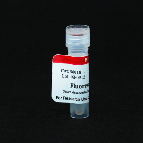
Fluorescein tyramide can be used for tyramide signal amplification (TSA) for increasing immunofluorescence sensitivity in multicolor immunocytochemistry (ICC), immunohistochemistry (IHC), or in situ hybridization (ISH).
Green fluorescent fluorescein tyramide conjugates are used for tyramide signal amplification (TSA), a method for high-density labeling of a target protein or nucleic acid in situ.
Also learn about TyraMax™ Amplification Dyes and Kits, Biotium's next generation tyramide dyes that offer brighter signal compared to the original CF® Dye Tyramides, and have advantages in brightness, photostability, and working solution stability compared to other TSA dyes. We also offer Ready-to-Use Tyramide Amplification Buffer, Tyramide Amplification Buffer Plus (an improved formulation for enhanced TSA sensitivity), and CF® Dye Tyramide Amplification Kits.
TSA is a highly sensitive method for differential gene or protein analysis or detection of low-abundance targets, in fluorescent ICC, IHC, and FISH applications. An antibody- or streptavidin-HRP conjugate catalyzes the deposition of fluorescent dye/biotin tyramides on tyrosine residues on and adjacent to a target protein or nucleic acid sequence in situ. This results in high-density labeling of the target and significantly improves the detection sensitivity up to 100-fold compared to conventional methods. TSA is particularly advantageous for fluorescence detection in human tissue, where conventional ICC or FISH often fails to provide adequate signal over autofluorescence background. In applications where increased sensitivity is not required, TSA enables the use of significantly lower antibody or probe concentrations for the same level of detection sensitivity thereby reducing issues of non-specific binding or cross-reactivity. Furthermore, since binding of the tyramide label is covalent, a large number of targets can be detected in the same sample using multiple rounds of sequential TSA, in which the availability of antibodies from different host species is not a limitation. TSA also can be easily integrated with conventional immunostaining. Learn more about Tyramide Signal Amplification.
Yes, while our Tyramide Amplification Buffer Plus has enhanced sensitivity for TSA resulting in exceptional brightness, specificity, and sensitivity, the TyraMax™ Dyes will work with any amplification buffer.
Multiplex immunohistofluorescence (mIHF) has become an essential tool for studying complex tissue biology, enabling researchers to visualize multiple protein targets within a single sample while preserving spatial context. However, many existing multiplexing platforms remain costly, inflexible, or dependent on proprietary reagents, limiting accessibility for broader research applications. To address these challenges, open and scalable workflows are needed to make robust, reproducible, and cost-effective multiplex imaging more widely available to researchers.
In a 2026 protocol from the Journal of Microscopy, Riggi et al. created an open and flexible 6-color immunohistofluorescence (Flex-6 mIHF) workflow to investigate protein co-localization within the breast cancer tumor microenvironment. To overcome the background from autofluorescence in FFPE tissue sections that limits detection with IF, the protocol leveraged tyramide signal amplification (TSA) in combination with secondary antibodies conjugated to peroxidase-labeled polymers. This enabled robust signal enhancement and precise spatial resolution of biomarkers. The authors also outlined a stepwise validation strategy and essential controls to ensure reliable multiplex staining.
Within this workflow, CF® Dye Tyramides (Biotium) generated bright, covalently bound signals that can withstand repeated cycles of antibody stripping, facilitating sequential multiplexing without signal loss. This approach enabled simultaneous detection of up to six protein markers plus a nuclear stain in a single tissue section without requiring extensive image processing or spectral unmixing. By performing TSA with careful selection of antibodies, fluorophores, and order of target detection, the protocol produced high signal-to-noise images that could be directly analyzed, significantly reducing time and computational burden.
This protocol highlights how Biotium’s CF® Dye–based TSA reagents allow researchers to build flexible, high-performance multiplex immunofluorescence workflows without reliance on closed systems. By delivering exceptional brightness, photostability, and spectral diversity, Biotium’s fluorescent solutions help enable scalable, reproducible imaging protocols for cancer biology and beyond while making sophisticated multiplexing approaches more accessible to the broader scientific community.
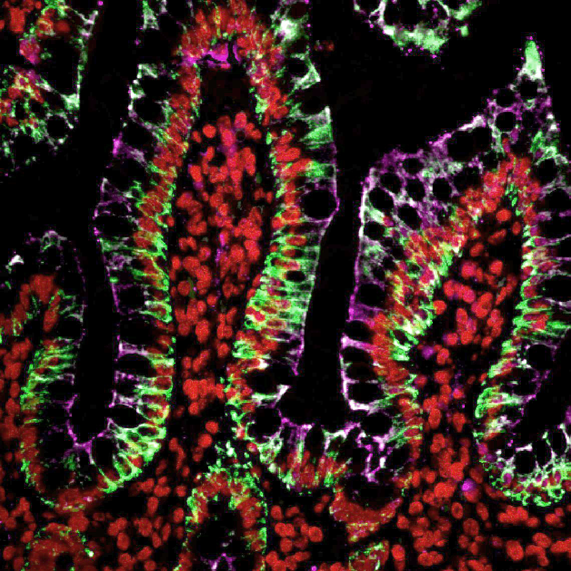
Sequential multiplex tyramide labeling of human colon FFPE section with three CF® Dye Tyramides. Cytokeratin (pan) was labeled with CF®488A Tyramide (green); Histone H1 was labeled with Cyanine 555 Tyramide (red); ZO1 was labeled with CF®640R Tyramide (magenta). Credit: Biotium.
Learn more about Biotium’s products for TSA, including our TyraMax™ Amplification Dyes which offer improved brightness, photostability, and chemical stability over CF® Dye Tyramides. Biotium also offers secondary antibodies conjugated to fluorophores or enzymes and a broad assortment of reagents for immunofluorescence microscopy.
Full Citation:
Riggi, J. A. M., Daumerie, A., Benhaddi, N., Berlière, M., Galant, C., González-Antelo, A., Nana, F. A., Van Bockstal, M. R., & Bouzin, C. (2026). A detailed protocol for open and low-cost six-plex immunofluorescence (Flex-6 mIHF) with a proof-of-concept study on breast cancer tissue. Journal of Microscopy, 1–19. https://doi.org/10.1111/jmi.70068
Our Tyramide Amplification Kits have been demonstrated to be robust and versatile for multi-color fluorescence imaging, compatible with dye-labeled antibodies and various cell staining methods (see Figure 1).
To use a Tyramide Amplification Kit in addition to one or more dye-labeled antibodies, follow the kit protocol to fix and block samples; label with primary antibodies; then detect primary antibodies using secondary antibodies. Dye labeled secondary antibodies can be co-incubated with the HRP-conjugated secondary or HRP-streptavidin from the tyramide kit. After washing, perform the CF® Dye tyramide reaction according to the kit protocol. The tyramide reaction does not interfere with the binding of dye-labeled antibodies or other fluorescent staining reagents.
Performing multi-color detection with more than one dye tyramide on the same sample requires sequential tyramide staining reactions, followed by HRP inactivation or antibody stripping between each step. See our tech tip:
Multi-Color Fluorescence Imaging Using Biotium's Tyramide Amplification Kits
