New Products
 Earth-Friendly Products
 Biotium Choice Antibodies
 Special Offers
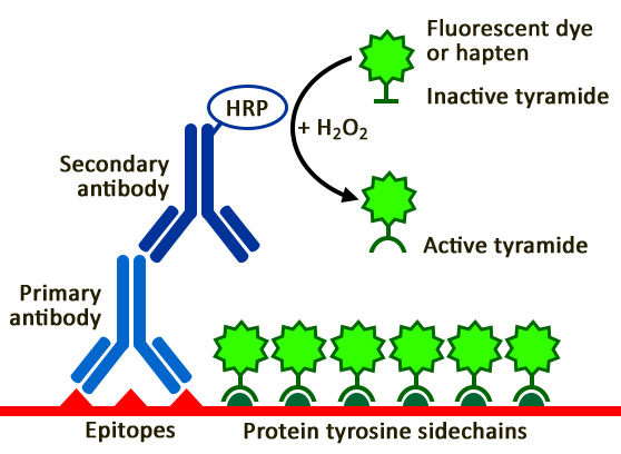
What is Tyramide Signal Amplification?
Boost Your Signal Up to 100-Fold
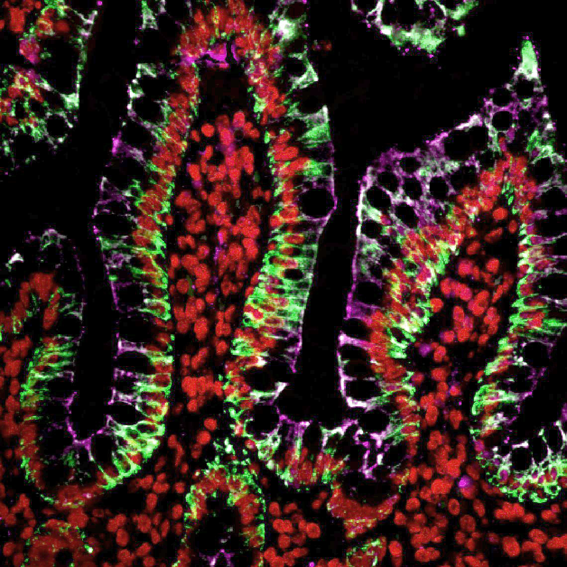
Tyramide signal amplification (TSA), sometimes referred to as Catalyzed Reporter Deposition (CARD), is a highly sensitive method capable of boosting signal up to 100-fold over conventional methods. This enables the detection of low-abundance targets for fluorescent immunocytochemistry (ICC), immunohistochemistry (IHC), and in situ hybridization (FISH) applications.
Advantages of Tyramide Signal Amplification
- Detect low-abundance targets
- Compatible with ICC, IHC and FISH
- Up to 100-fold more sensitive than conventional methods
- Similar workflow to conventional ICC, IHC and FISH
- Use less primary antibody
- Simplified primary antibody panel design for multiplex IHC
How TSA Works
Why Use TSA?
Well-Suited for FFPE Tissue
Formalin fixation crosslinks proteins and progressively degrades antigenicity, reducing how efficiently antibodies can bind their targets. Signal amplification by TSA compensates for this loss, making it effective in FFPE samples where standard detection methods often fall short. This is particularly valuable in clinical and translational research settings, where fresh tissue is rarely available.
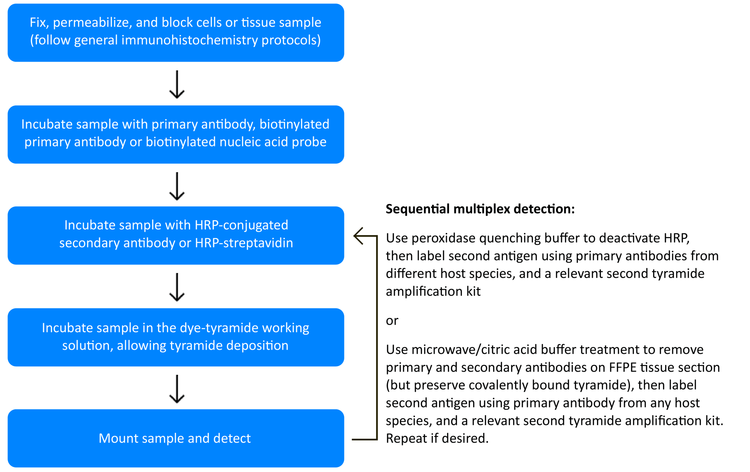
Covalent Signal Stability for Sequential Probing
Tyramide deposits covalently onto tissue, so the fluorescent signal remains intact even after HRP inactivation or antibody stripping. This signal durability makes TSA well-suited for sequential multiplex probing, a workflow that is incompatible with non-covalent detection methods.
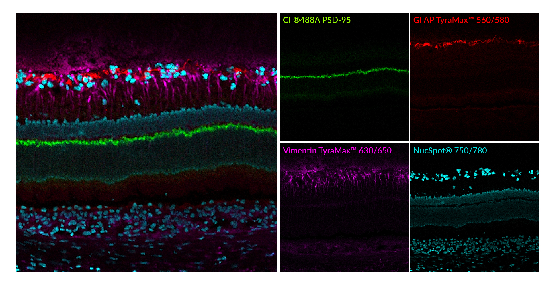
Simplifies CycIF Panels
Multiple rounds of tyramide signal amplification can be performed for high-plex imaging (see our Tech Tip: Multicolor Fluorescence Imaging using Tyramide Amplification Kits). When TSA followed by antibody removal is used for multiplex fluorescent IHC, TSA not only facilitates detection of low-abundance targets, but also simplifies antibody panel design because primary antibodies of choice may be used, irrespective of host species or isotype.
Serial TSA Workflow for Multiplexing
TyraMax™: Next-Generation TSA Reagents
TSA Dyes for Superior Spatial Imaging
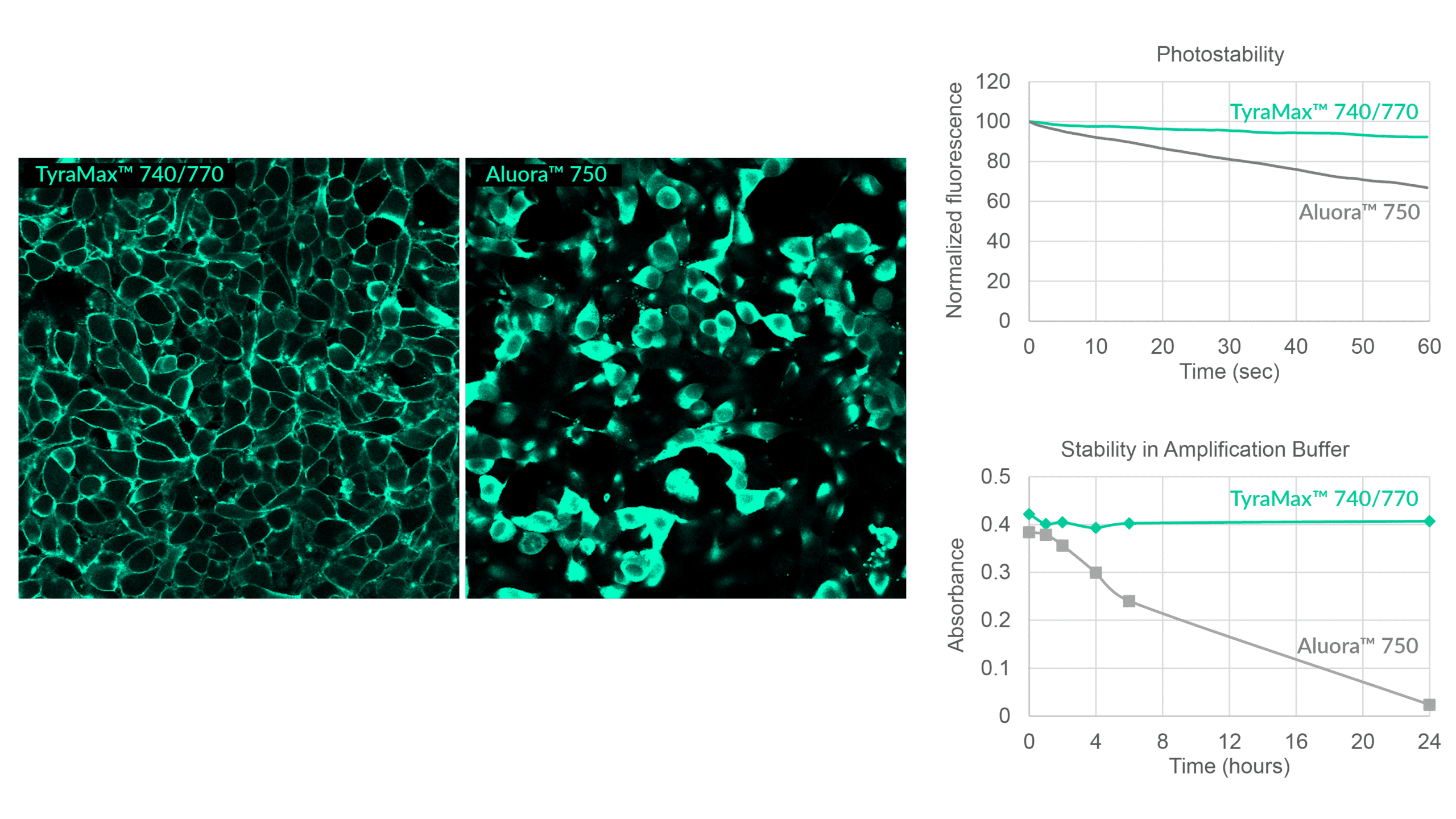
Biotium's TyraMax™ line offers the widest selection of dye colors on the market, with unique options spanning from blue to near-infrared for unmatched multiplexing flexibility and spectral panel design. All TyraMax™ dyes, including near-IR dyes, are chemically stable in amplification buffer for at least 24 hours. This makes them ideal for automated staining workflows in spatial biology platforms with more resistance to oxidation unlike other TSA dyes.
Compared to conventional immunofluorescence, TyraMax™ Dyes provide up to 100-fold higher detection sensitivity when used with TSA while maintaining sharp, photostable signals for multiplex imaging workflows like cyclic immunofluorescence (CycIF).
Key Features
- Largest selection of tyramide dye colors available, from blue to near-IR
- Stable in amplification buffer for at least 24 hours, including near-IR dyes
- Brighter and more photostable than competitor TSA dyes
- Available as standalone dyes, trial sizes, or multiplex dye sets
- Ideal for automated workflows
- Recommend to use with Tyramide Amplification Buffer Plus for optimal results
View Product Page

TyraMax™ Amplification Dyes
| Dye | Abs/Em (nm) | Laser Line | Detection channel | Dye Features | Size | Catalog No. |
|---|---|---|---|---|---|---|
| TyraMax™ 410/450 | 408/452 | 405 nm | DAPI/Alexa Fluor® 405 | Unique tyramide color, spectrally similar to Alexa Fluor® 405 | 20 uL | 96134-20UL |
| 100 uL | 96134-100UL | |||||
| TyraMax™ 430/500 | 421/497 | 405 nm | FITC | Brighter than Aluora® 430 and Opal® 480 | 20 uL | 96135-20UL |
| 100 uL | 96135-100UL | |||||
| TyraMax™ 400/550 | 394/547 | 405 nm | FITC | Unique tyramide color, spectrally similar to Pacific Orange® | 20 uL | 96136-20UL |
| 100 uL | 96136-100UL | |||||
| TyraMax™ 490/520 | 497/518 | 488 nm | FITC | Brighter than Opal® 520, replacement for Aluora® 488 | 20 uL | 96137-20UL |
| 100 uL | 96137-100UL | |||||
| TyraMax™ 555/565 | 552/569 | 555 nm or 561 nm | TRITC | Brighter than Opal® 570, replacement for Aluora® 555 | 20 uL | 96138-20UL |
| 100 uL | 96138-100UL | |||||
| TyraMax™ 560/580 | 562/584 | 555 nm or 561 nm | TRITC | Alternative to Aluora® 555, Opal® 570 with superior photostability | 20 uL | 96139-20UL |
| 100 uL | 96139-100UL | |||||
| TyraMax™ 630/650 | 631/650 | 633 nm or 640 nm | Cy®5 | Bright and photostable alternative to Aluora® 647, Opal® 650 | 20 uL | 96140-20UL |
| 100 uL | 96140-100UL | |||||
| TyraMax™ 647/670 | 650/670 | 633 nm or 640 nm | Cy®5 | Brighter than Opal® 650, replacement for Aluora® 647 | 20 uL | 96141-20UL |
| 100 uL | 96141-100UL | |||||
| TyraMax™ 660/680 | 663/683 | 633 nm or 640 nm | Alexa Fluor® 680 | Unique tyramide color, spectrally similar to Alexa Fluor® 660. Brighter and more photostable Opal® 690 when excited at 640 nm | 20 uL | 96142-20UL |
| 100 uL | 96142-100UL | |||||
| TyraMax™ 680/700 | 680/701 | 685 nm (detectable with 640 nm excitation) | Alexa Fluor® 680 | Brighter and more photostable than Opal® 690 when excited at 685 nm | 20 uL | 96143-20UL |
| 100 uL | 96143-100UL | |||||
| TyraMax™ 710/740 | 711/736 | 685 nm | Alexa Fluor® 700 | Brighter and more photostable than Aluora® 700 | 20 uL | 96144-20UL |
| 100 uL | 96144-100UL | |||||
| TyraMax™ 740/770 | 742/768 | 730 nm | Alexa Fluor® 750 | Single-step detection, unlike Opal® 780. Stable in amplification buffer for up to 24 hours, unlike Aluora® 750 | 20 uL | 96145-20UL |
| 100 uL | 96145-100UL |
Amplification Buffers & Other Tyramide Dyes
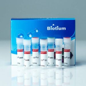
Standalone CF® Dye Tyramides
Biotium offers a wide selection of bright and photostable CF® Dye Tyramides from blue to near-infrared. Other classic dye tyramides are available as well as biotin-XX and DNP tyramides.
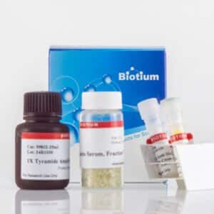
Tyramide Signal Amplification Kits
- Our kits provide all critical reagents for tyramide labeling
- Choose your tyramide: biotin tyramide or one of six CF® Dye tyramides
- Choose your HRP conjugate: goat anti-mouse, goat anti-rabbit, or streptavidin
- Kits also include Amplification Buffer and BSA for blocking
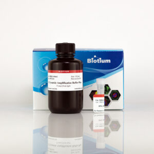
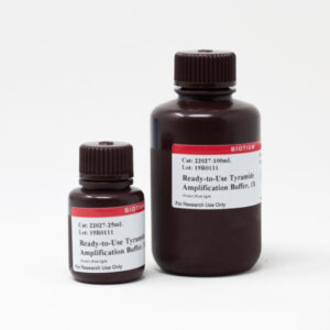
Tyramide Amplification Buffers
- Tyramide Amplification Buffer Plus has enhanced sensitivity for TSA, with improved brightness, specificity, and sensitivity over our original buffer.
- Ready-to-Use Tyramide Amplification Buffer was our original buffer, and has been replaced with the above buffer. The advantage of this buffer is that there is no need to add hydrogen peroxide.

Tyramide Signal Amplification Kits
| Tyramide Label | Ex/Em | Secondary conjugate | Catalog no. |
|---|---|---|---|
| CF®488A | 490/515 nm | Goat anti-mouse HRP | 33000 |
| Goat anti-rabbit HRP | 33001 | ||
| Streptavidin HRP | 33002 | ||
| CF®543 | 541/560 nm | Goat anti-mouse HRP | 33003 |
| Goat anti-rabbit HRP | 33004 | ||
| Streptavidin HRP | 33005 | ||
| CF®568 | 562/583 nm | Goat anti-mouse HRP | 33006 |
| Goat anti-rabbit HRP | 33007 | ||
| Streptavidin HRP | 33008 | ||
| CF®594 | 593/614 nm | Goat anti-mouse HRP | 33009 |
| Goat anti-rabbit HRP | 33010 | ||
| Streptavidin HRP | 33011 | ||
| CF®640R | 642/662 nm | Goat anti-mouse HRP | 33012 |
| Goat anti-rabbit HRP | 33013 | ||
| Streptavidin HRP | 33014 | ||
| CF®680R | 680/701 nm | Goat anti-mouse HRP | 33015 |
| Goat anti-rabbit HRP | 33016 | ||
| Streptavidin HRP | 33017 | ||
| Biotin-XX | N/A | Goat anti-mouse HRP | 33018 |
| Goat anti-rabbit HRP | 33019 | ||
| Streptavidin HRP | 33020 |
Standalone Dye and Hapten Labeled Tyramides
| Tyramide label | Ex/Em | Size | Catalog no. |
|---|---|---|---|
| CF®350 | 347/448 nm | 0.5 mg | 92170 |
| CF®405L | 395/545 nm | 0.5 mg | 92198 |
| CF®405S | 404/431 nm | 0.5 mg | 92197 |
| CF®405M | 408/452 nm | 0.5 mg | 96057 |
| CF®430 | 426/498 nm | 0.5 mg | 96053 |
| CF®488A | 490/515 nm | 0.5 mg | 92171 |
| FITC | 492/514 nm | 0.5 mg | 96018 |
| CF®514 | 516/548 nm | 0.5 mg | 92199 |
| CF®532 | 527/558 nm | 0.5 mg | 96066 |
| CF®543 | 541/560 nm | 0.5 mg | 92172 |
| CF®550R | 551/577 nm | 0.5 mg | 96077 |
| CF®555 | 555/565 nm | 0.5 mg | 96021 |
| Cyanine 555 | 555/565 nm | 0.5 mg | 96020 |
| CF®568 | 562/583 nm | 0.5 mg | 92173 |
| CF®583R | 586/609 nm | 0.5 mg | 96085 |
| CF®594 | 593/614 nm | 0.5 mg | 92174 |
| CF®620R | 617/639 nm | 0.5 mg | 92194 |
| CF®640R | 642/662 nm | 0.5 mg | 92175 |
| CF®647 | 650/665 nm | 0.5 mg | 96022 |
| CF®660R | 663/682 nm | 0.5 mg | 92195 |
| CF®680R | 680/701 nm | 0.5 mg | 92196 |
| CF®710 | 712/736 nm | 0.5 mg | 96127 |
| CF®725 | 729/755 nm | 0.5 mg | 96128 |
| CF®740 | 742/767 nm | 0.5 mg | 96124 |
| CF®750 | 755/779 nm | 0.5 mg | 96052 |
| CF®754 | 748/793 nm | 0.5 mg | 96090 |
| Biotin-XX | N/A | 0.5 mg | 92176 |
| DNP | N/A | 0.5 mg | 96019 |