 New Products
 Earth-Friendly Products
 Biotium Choice Antibodies
 Special Offers


 Powered by Bioz


Content #1
Content #1
Content #1
Fluorescent CF® Dye tyramides are used for tyramide signal amplification (TSA) for increasing immunofluorescence sensitivity in multicolor immunocytochemistry (ICC), immunohistochemistry (IHC), or in situ hybridization (ISH).
CF® Dye tyramide conjugates are used for tyramide signal amplification (TSA), a method for high-density labeling of a target protein or nucleic acid in situ.
Also learn about TyraMax™ Amplification Dyes and Kits, Biotium's next generation tyramide dyes that offer brighter signal compared to the original CF® Dye Tyramides, and have advantages in brightness, photostability, and working solution stability compared to other TSA dyes. We also offer Ready-to-Use Tyramide Amplification Buffer, Tyramide Amplification Buffer Plus (an improved formulation for enhanced TSA sensitivity), and CF® Dye Tyramide Amplification Kits.
Biotium’s next-generation CF® Dyes were designed to be highly water-soluble with advantages in brightness and photostability compared to other commercially available fluorescent dyes. Our CF® Dye Tyramide conjugates are available in 22 colors. Learn more about CF® Dyes.
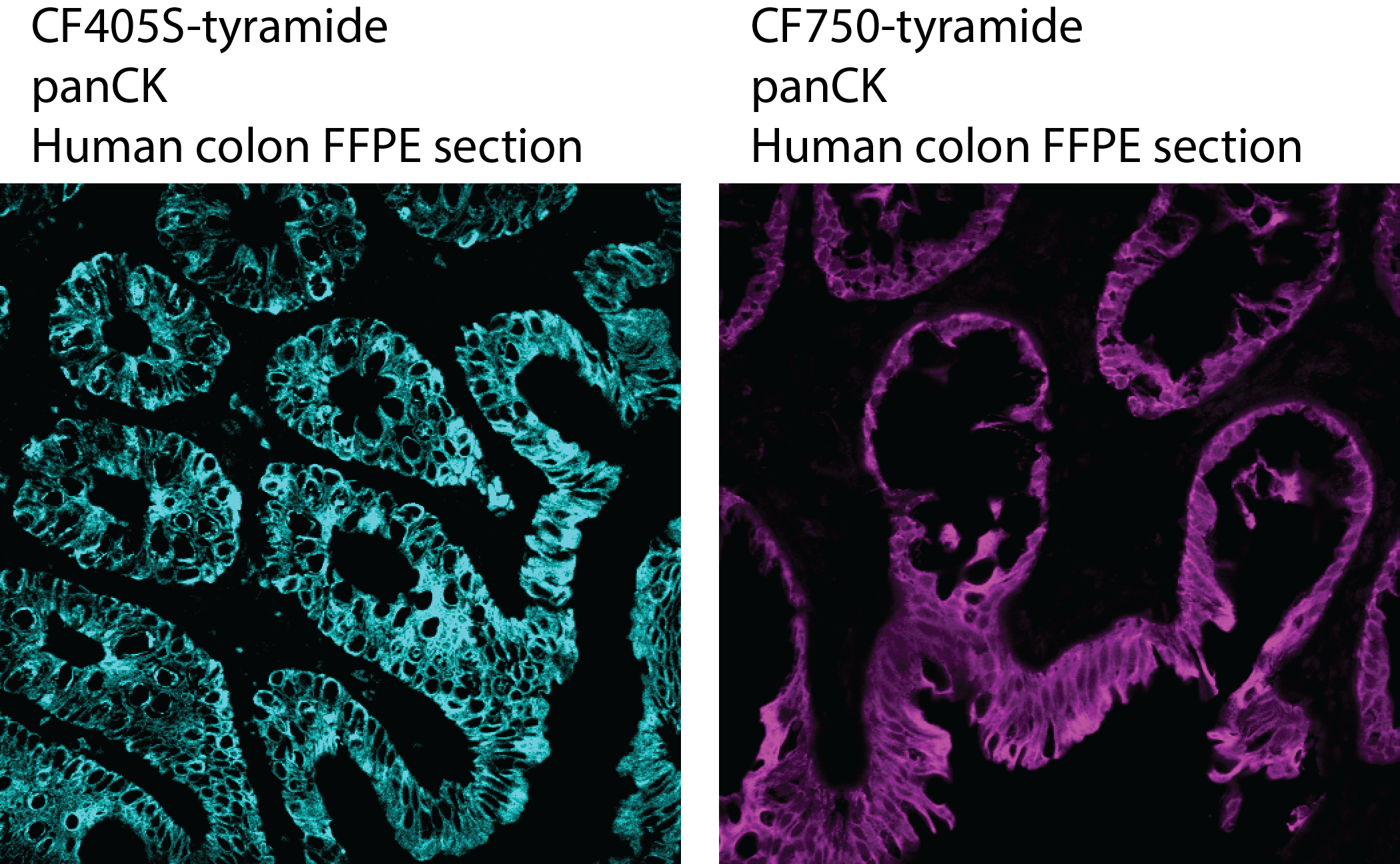
For researchers considering near-infrared detection, we recommend CF®740 tyramide over CF®750 and CF®754. This is because CF®750 is unstable in oxidizing amplification buffer and should be added to the buffer immediately before performing the staining reaction. The poor stability in oxidizing amplification buffer makes the dye challenging to use for automated staining platforms (ie. BOND RX) that require longer periods with the dye in buffer. CF®754 tyramide is stable in oxidizing amplification buffer, but the dye has a broad absorption peak that can cause channel spillover. CF®740 tyramide was developed to be stable in oxidizing amplification buffer when compared to Alexa Fluor® 750 and CF®750. In addition, CF®740 tyramide has a narrower absorption peak that minimizes spillover and therefore a superior option to CF®754 tyramide. Near-IR tyramide background staining may be tissue dependent and not suitable for all targets. In general, we would recommend using near-IR staining for more abundant targets in your panel.
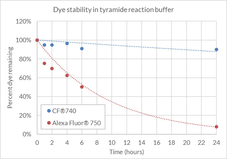
CF®740 free acid or Alexa Fluor® 750 free acid was diluted to 2 uM in 1X Tyramide Amplification Buffer with 0.0015% hydrogen peroxide and incubated at room temperature, protected from light. Dye concentration was measured by absorbance at the time points shown. CF®740 remained stable in the presence of hydrogen peroxide over 24 hours (≤90% of the starting dye concentration), while Alexa Fluor® 750 degraded to less than 10% of the starting concentration after 24 hours.
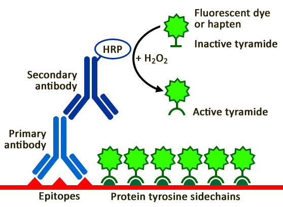
TSA is a highly sensitive method for differential gene or protein analysis or detection of low-abundance targets, in fluorescent ICC, IHC, and FISH applications. An antibody- or streptavidin-HRP conjugate catalyzes the deposition of fluorescent dye/biotin tyramides on tyrosine residues on and adjacent to a target protein or nucleic acid sequence in situ. This results in high-density labeling of the target and significantly improves the detection sensitivity up to 100-fold compared to conventional methods. TSA is particularly advantageous for fluorescence detection in human tissue, where conventional ICC or FISH often fails to provide adequate signal over autofluorescence background. In applications where increased sensitivity is not required, TSA enables the use of significantly lower antibody or probe concentrations for the same level of detection sensitivity thereby reducing issues of non-specific binding or cross-reactivity. Furthermore, since binding of the tyramide label is covalent, a large number of targets can be detected in the same sample using multiple rounds of sequential TSA, in which the availability of antibodies from different host species is not a limitation. TSA also can be easily integrated with conventional immunostaining. Learn more about Tyramide Signal Amplification.
| Product | Ex/Em | MW (g/mol) | Size | Catalog No. | Dye Features |
|---|---|---|---|---|---|
| CF®350 Tyramide | 347/448 nm | ~614 | 0.5 mg | 92170 | CF®350 Features |
| CF®405S Tyramide | 404/431 nm | ~689 | 0.5 mg | 92197 | CF®405S Features |
| CF®405M Tyramide | 408/452 nm | ~621 | 0.5 mg | 96057 | CF®405M Features |
| CF®405L Tyramide | 395/545 nm | ~1692 | 0.5 mg | 92198 | CF®405L Features |
| CF®430 Tyramide | 426/498 nm | ~707 | 0.5 mg | 96053 | CF®430 Features |
| CF®488A Tyramide | 490/515 nm | ~666 | 0.5 mg | 92171 | CF®488A Features |
| CF®514 Tyramide | 516/548 nm | ~1337 | 0.5 mg | 92199 | CF®514 Features |
| CF®532 Tyramide | 527/558 nm | ~804 | 0.5 mg | 96066 | CF®532 Features |
| CF®543 Tyramide | 541/560 nm | ~1006 | 0.5 mg | 92172 | CF®543 Features |
| CF®550R Tyramide | 551/577 nm | ~806 | 0.5 mg | 96077 | CF®550R Features |
| CF®555 Tyramide | 555/565 nm | ~1120 | 0.5 mg | 96021 | CF®555 Features |
| CF®568 Tyramide | 562/583 nm | ~833 | 0.5 mg | 92173 | CF®568 Features |
| CF®583R Tyramide | 586/609 nm | ~892 | 0.5 mg | 96085 | CF®583R Features |
| CF®594 Tyramide | 593/614 nm | ~848 | 0.5 mg | 92174 | CF®594 Features |
| CF®620R Tyramide | 617/639 nm | ~857 | 0.5 mg | 92194 | CF®620R Features |
| CF®640R Tyramide | 642/662 nm | ~951 | 0.5 mg | 92175 | CF®640R Features |
| CF®647 Tyramide | 650/665 nm | ~1104 | 0.5 mg | 96022 | CF®647 Features |
| CF®660R Tyramide | 663/682 nm | ~1007 | 0.5 mg | 92195 | CF®660R Features |
| CF®680R Tyramide | 680/701 nm | ~1031 | 0.5 mg | 92196 | CF®680R Features |
| CF®710 Tyramide | 712/736 nm | ~977 | 0.5 mg | 96127 | CF®710 Features |
| CF®725 Tyramide | 729/750 nm | ~1005 | 0.5 mg | 96128 | CF®725 Features |
| CF®740 Tyramide | 742/767 nm | ~1005 | 0.5 mg | 96124 | CF®740 Features |
| CF®750 Tyramide* | 755/779 nm | ~3040 | 0.5 mg | 96052 | CF®750 Features |
| CF®754 Tyramide | 748/793 nm | ~1000 | 0.5 mg | 96090 |
* CF®750 Tyramide is not stable in TSA buffer, and should be added to the buffer immediately before performing the staining reaction.
1. Journal of Immunology Research (2020), 2020:2328675. DOI:10.1155/2020/2328675
2. Scientific Reports (2019), 9:1144. DOI:10.1038/s41598-018-38171-5
3. bioRxiv (2019) preprint. DOI:10.1101/651083
4. OncoImmunology (2019), 8(6):e1581528-10. DOI:10.1080/2162402X.2019.1581528
5. Microbiological Research (2018), 217: 69-80. DOI:10.1016/j.micres.2018.08.017
6. Mol Neurodegeneration (2017), 12:68. DOI:10.1186/s13024-017-0202-z
7. Biology Open (2017), 6: 891-896. DOI:10.1242/bio.025809
8. The American Journal of Pathology (2016), 186(10):2650-2664. DOI:10.1016/j.ajpath.2016.06.020
Download a list of CF® Dye references.
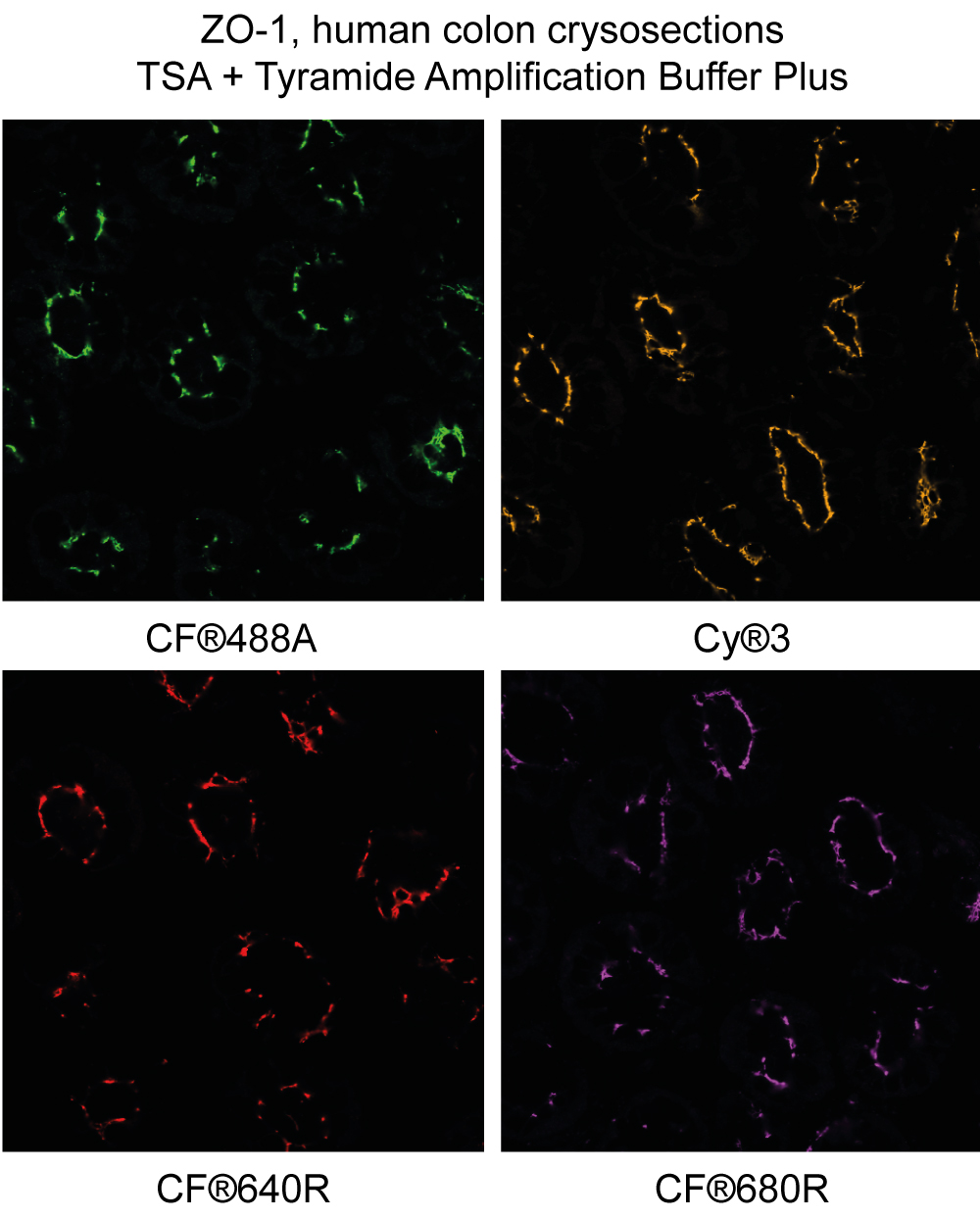
Yes, while our Tyramide Amplification Buffer Plus has enhanced sensitivity for TSA resulting in exceptional brightness, specificity, and sensitivity, the TyraMax™ Dyes will work with any amplification buffer.
Multiplex immunohistofluorescence (mIHF) has become an essential tool for studying complex tissue biology, enabling researchers to visualize multiple protein targets within a single sample while preserving spatial context. However, many existing multiplexing platforms remain costly, inflexible, or dependent on proprietary reagents, limiting accessibility for broader research applications. To address these challenges, open and scalable workflows are needed to make robust, reproducible, and cost-effective multiplex imaging more widely available to researchers.
In a 2026 protocol from the Journal of Microscopy, Riggi et al. created an open and flexible 6-color immunohistofluorescence (Flex-6 mIHF) workflow to investigate protein co-localization within the breast cancer tumor microenvironment. To overcome the background from autofluorescence in FFPE tissue sections that limits detection with IF, the protocol leveraged tyramide signal amplification (TSA) in combination with secondary antibodies conjugated to peroxidase-labeled polymers. This enabled robust signal enhancement and precise spatial resolution of biomarkers. The authors also outlined a stepwise validation strategy and essential controls to ensure reliable multiplex staining.
Within this workflow, CF® Dye Tyramides (Biotium) generated bright, covalently bound signals that can withstand repeated cycles of antibody stripping, facilitating sequential multiplexing without signal loss. This approach enabled simultaneous detection of up to six protein markers plus a nuclear stain in a single tissue section without requiring extensive image processing or spectral unmixing. By performing TSA with careful selection of antibodies, fluorophores, and order of target detection, the protocol produced high signal-to-noise images that could be directly analyzed, significantly reducing time and computational burden.
This protocol highlights how Biotium’s CF® Dye–based TSA reagents allow researchers to build flexible, high-performance multiplex immunofluorescence workflows without reliance on closed systems. By delivering exceptional brightness, photostability, and spectral diversity, Biotium’s fluorescent solutions help enable scalable, reproducible imaging protocols for cancer biology and beyond while making sophisticated multiplexing approaches more accessible to the broader scientific community.
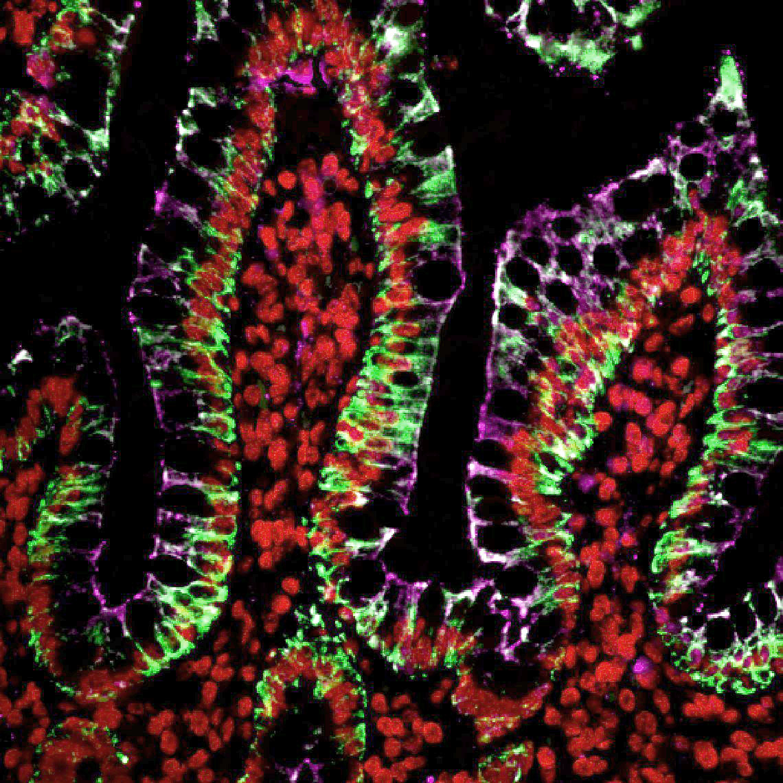
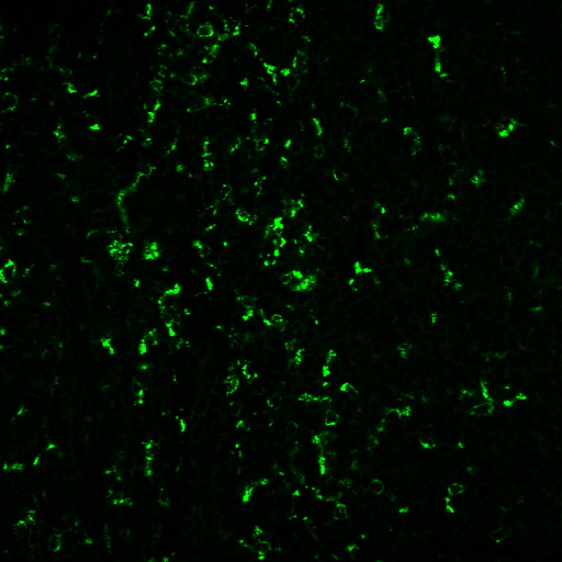
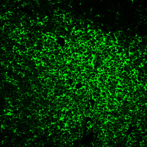
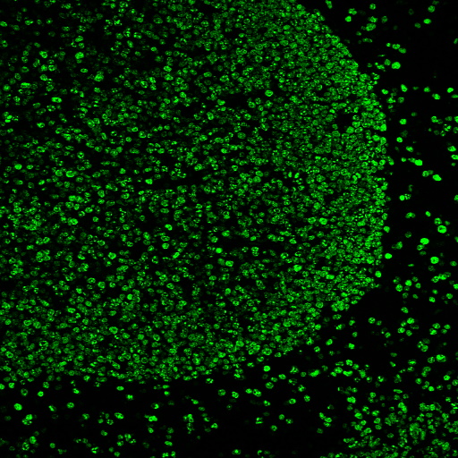
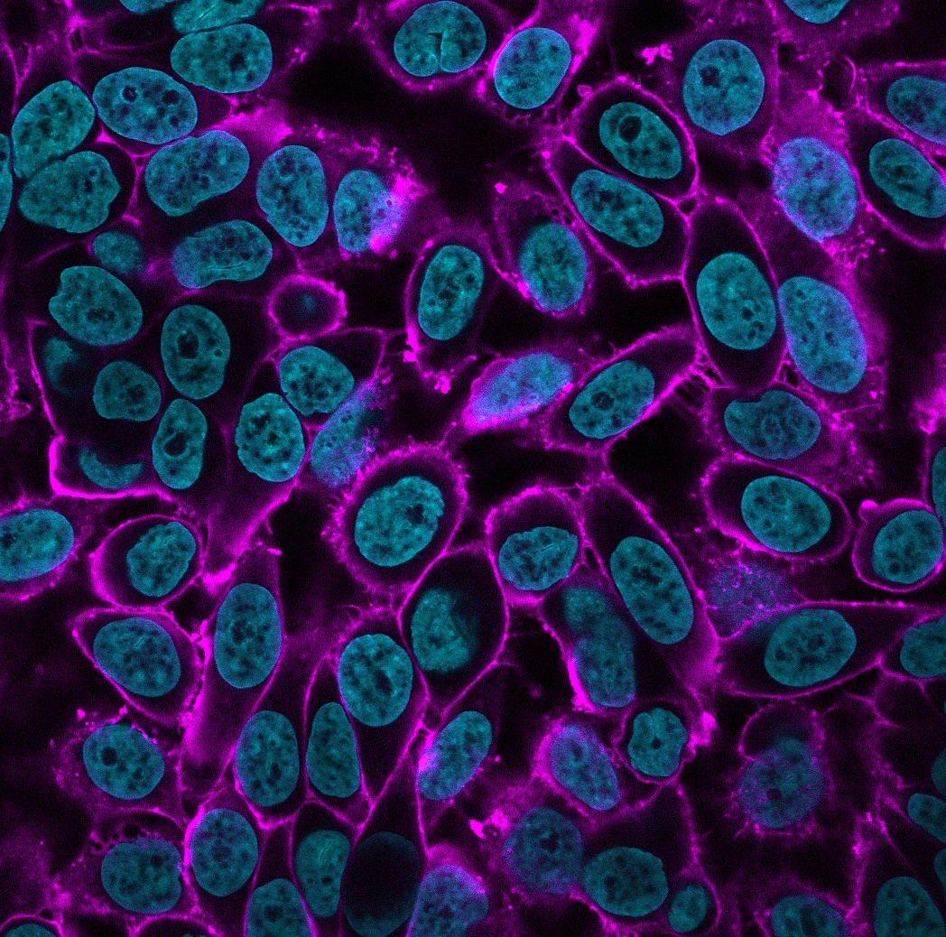
Sequential multiplex tyramide labeling of human colon FFPE section with three CF® Dye Tyramides. Cytokeratin (pan) was labeled with CF®488A Tyramide (green); Histone H1 was labeled with Cyanine 555 Tyramide (red); ZO1 was labeled with CF®640R Tyramide (magenta). Credit: Biotium.
Learn more about Biotium’s products for TSA, including our TyraMax™ Amplification Dyes which offer improved brightness, photostability, and chemical stability over CF® Dye Tyramides. Biotium also offers secondary antibodies conjugated to fluorophores or enzymes and a broad assortment of reagents for immunofluorescence microscopy.
Full Citation:
Riggi, J. A. M., Daumerie, A., Benhaddi, N., Berlière, M., Galant, C., González-Antelo, A., Nana, F. A., Van Bockstal, M. R., & Bouzin, C. (2026). A detailed protocol for open and low-cost six-plex immunofluorescence (Flex-6 mIHF) with a proof-of-concept study on breast cancer tissue. Journal of Microscopy, 1–19. https://doi.org/10.1111/jmi.70068
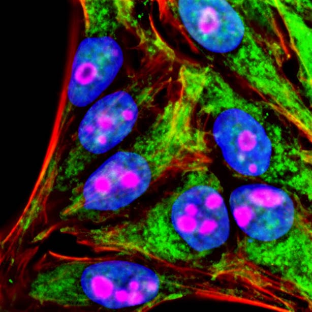
Our Tyramide Amplification Kits have been demonstrated to be robust and versatile for multi-color fluorescence imaging, compatible with dye-labeled antibodies and various cell staining methods (see Figure 1).
To use a Tyramide Amplification Kit in addition to one or more dye-labeled antibodies, follow the kit protocol to fix and block samples; label with primary antibodies; then detect primary antibodies using secondary antibodies. Dye labeled secondary antibodies can be co-incubated with the HRP-conjugated secondary or HRP-streptavidin from the tyramide kit. After washing, perform the CF® Dye tyramide reaction according to the kit protocol. The tyramide reaction does not interfere with the binding of dye-labeled antibodies or other fluorescent staining reagents.
Performing multi-color detection with more than one dye tyramide on the same sample requires sequential tyramide staining reactions, followed by HRP inactivation or antibody stripping between each step. See our tech tip:
Multi-Color Fluorescence Imaging Using Biotium's Tyramide Amplification Kits
Targeted activation of immune checkpoints is an emerging strategy for treating Type 1 Diabetes (T1D), where autoreactive T cells destroy insulin-producing beta cells. Autoimmune diabetes has been linked to disruption of the PD-1/PD-L1 pathway, which normally suppresses T cell activity to maintain immune tolerance. To address the limitations of systemic immunosuppression, an immune modulating monoclonal TCR against autoimmunity (ImmTAAI) molecule was created to selectively activate PD-1 signaling at the beta cell surface. This is a bispecific molecule that combines a T cell receptor targeting a preproinsulin peptide presented by HLA-A2 with a PD-1 agonist domain, enabling localized suppression of autoreactive T cells.
In a 2026 Science Advances publication, Becker et al. evaluated ImmTAAI function in live human pancreas tissue slices through confocal imaging, binding assays, and functional coculture systems using engineered T cell “avatars” designed to mimic the behavior of autoreactive T cells in T1D. Biotium’s CF®647 succinimidyl ester dye was used to label and visualize ImmTAAI molecules in live and fixed human pancreas slices. This labeling enabled precise tracking of ImmTAAI localization and quantification of its binding to beta cells via confocal microscopy without affecting binding affinity, specificity, or functional potency of ImmTAAI.
The researchers found that ImmTAAI binds specifically and dose-dependently to beta cells in a human leukocyte antigen (HLA)-dependent manner, with increased binding under inflammatory conditions. ImmTAAI treatment increased T cell movement, reduced T cell–beta cell interactions, and suppressed cytotoxic activity of target beta cells. Furthermore, ImmTAAI conferred protection of beta cells from immune attack in cell culture and in tissue slices, and helped preserve insulin secretion in live pancreas slices from a donor recently diagnosed with type 1 diabetes.
CF®647 enabled colocalization studies in a PD/PD-L1 therapeutic model and confirmed selective targeting within complex tissue environments. These findings highlight the value of bright, photostable far-red dyes like CF®647 for imaging-driven validation of targeted immunotherapies.
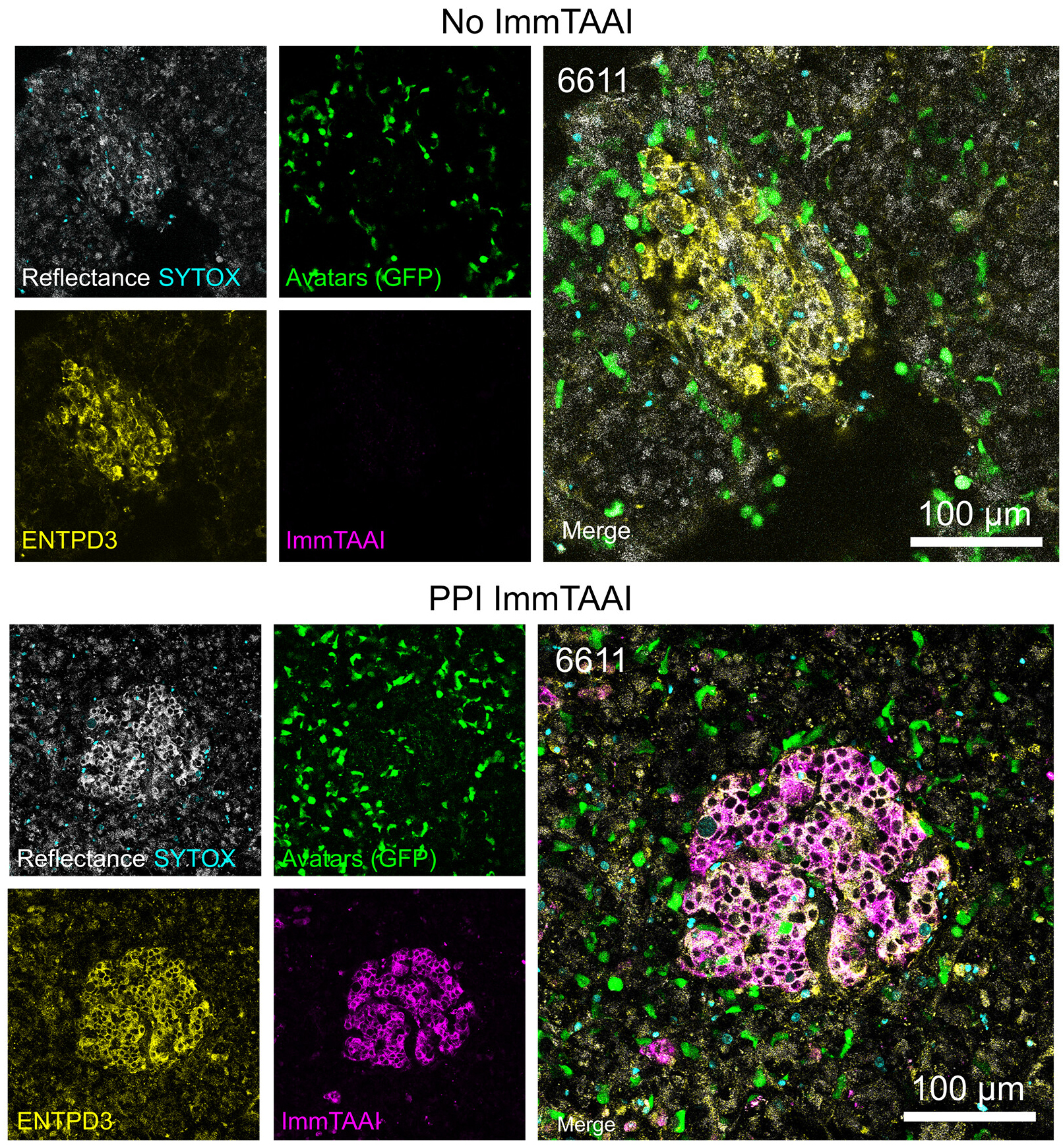
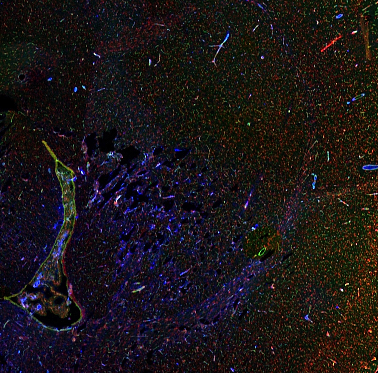
Live-cell confocal imaging (18 h) of pancreas slices treated with or without PPI ImmTAAI after addition of 200,000 IGRP-specific T cells per slice shows CF®647-labeled ImmTAAI colocalization with ENTPD3. Adapted from Becker et. al. Reproduced under CC BY 4.0.
Learn more about Biotium’s next-generation CF® Dye probes featuring exceptional brightness, photostability, and signal-to-noise, available in over 40 colors from blue to near-IR. CF® Dyes are also available in convenient Mix-n-Stain™ Labeling Kits for quick and efficient antibody labeling.
Full Citation
Matthew W. Becker et al. Beta cell–targeted PD-1 agonist inhibits cell-mediated autoimmunity in pancreas tissue slices. Sci. Adv. 12, eaec9029(2026). DOI:10.1126/sciadv.aec9029
Bioscience kits
The guaranteed shelf life from date of receipt for bioscience kits is listed on the product information sheet. Some kits have an expiration date printed on the kit box label, this is the guaranteed shelf life date calculated from the day that the product shipped from our facility. Kits often are functional for significantly longer than the guaranteed shelf life. If you have an older kit in storage that you wish to use, we recommend performing a small scale positive control experiment to confirm that the kit still works for your application before processing a large number of samples or precious samples.
Antibodies and other conjugates
The guaranteed shelf life from date of receipt for antibodies and conjugates is listed on the datasheet sheet which can be downloaded on the product page. Antibodies and other conjugates often are functional for significantly longer than the guaranteed shelf life. If you have an older conjugate in storage that you wish to use, we recommend performing a small scale positive control experiment to confirm that the product still works for your application before processing a large number of samples or precious samples.
For lyophilized antibodies, we recommend reconstituting the antibody with glycerol and antimicrobial preservative like sodium azide for the longest shelf life (note that sodium azide is not compatible with HRP-conjugates).
Chemicals, dyes, and gel stains
Biotium guarantees the stability of chemicals, dyes, and gel stains for at least a year from the date you receive the product. However, the majority of these products are highly stable for many years, as long as they are stored as recommended. Storage conditions can be found on the product information sheet or product safety and data sheet, material safety data sheet, and on the product label. Fluorescent compounds should be protected from light for long term storage.
If you have a Biotium compound that has been in storage for longer than one year that you wish to use, we recommend performing a small scale positive control experiment to confirm that the compound still works for your application before processing a large number of samples or precious samples.
Expiration date based on date of manufacture (DOM)
If your institution requires you to document expiration date based on date of manufacture for reagents, please contact [email protected] for assistance.
Chemical products with special stability considerations:
Esters
Ester compounds include the following:
Ester dyes are stable in solid form as long as they are protected from light and moisture. Esters are not stable in aqueous solution. Concentrated stock solutions should be prepared in anhydrous DMSO (see Biotium catalog no. 90082). Stock solutions in anhydrous DMSO can be stored desiccated at -20°C for one month or longer. Esters should be diluted in aqueous solution immediately before use. Succinimidyl esters (SE) should be dissolved in a solution that is free of amine-containing compounds like Tris, glycine, or protein, which will react with the SE functional group. AM esters and diacetate compounds should be dissolved in a solution that is free of serum, because serum could contain esterases that would hydrolyze the compound.
A note on CF® Dye succinimidyl ester stability
Succinimidyl esters (SE) are generally susceptible to hydrolysis, which can result in lower labeling efficiency. Many commercially available fluorescent dyes used for life science research are heavily sulfonated dyes which makes them particularly hygroscopic, worsening the hydrolysis problem. In addition, for several commercially available SE reactive dyes, the SE group is derived from an aromatic carboxylic acid, while the SE group in all of Biotium’s CF® Dyes is prepared from an aliphatic carboxylic acid. This structural difference reduces the susceptibility of CF® Dye SE reactive groups to hydrolysis, resulting in relatively stable reactive dyes with consistently higher labeling efficiency compared to other SE derivatives of other fluorescent dyes.
Maleimides, MTS and thiosulfate dyes
Like the succinimidyl ester dyes, these dyes are also susceptible to hydrolysis, although generally to a much lower degree. Thus, for long term storage, anhydrous DMSO is recommended for making stock solutions.
Other reactive dyes
Amines, aminooxy (also known as oxylamine), hydrazide, azide, alkyne, BCN, and tyramide reactive dyes, as well as dye free acids, are generally stable in aqueous solution when stored at -20°C for 6-12 months or longer, as long as no compounds are present that may react with the dye’s functional group. See the product information sheets for specific reactive dyes more information.
Coelenterazines and D-luciferin
Coelenterazines are stable in solid form when stored as recommended; they are not stable in aqueous solution. Concentrated coelenterazine stock solutions (typically 1-100 mg/mL) should be prepared in ethanol or methanol; do not use DMSO or DMF to dissolve coelenterazines, because these solvents will oxidize the compounds. Ethanol or methanol stocks of coelenterazine can be stored at -20°C or below for six months or longer; alcohol stocks may evaporate during storage, so use tightly sealing screw cap vials and wrap the vials with Parafilm for long term storage. Propylene glycol also can be used as a solvent to minimize evaporation. If the solvent evaporates, the coelenterazine will still be present in the vial, so note the volume in the vial prior to storage so that you can adjust the solvent volume to correct for evaporation if needed. Prepare working solutions in aqueous buffers immediately before use. Coelenterazines are stable for up to five hours in aqueous solution.
Aquaphile™ coelenterazines are water soluble formulations of coelenterazines. They are stable in solid form when stored as recommended. Aquaphile™ coelenterazines should be dissolved in aqueous solution immediately before use. They are stable for up to five hours in aqueous solution.
Note that coelenterazines are predominantly yellow solids, but may contain dark red or brown flecks. This does not affect product stability or performance. If your coelenterazine is uniformly brown, then it is oxidized and needs to be replaced.
D-luciferin is stable in solid form and as a concentrated stock solution when stored as recommended; it is not stable at dilute working concentrations in aqueous solution. Prepare concentrated D-luciferin stock solutions (typically 1-100 mg/mL) in water, and store in aliquots at -20°C or below for six months or longer. Prepare working solutions immediately before use.
Dyes that carry multiple negative charges can introduce background. Usually, this is more of a concern with labeled antibodies that carry many dyes, as opposed to a small toxin like bungarotoxin. When staining tissues, the endogenous autofluorescence of the tissue itself is often the most significant source of background. Endogenous fluorescence background in tissue is usually highest in the blue wavelengths (DAPI channel) and lowest in the far-red (Cy®5 channel). Our CF®633 bungarotoxin (catalog no. 00009) is a far-red conjugate for the Cy®5 channel with a low negative charge that should have low background from either the dye or autofluorescence.
We test fluorescent bungarotoxin on rat skeletal muscle sections. While the tissue shows autofluorescence, the bungarotoxin staining of motor endplates is usually much brighter than the background for all of the dye colors we've tested. However, if you are staining human tissue (especially brain), lipofuscin autofluorescence may be bright in all channels. This usually shows up as bright, punctate dots around cell nuclei. While we would usually recommend our TrueBlack® lipofuscin quenchers for human brain tissue, they are not compatible with bungarotoxin staining. We have, however, found that EverBrite TrueBlack® Mounting Medium (cat. no. 23017) can be used to mount skeletal muscle sections stained with bungarotoxin.
Cy Dye is a registered trademark of Cytiva.
Bioscience kits
The guaranteed shelf life from date of receipt for bioscience kits is listed on the product information sheet. Some kits have an expiration date printed on the kit box label, this is the guaranteed shelf life date calculated from the day that the product shipped from our facility. Kits often are functional for significantly longer than the guaranteed shelf life. If you have an older kit in storage that you wish to use, we recommend performing a small scale positive control experiment to confirm that the kit still works for your application before processing a large number of samples or precious samples.
Antibodies and other conjugates
The guaranteed shelf life from date of receipt for antibodies and conjugates is listed on the datasheet sheet which can be downloaded on the product page. Antibodies and other conjugates often are functional for significantly longer than the guaranteed shelf life. If you have an older conjugate in storage that you wish to use, we recommend performing a small scale positive control experiment to confirm that the product still works for your application before processing a large number of samples or precious samples.
For lyophilized antibodies, we recommend reconstituting the antibody with glycerol and antimicrobial preservative like sodium azide for the longest shelf life (note that sodium azide is not compatible with HRP-conjugates).
Chemicals, dyes, and gel stains
Biotium guarantees the stability of chemicals, dyes, and gel stains for at least a year from the date you receive the product. However, the majority of these products are highly stable for many years, as long as they are stored as recommended. Storage conditions can be found on the product information sheet or product safety and data sheet, material safety data sheet, and on the product label. Fluorescent compounds should be protected from light for long term storage.
If you have a Biotium compound that has been in storage for longer than one year that you wish to use, we recommend performing a small scale positive control experiment to confirm that the compound still works for your application before processing a large number of samples or precious samples.
Expiration date based on date of manufacture (DOM)
If your institution requires you to document expiration date based on date of manufacture for reagents, please contact [email protected] for assistance.
Chemical products with special stability considerations:
Esters
Ester compounds include the following:
Ester dyes are stable in solid form as long as they are protected from light and moisture. Esters are not stable in aqueous solution. Concentrated stock solutions should be prepared in anhydrous DMSO (see Biotium catalog no. 90082). Stock solutions in anhydrous DMSO can be stored desiccated at -20°C for one month or longer. Esters should be diluted in aqueous solution immediately before use. Succinimidyl esters (SE) should be dissolved in a solution that is free of amine-containing compounds like Tris, glycine, or protein, which will react with the SE functional group. AM esters and diacetate compounds should be dissolved in a solution that is free of serum, because serum could contain esterases that would hydrolyze the compound.
A note on CF® Dye succinimidyl ester stability
Succinimidyl esters (SE) are generally susceptible to hydrolysis, which can result in lower labeling efficiency. Many commercially available fluorescent dyes used for life science research are heavily sulfonated dyes which makes them particularly hygroscopic, worsening the hydrolysis problem. In addition, for several commercially available SE reactive dyes, the SE group is derived from an aromatic carboxylic acid, while the SE group in all of Biotium’s CF® Dyes is prepared from an aliphatic carboxylic acid. This structural difference reduces the susceptibility of CF® Dye SE reactive groups to hydrolysis, resulting in relatively stable reactive dyes with consistently higher labeling efficiency compared to other SE derivatives of other fluorescent dyes.
Maleimides, MTS and thiosulfate dyes
Like the succinimidyl ester dyes, these dyes are also susceptible to hydrolysis, although generally to a much lower degree. Thus, for long term storage, anhydrous DMSO is recommended for making stock solutions.
Other reactive dyes
Amines, aminooxy (also known as oxylamine), hydrazide, azide, alkyne, BCN, and tyramide reactive dyes, as well as dye free acids, are generally stable in aqueous solution when stored at -20°C for 6-12 months or longer, as long as no compounds are present that may react with the dye’s functional group. See the product information sheets for specific reactive dyes more information.
Coelenterazines and D-luciferin
Coelenterazines are stable in solid form when stored as recommended; they are not stable in aqueous solution. Concentrated coelenterazine stock solutions (typically 1-100 mg/mL) should be prepared in ethanol or methanol; do not use DMSO or DMF to dissolve coelenterazines, because these solvents will oxidize the compounds. Ethanol or methanol stocks of coelenterazine can be stored at -20°C or below for six months or longer; alcohol stocks may evaporate during storage, so use tightly sealing screw cap vials and wrap the vials with Parafilm for long term storage. Propylene glycol also can be used as a solvent to minimize evaporation. If the solvent evaporates, the coelenterazine will still be present in the vial, so note the volume in the vial prior to storage so that you can adjust the solvent volume to correct for evaporation if needed. Prepare working solutions in aqueous buffers immediately before use. Coelenterazines are stable for up to five hours in aqueous solution.
Aquaphile™ coelenterazines are water soluble formulations of coelenterazines. They are stable in solid form when stored as recommended. Aquaphile™ coelenterazines should be dissolved in aqueous solution immediately before use. They are stable for up to five hours in aqueous solution.
Note that coelenterazines are predominantly yellow solids, but may contain dark red or brown flecks. This does not affect product stability or performance. If your coelenterazine is uniformly brown, then it is oxidized and needs to be replaced.
D-luciferin is stable in solid form and as a concentrated stock solution when stored as recommended; it is not stable at dilute working concentrations in aqueous solution. Prepare concentrated D-luciferin stock solutions (typically 1-100 mg/mL) in water, and store in aliquots at -20°C or below for six months or longer. Prepare working solutions immediately before use.
For dyes or reagents that are supplied lyophilized (as solids), it is hard to compare quantities based on appearance of the dye in the tube, because during the lyophilization process the dye can dry down in different ways, either spread out all over the tube, clumped together, or coating the sides or bottom of the tube. Centrifugation of the tube may not help in collecting the dye solid to the bottom of the tube as this generally works for solutions. However, lyophilized solids are packaged based on highly accurate absorbance measurement of the reagent solution prior to drying, so the vial will contain the correct amount of dye.
Biotium ships all antibodies (primary, secondary and conjugates) at room temperature. We guarantee their quality and performance under these conditions based upon our stability testing. Antibodies were subjected to accelerated stability testing by storing them at various temperatures (4°C, room temperature, or 37°C) for 1 week to mimic simulated shipping conditions and tested in immunostaining experiments. All antibodies showed the expected brightness and specificity, even after storage at sub-optimal temperatures for a week or longer. You can also download our Product Storage Statement here.
In line with our goal to be more environmentally friendly by reducing the use of excess packaging, and lowering shipping costs for our customers, products that have passed our stability testing are shipped at room temperature.
Once you have received the antibody vial, please follow the long-term storage instructions on the product information (PI) sheet.
