 New Products
 Earth-Friendly Products
 Biotium Choice Antibodies
 Special Offers


Content #1
Content #1
Content #1
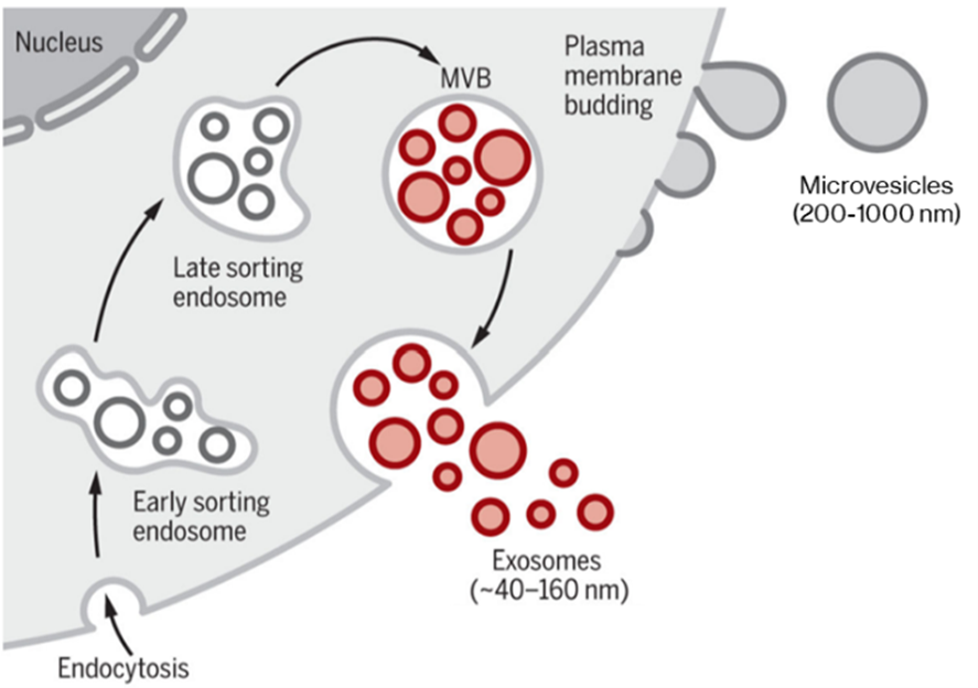
Classical modes of EV generation, producing microvesicles directly from plasma membrane (PM) and exosomes via the late sorting endosome and multivesicular body (MVB). Certain cell types can also produce exosomes by direct PM budding. Adapted from Kalluri & LeBleu (2020).5 Reproduced under the Creative Commons license.
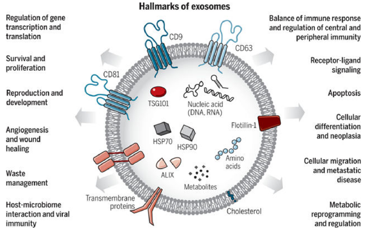
Exosomes are varied in size and composition, carrying diverse cargoes including membrane proteins (particularly CD63 and CD81). Exosomes are involved in a variety of functions. From Kalluri & LeBleu (2020).5 Reproduced under the Creative Commons license.
EVs carry cargoes of proteins, RNA, and DNA that function in the recipient cells.1,2,5,6 Microvesicles secreted by cancer cells (sometimes called oncosomes) can transport oncogenic miRNA that promotes invasion and metastasis in breast cancer cells,8 and exosomes from pancreatic cancer cells initiate transformation in normal cells.9 EVs can also contribute to immunity and immunosuppression: antigen presenting cells secrete exosomes containing major histocompatibility complex class II bound with antigen, which can promote adaptive immunity over a distance,5 and tumor-derived EVs can promote immunosuppression within the tumor microenvironment.5 Viruses can use EVs to escape from cells and infect others; for example, rotavirus capsids hide within large secreted microvesicles and HIV Gag proteins use T-cell derived exosomes.4,10
Exosomes have also been associated with neurodegeneration. Prion protein is present in brain-derived exosomes, where it plays roles in immunomodulation, plasticity, and normal brain function. Pathogenic prion protein in spongiform encephalopathies also gets carried by exosomes along with other factors—microRNAs in particular—that potentiate its neurotoxic effects.11 MicroRNAs associated with the progression of Alzheimer’s Disease and Parkinson’s Disease have also been found in brain-derived exosomes. Exosomes’ roles in the pathogenesis of these and other neurodegenerative diseases remain to be determined.
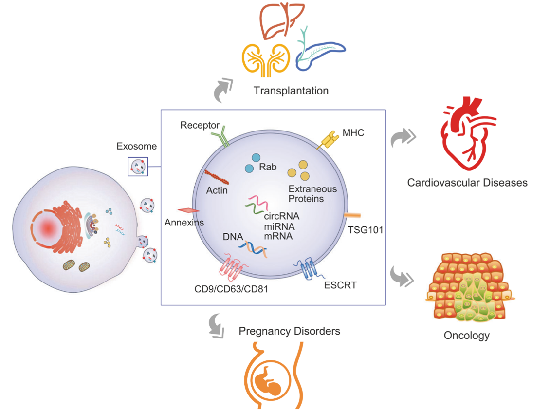
Circulating EVs make non-invasive liquid biopsies possible. Figure from Zhou et al. 2020.12 Reproduced under the Creative Commons license.
As small heterogeneous extracellular particles, EVs pose challenges to characterize; a principal confounder is the lack of standardized isolation and analysis methods.5,15,16 EVs have classically been isolated by ultracentrifugation or sucrose gradient ultracentrifugation,2,3 but the purity of isolated EVs is poor.15 New isolation techniques such as acoustic nanofiltering and microfluidic immuno-isolation improve purity, but require specialized equipment.
Scanning and transmission electron microscopy can readily visualize EVs, but sample preparation creates artifacts and, depending on the preparation method, can cause EV sampling bias.2,4,16 Fluorescence imaging with lipophilic dyes in culture systems are confounded by other small particles in biofluids and cell culture media like serum proteins and lipoproteins.17 Mass spectroscopy, NGS and proteomics are commonly used to analyze EV content, but heterogeneity and purity are lingering concerns.
Analyzing exosomes with flow cytometry is a new field; one challenge is distinguishing exosomes from cellular debris. Fortunately, EVs have well-defined markers and properties that can be exploited:1,16 CD9, CD63, and CD81 are markers for EVs, with CD63 being specific to exosomes. In certain types of EVs, Ep-CAM and HSP70 are useful targets.1,5 For flow cytometry of EVs, it is crucial to avoid forming aggregates as the loss of individual resolution is disastrous for such a heterogeneous population.15 Antibodies validated for EV detection by flow cytometry are a key starting point. Membrane stains validated for flow cytometry are useful for measuring EV size by fluorescence intensity.
EVs represent an exciting and booming field of cell biology with life-changing diagnostic and therapeutic potential. Better understanding and characterization of EVs is critical in making this potential into reality. Biotium’s curated EV technology resource will assemble the latest validated tools for EV research. Without a doubt, EV research will drive advances in cell biology and medicine.

Christopher Pratt earned his PhD in cell biology and neuroscience from Carnegie Mellon University in 2016 under the guidance of Marcel Bruchez, PhD. For his thesis, he developed and applied novel fluorescent probes to understand synaptic vesicle and ion channel trafficking in the brain, making him a ardent microscopist. With a passion for communicating science and data, Christopher is a regular contributor to Biotium’s Full Spectrum Blog and is a freelance science and medical writer, analyst, and research consultant based in Chicago, Illinois. Visit his website, twitter, or LinkedIn and get in touch!

Content #1
Content #1
Content #1
Content #2
Content #3