 New Products
 Earth-Friendly Products
 Biotium Choice Antibodies
 Special Offers


Content #1
Content #1
Content #1
Antibodies have been indispensable tools in biology since Albert Coons and his colleagues at Harvard first conjugated fluorescein isothiocyanate, a fluorescent dye, to antibodies in 1941.1 Following that foundational work, Georges Köhler and César Milstein’s development of hybridoma-based monoclonal antibodies in 1975 marked the next major leap in antibody technology, forging a clear progression from polyclonal to monoclonal to single-domain antibodies.2 In the decades that followed, fluorescent antibodies have powered discoveries in cell signaling, neuroscience, immunology, and molecular imaging. However, as imaging techniques advance toward higher resolution and quantitative precision, the limitations of conventional antibodies grow increasingly apparent. These limitations include their large size, steric hindrance, and increased background from nonspecific binding when using secondary antibodies. To meet the demands of powerful new labeling and imaging techniques, researchers are turning to single-domain antibodies (SdAbs), also known as Nanobodies®. Many consider SdAbs to be the next generation of compact, high-performance antibody reagents.
SdAbs are the smallest functional fragments of an antibody that still retain full antigen-binding capability. They’re most commonly derived from certain types of antibodies found in cartilaginous fish and camelids, a group of animals including camels, llamas, and alpacas. In the early 1990s, researchers studying dromedary camels discovered that they produce a distinct class of heavy-chain-only antibodies, which lack the light chains present in conventional antibodies (Fig. 1).2 The single antigen-binding domain from these antibodies, the VHH, was found to function independently, marking the development of what we now call SdAbs.
Explored initially for therapeutic use due to their high tissue penetration and low immunogenicity, SdAbs are now used in basic research for immunofluorescence, super-resolution microscopy, protein purification, and live-cell imaging.2-4 Their simplicity, stability, and modularity make them ideal tools for a variety of imaging and other immunodetection applications.
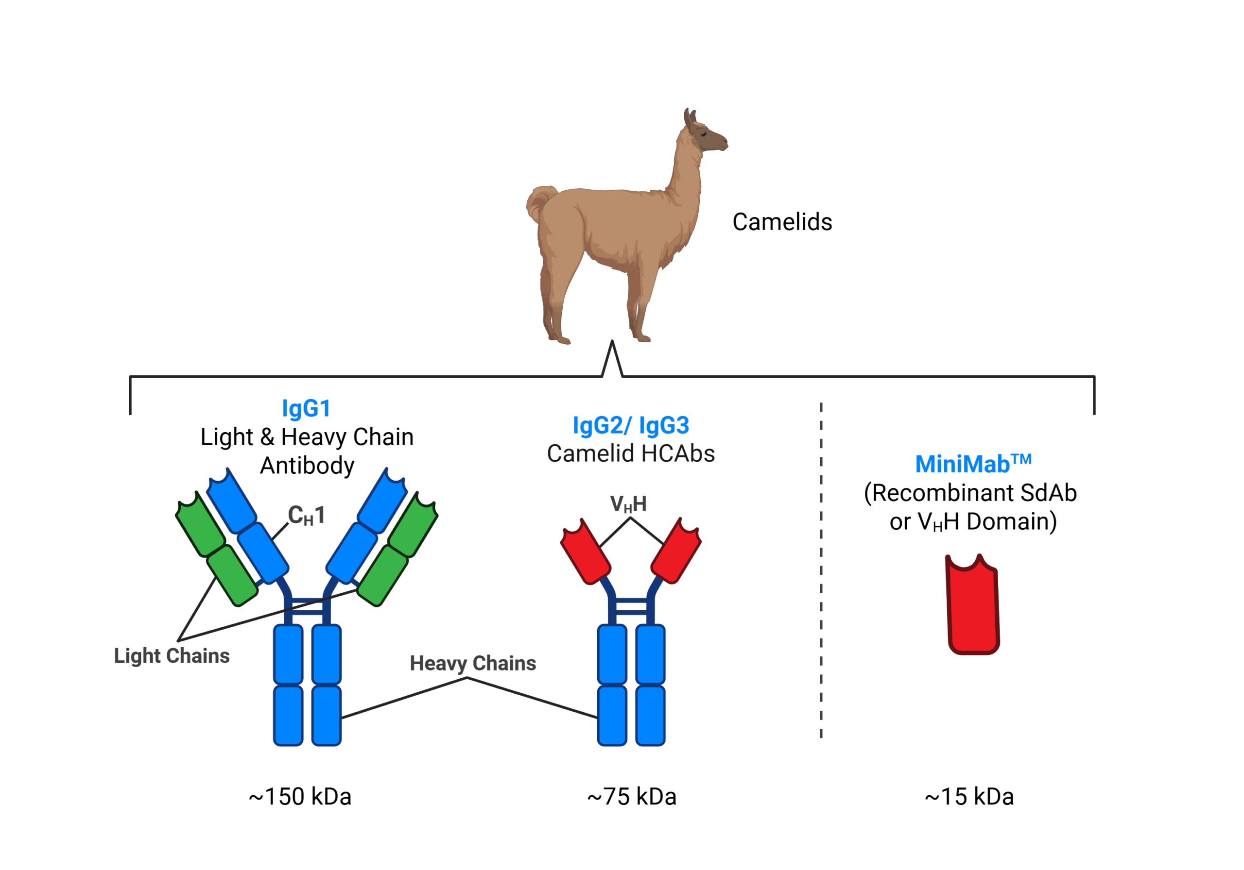
Figure 1. Alpacas produce three IgG subclasses: IgG1, IgG2, and IgG3. Camelid IgG1 has both heavy and light chains and is most similar to the widely used IgG antibodies from species such as mouse or rabbit. Camelid IgG2 and IgG3 are heavy-chain-only antibodies (HCAbs) lacking light chains and the CH1 domain. MiniMab™ SdAbs are the recombinant form of the VHH variable domain of these HCAbs. Created in BioRender. Criado, M. (2025) https://BioRender.com/.
SdAbs stand apart from conventional antibodies not only in size but in performance. They consist of a single variable domain with a diameter of ~2.5 nm and a molecular weight of roughly 15 kDa, about one-tenth that of a typical immunoglobulin G (IgG) antibody, and are still readily conjugated or fused to fluorophores, enzymes, or affinity tags.3 Their compact structure allows them to reach epitopes in cavities or grooves that are otherwise inaccessible and enables deeper tissue penetration for more uniform labeling in dense or fixed samples.2-4 They are also highly soluble and are more stable under conditions that would denature most antibodies, such as high temperatures, detergents, and fixation treatments, making them compatible with a wide range of workflows.4-5
Nanobodies can be efficiently produced recombinantly in prokaryotic systems such as E. coli, bypassing the need for complex eukaryotic expression systems.11 Their small, simple structure allows easy expression, purification, and large-scale production, making them valuable for biosensors, diagnostics, and therapeutics.11 High-yield, cost-effective, and easily scalable production supports both clinical applications and research, broadening access for scientists with limited resources.11 Additionally, their recombinant nature also makes them highly engineerable. The result is faster, cleaner labeling with minimal background.4
One potentially transformative application for SdAbs is their use in super-resolution microscopy. In widefield or confocal imaging, which have a resolution of ∼250 nm at best, the small uncertainty (~20 nm) introduced by labeling targets with primary and secondary antibodies is negligible. However, as imaging resolution increases with techniques such as STED, SIM, and dSTORM, the physical size of IgG antibodies, combined with the multiple dye labels on secondary antibodies, introduces significant localization error in the target position.6 This “linkage error” can add 20-30 nm between the fluorophore and the epitope when using secondary antibodies and 10-15 nm when using primary antibodies, reducing effective resolution. SdAbs largely eliminate linkage error, therefore, significantly improving localization accuracy and resolution.7
SdAbs are also enhancing signal quality across many assays. In immunofluorescence, they penetrate tissue more efficiently and produce cleaner labeling with reduced Fc receptor-mediated background.3-5 The Fc portion of conventional antibodies, usually IgG, binds Fc receptors on cells (macrophages, microglia, dendritic cells, neutrophils, some endothelial cells, etc.), producing unwanted staining that looks like real antigen signal. Because sdAbs lack an Fc region, they largely avoid that artifact. In Western blotting, SdAbs HRP or fluorescent conjugates provide sharper bands and improved signal-to-noise.12 Their small size also benefits flow cytometry, where reduced steric hindrance enables more accurate detection of densely expressed surface markers.4
Beyond their ability to improve imaging precision, SdAbs also offer significant practical advantages in assay performance. Their rapid binding kinetics, driven by a smaller molecular size and reduced diffusion barriers, allow them to reach equilibrium faster than conventional IgGs, enabling shorter incubation times. SdAbs are also exceptionally stable, retaining their structure and antigen-binding activity under elevated temperatures, low pH, and in the presence of detergents that can denature conventional antibodies.2 In addition, their high solubility and low aggregation propensity make them easier to handle and more consistent in demanding workflows such as multiplex labeling, harsh wash conditions, or long-term storage.2 Together, these features make SdAbs robust and efficient tools for diverse imaging and analytical applications.
Additionally, SdAbs can be genetically encoded to be expressed inside cells as “intrabodies,” which bind endogenous proteins in real time, enabling live-cell visualization of dynamic processes.8 Emerging computational design and AI-assisted SdAbs discovery methods are expected to accelerate their customization for intracellular targets.9-11
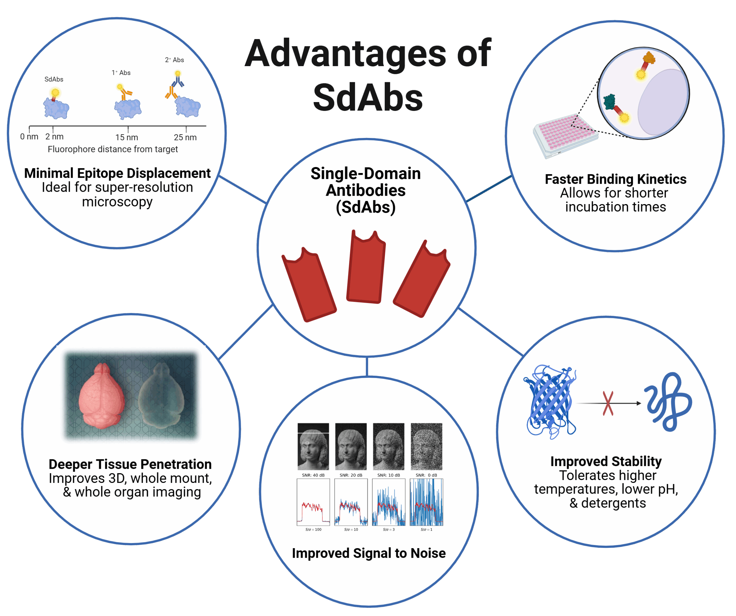
Figure 2. Key advantages of single-domain antibodies (SdAbs). Due to their small size and robust nature, SdAbs exhibit minimal epitope displacement for precise localization, faster binding kinetics, enhanced stability under harsh conditions, improved signal-to-noise ratios, and superior tissue penetration, making them powerful tools for advanced imaging and detection applications. Created in BioRender. Davis, J. (2025) https://BioRender.com/.
At Biotium, we’ve developed MiniMab™ SdAbs reagents to bring these advantages to everyday research. MiniMab™ SdAbs are directly conjugated to our CF® Dyes to deliver exceptional brightness, stability, and target specificity.
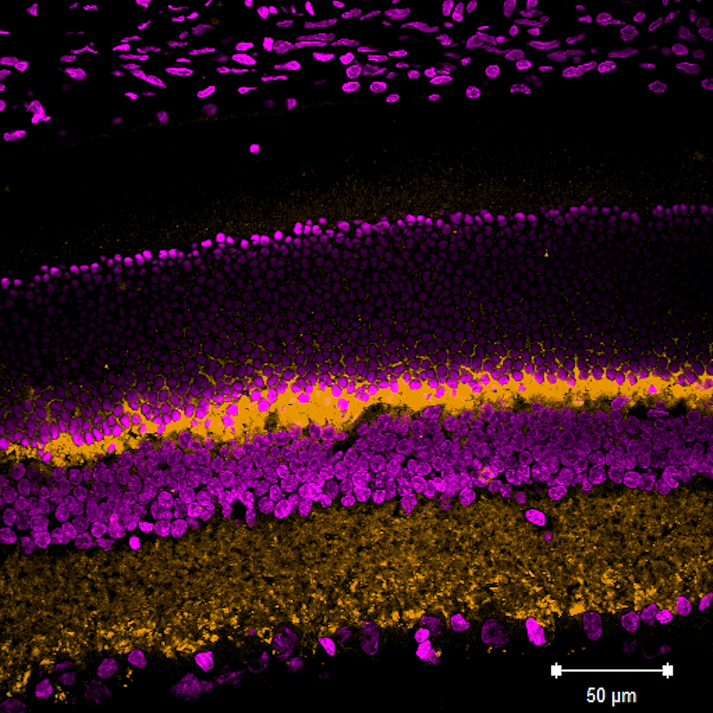
Figure 3. Rat eye cryosection stained with CF®568 VGLUT1 rVHH (SdAb2412.VGLUT1) MiniMab™ (orange). Nuclei are stained with NucSpot® 680/700 (magenta). Scale bar: 50 μm.
Because MiniMab™ SdAbs are much smaller than conventional antibodies, they penetrate tissue samples more efficiently and minimize linkage error in super-resolution imaging. Each MiniMab™ conjugate is validated for immunofluorescence, and a subset is validated for Western blotting and flow cytometry, offering scientists a plug-and-play solution for clear, reproducible results.
MiniMab™ SdAbs are particularly advantageous for neuroscience, where fine structural detail and low background are essential for mapping synaptic and cellular architecture. By combining the precision of SdAbs design with the brightness and photostability of CF® Dyes, MiniMab™ reagents enable researchers to visualize targets with exceptional clarity, helping bridge the gap between standard immunofluorescence and true molecular-resolution imaging. MiniMab™ SdAbs are also available labeled with our top-performing CF® Dyes for STORM.
As the demand for high-precision imaging and quantitative protein analysis grows, SdAbs are redefining what’s possible in the lab. Their small size, exceptional stability, and direct labeling capability make them indispensable for cutting-edge applications, from super-resolution microscopy to live-cell imaging.4-7
Whether you’re mapping intricate neuronal structures, analyzing protein complexes, or simplifying your immunofluorescence workflow, SdAbs offer a smarter, more efficient path forward. Biotium’s MiniMab™ SdAbs bring this power to your bench, delivering brighter signal, lower background, and higher resolution for your research.
References
Nanobody is a registered trademark of Ablynx N.V.

Julianne Davis earned an MSc in Behavioral Neuroscience from the University of Washington, Seattle, where she examined the role of memory in cost-based decision-making. She has also studied sensory integration at the Allen Institute and the neural basis of feeding, thirst, and metabolism at the University of California, San Francisco as a research scientist. Currently, she is a Technical Writer and Support Scientist at Biotium.

Content #1
Content #1
Content #1
Content #2
Content #3