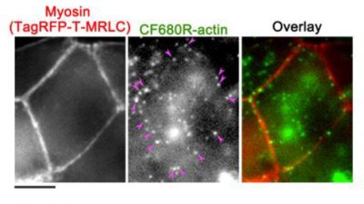
Single-molecule microscopic imaging is a powerful tool for studying cellular processes. However there are technical challenges that need to be overcome, such as background from cellular autofluorescence and fluorophores that are too dim or not photostable. In their recent article in the journal Sensors, authors Yamashiro and Watanabe address these challenges by developing a new actin probe labeled with the near-infrared dye CF™680R, which they used in electroporation-mediated single-molecule speckle (eSiMS) microscopy. Because cells tend to exhibit less autofluorescence in the infrared range, CF™680R was chosen over DyLight® 549 due to its excitation and emission in the near-infrared region of the spectrum.
They found that the CF™680R-actin probe was brighter and more photostable than other dye-actin conjugates that they tested. It is also able to be used in 3-color SiMS together with green and red fluorescent proteins. The CF™680R-actin probe was shown to move with the retrograde actin flow in lamellipodia, demonstrating that this actin probe behaves as expected. In addition, the CF™680R-actin probe allowed the authors to measure the turnover rate of actin in adherens junctions for the first time.
To read the original article, click here.
Yamashiro and Watanabe. An infrared actin probe for deep-cell electroporation-based single-molecule speckle (eSiMS) microscopy. Sensors. 17(7), 1545 (2017). Doi: 10.3390/s17071545
To learn more about bright and photostable CF™ Dyes, including our unrivaled selection of near-infrared dyes, click here.
DyLight is a registered trademark of Thermo Fisher Scientific.