A key application of recently developed super-resolution microscopy methods is multicolor detection of different targets, to dissect molecular interactions at the nanoscale level. However, the introduction of more than two colors with current methods requires difficult optical and statistical techniques. Now, a report from Winter and colleagues in Stefan Hell’s group at the Max Planck Institute describes hyperSTED, a simplified approach to multiplex super-resolution microscopy using just two lasers. They report the first instance of live cell STED microscopy using three and four different dyes, including Biotium’s CF™680R.
To read the original article, click here.
Winter, F. R. et al. Multicolour nanoscopy of fixed and living cells with a single STED beam and hyperspectral detection. Sci. Rep. 7, 46492; doi: 10.1038/srep46492 (2017).
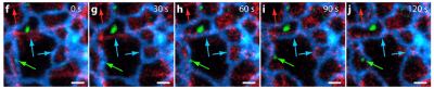
Three-color live cell hyperSTED imaging with CF680R. Figure 5 of Winter et al. 2017 Sci. Rep.
Learn more about Biotium’s CF™ dyes for super-resolution microscopy.