 New Products
 Earth-Friendly Products
 Biotium Choice Antibodies
 Special Offers


Content #1
Content #1
Content #1
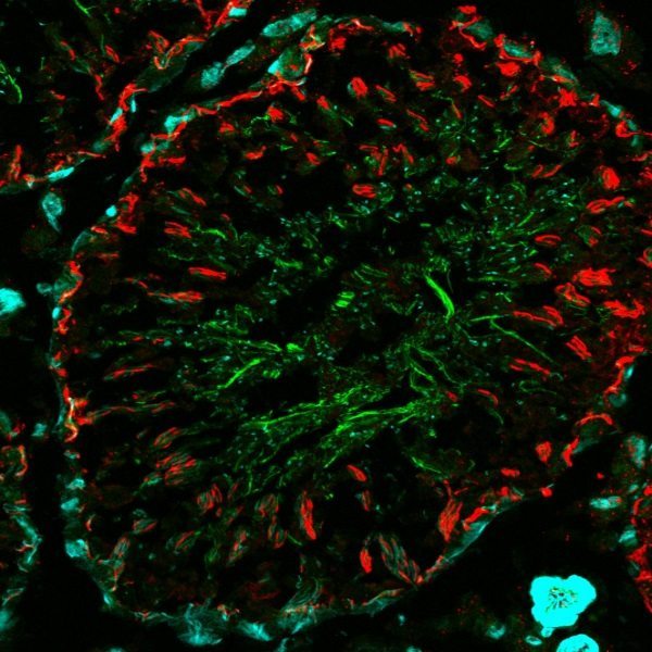
Expansion microscopy (ExM) is a new technique that allows for super-resolution imaging by expanding tissues through in-situ synthesis of swellable hydrogels. This expansion enables immunofluorescent detection of fine molecular details with 60 nm lateral resolution. However, due to the dramatic bleaching effect caused by expansion and free radical chemistry with the fluorophore, bright and stable fluorophores are necessary for producing high-quality images.
In a recent issue of Elsevier Methods, Min et. al. studied 22 CF® dyes for their compatibility with ExM in African green monkey kidney (BS-C-1) cells before and after expansion. The CF® dyes were conjugated to an antibody to stain the nuclear lamina using Mix-n-Stain™ CF® Antibody Labeling Kits. The selected dyes cover a wide emission spectrum and were tested using four conventional laser wavelengths (405, 488, 560 and 640 nm). Results indicate that CF®405S, CF®488A, CF®568 and CF®660R exhibit the brightest signal for each respective laser, with nearly 50% signal retention after expansion. The brightness of each dye was then compared to the popular Alexa Fluor® dyes. The authors observe significantly greater fluorescence of CF®405S and CF®568 when compared to their Alexa Fluor® counterparts. Meanwhile, CF®488A and Alexa Fluor® 488 showed similar fluorescence. The same CF® dyes were also applied toward 4-color ExM imaging of a 150-um thick mouse-brain section. The fluorescence signal was well-retained after expansion while accurately resolving finer details of the neurons, dendrites and myelin sheath. These results demonstrate the suitability and superiority of CF® dyes for ExM super-resolution imaging.
Learn more about our Mix-n-Stain™ CF® dye antibody labeling kits, and our other products for super-resolution microscopy.
Alexa Fluor® is a registered trademark of Thermo Fisher Scientific.
Full Citation
Min, K., Cho, I., Choi, M., & Chang, J.-B. (2019). Multiplexed expansion microscopy of the brain through fluorophore screening. Methods. https://doi.org/10.1016/j.ymeth.2019.07.017
