 New Products
 Earth-Friendly Products
 Biotium Choice Antibodies
 Special Offers


Content #1
Content #1
Content #1
Nerve terminal dyes like SynaptoGreen™ (a.k.a. FM®1-43), invented more than 20 years ago, have been instrumental in shaping our current view of synaptic function. Nerve terminal dye studies have generated several still debated hypotheses for synaptic vesicle release and recycling during neurotransmission.
Due to the unique properties of these styryl dyes - amphipathic nature, lipid-sensitive emission, and charged head group - they preferentially partition to outer membranes and recycling vesicles. Their dynamic interaction with membranes allows measurement of endocytosis-exocytosis rates via dye uptake and release. Nerve terminal dyes have also been used to study single vesicle release events. A recent paper describes a detailed protocol of FM® dye-based imaging of synaptic vesicle dynamics in primary cultures of rodent hippocampal neurons and compares them to other fluorescent vesicle labels.
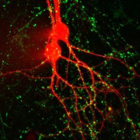
Cultured rat hippocampal neurons microinjected with CF®647 hydrazide (red) and stained with SynaptoGreen™ (FM1-43, green, synaptic vesicles). Image courtesy of Professor Guosong Liu, Tsinghua University, Beijing, China.
To read the original article, click here.
Lazarenko, R. M., DelBove, C. E. and Zhang, Q. (2018). Fluorescent Measurement of Synaptic Activity Using FM Dyes in Dissociated Hippocampal Cultured Neurons. Bio-protocol 8(2): e2690. DOI: 10.21769/BioProtoc.2690.
Learn more about Biotium's high-purity grade nerve terminal probes SynaptoGreen™ and SynaptoRed™ (originally FM® dyes) and nerve terminal staining kits.
