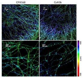
It was recently discovered that the membrane cytoskeleton in neurons is highly structured and periodic, and that these nanostructures are markedly different from the dense filament bundles occurring in cells such as fibroblasts, or the two-dimensional (2D) triangular lattices found in erythrocytes. In a recent publication in Cell Reports, Hauser and associates shed further light on these structures, by using 3D STORM super-resolution microscopy to resolve the spectrin-actin-based membrane cytoskeleton of neural stem cells (NSCs) and NSC-derived cells. Two-color 3D STORM was performed using antibodies conjugated to dyes with photoswitching properties, including Biotium’s CF®568.
Hauser et al. showed that undifferentiated NSCs contain areas of locally periodic, 1D membrane cytoskeleton structures with ∼180 nm to 190 nm periodicity. These structures become more ordered as the NSCs mature into terminally differentiated neuronal and glial cell types, and the 1D periodic “strips” were often found to dominate the flat 2D cell membranes. Interestingly, the authors showed a remarkable alignment of the cytoskeletons between neighboring cells at axon-axon and axon-oligodendrocyte contacts, suggesting that the cytoskeletal motif serves as a scaffold and a molecular ‘‘ruler’’ during NSC interactions, mediating cell selection and binding position at the nanoscale.
Learn more about CF® dyes for super-resolution microscopy, and CF® dye single-labeled secondary antibodies for STORM.