CytoLinerTM Fixed Cell Membrane Stains are unique lipophilic fluorescent dyes that provide selective and uniform plasma membrane staining in formaldehyde-fixed cells, ensuring consistent results. Unlike other lipophilic dyes, such as DiO, Dil, Vybrant® dyes, or CellMaskTM Deep Red, CytoLinerTM Dyes have optimized chemical structures that balance solubility with lipophilicity, which permits more homogenous and reliable cell surface membrane staining.
While our original CellBrite® Cytoplasmic Membrane Dyes can be used to stain formaldehyde-fixed cells, they are challenging to work with and display high variability. Instead, we recommend using CytoLinerTM Dyes as they are easier to use and offer superior uniformity for staining fixed cells (Figure 1). CytoLinerTM is compatible with mild detergent permeabilization before staining and with blocking agents used during immunofluorescence protocols, allowing subsequent staining with antibodies or other probes. However, these stains cannot be used with cells fixed by solvents or with samples that require deparaffinization (e.g., FFPE tissue), because these treatments remove lipids required for CytoLinerTM staining.
CytoLinerTM staining can be performed on adherent cells or cells in suspension, and is compatible with poly-L-lysine coated cultureware or Transwell® filters. We do not recommend using the dyes to stain cryosections, since they will stain intracellular structures and may not stain all cell membranes uniformly. In addition, CytoLinerTM staining does not tolerate mounting medium or clearing agents, and cells should be imaged in PBS. General protocols for CytoLinerTM are outlined below, and the complete CytoLinerTM Fixed Cell Membrane Stains Product Information Sheet is available for download.
Procedure for adherent cells:
- Remove culture medium from live cells and rinse cells twice with HBSS buffer
with Ca2+/Mg2+
- Fix cells with 4% paraformaldehyde in PBS for 15 minutes at room temperature.
Note: Fixation concentration, time, and temperature may require optimization for different cell types or co-stains.
- Rinse cells 3 times with PBS.
- Permeabilize cells with 0.1% Triton® X-100 in PBS for 10 minutes at room temperature.
Note: Permeabilization time, temperature, and detergent concentration may require optimization for different cell types or culture conditions. Using higher concentrations of Triton® X-100 or permeabilizing for more than 10 minutes at room temperature may result in dimmer and less specific staining.
- Rinse cells 3 times with PBS.
- Prepare 1X CytoLinerTM Staining Buffer by diluting the 100X buffer with water at a ratio of 1:100.
Note: Staining may be done in other buffers, such as PBS, but the staining may be less specific. For best results, use 1X CytoLinerTM Staining Buffer.
- Prepare CytoLinerTM Dye staining solution by diluting the 500X CytoLinerTM Dye in 1X CytoLinerTM Staining Buffer at a ratio of 1:500 and immediately vortex to mix completely.
Note: Prepare the staining solution within 10 minutes of use and mix well before adding to cells. Do not add concentrated CytoLinerTM Dye directly to cells in buffer because this may result in uneven staining. Dye concentration may require optimization for different cell types.
- Remove PBS from cells and add the 1X staining solution with enough volume to cover the cells completely.
- Incubate for 10 minutes at room temperature, protected from light.
Note: Staining time may require optimization for different cell types.
- Remove the staining solution and rinse cells 3 times with PBS.
- Proceed with imaging, or continue to blocking and staining with antibodies or other probes.
Note: After the initial permeabilization step and CytoLinerTM staining, all buffers used for blocking, antibody incubation, and washing must be detergent-free.
Note: Staining with CytoLinerTM after antibody staining will work, however, the background may be higher.
Procedure for cells in suspension:
- Transfer up to 106 cells to a microcentrifuge tube.
- Pellet cells by centrifuging at 350 x g for 3 minutes.
- Remove supernatant and resuspend pellet in 500 uL of PBS to wash.
- Pellet cells and repeat step 3 for two washes total.
- Pellet cells and resuspend in 100 uL of 4% paraformaldehyde in PBS. Incubate for 15 minutes at room temperature.
Note: Fixation concentration, time, and temperature may require optimization for different cell types or co-stains.
- Pellet cells and remove supernatant. Wash twice with 500 uL of PBS as in step 3.
- Pellet cells and resuspend in 100 uL 0.1% Triton® X-100 in PBS. Incubate for 10 minutes at room temperature.
Note: Permeabilization time, temperature, and detergent concentration may require optimization for different cell types or culture conditions. Using higher concentrations of Triton® X-100 or permeabilizing for more than 10 minutes at room temperature may result in dimmer and less specific staining.
- Pellet cells and wash twice in 500 uL PBS as in step 3.
- Prepare 1X CytoLinerTM Staining Buffer by diluting the 100X buffer with water at a ratio of 1:100.
Note: Staining may be done in other buffers, such as PBS, but the staining may be less specific. For best results, use 1X CytoLinerTM Staining Buffer.
- Prepare CytoLinerTM Dye staining solution by diluting the 500X CytoLinerTM Dye in 1X CytoLinerTM Fixed Cell Staining Buffer at a ratio of 1:500 and immediately vortex to mix completely.
Note: Prepare the staining solution within 10 minutes of use and mix well before adding to cells. Dye concentration may require optimization for different cell types.
- Pellet cells and remove supernatant. Add 100 uL of CytoLinerTM Dye staining solution and resuspend cells by gently pipetting up and down.
- Incubate for 10 minutes at room temperature, protected from light.
Note: Staining time may require optimization for different cell types.
- Pellet cells and wash twice with 500 uL PBS as in step 3.
- Resuspend cells in 100 uL PBS.
- Proceed with imaging, or continue to blocking and staining with antibodies or other probes.
Note: After the initial permeabilization step and CytoLinerTM staining, all buffers used for blocking, antibody incubation, and washing must be detergent-free
Note: Staining with CytoLinerTM after antibody staining will work, however, the background may be higher.
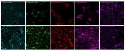
Figure 1. Comparison of fixed cell staining with CytoLinerTM Fixed Cell Membrane Stains versus the classic CellBrite® Cytoplasmic Membrane Dyes. In comparison to the CellBrite® lipophilic carbocyanine dyes, CytoLinerTM Dyes show more uniform and reliable staining of paraformaldehyde-fixed, mildly-permeabilized cell membranes. Paraformaldehyde-fixed cells were permeabilized for 10 minutes at room temperature with 0.1% Triton® X-100 in PBS, then rinsed with PBS and stained with CellBrite® Dyes in PBS or CytoLinerTM Stains in 1X CytoLinerTM Buffer for 10 minutes at room temperature, then rinsed with PBS. Imaged by confocal microscopy; scale bar: 20 um.
VyBrant is a registered trademark of Thermo Fisher Scientific; CellMask is a trademark of Thermo Fisher Scientific; Transwell is a registered trademark of Corning Incorporated; Triton is a registered trademark of The Dow Chemical Company.
 New Products
 Earth-Friendly Products
 Biotium Choice Antibodies
 Special Offers


