 New Products
 Earth-Friendly Products
 Biotium Choice Antibodies
 Special Offers


 Powered by Bioz


Content #1
Content #1
Content #1
Dye solutions of lipophilic carbocyanine dyes DiB, Neuro-DiO, DiI, and DiD, as well as novel near-infrared lipophilic dyes for non-toxic labeling of cytoplasmic membranes.
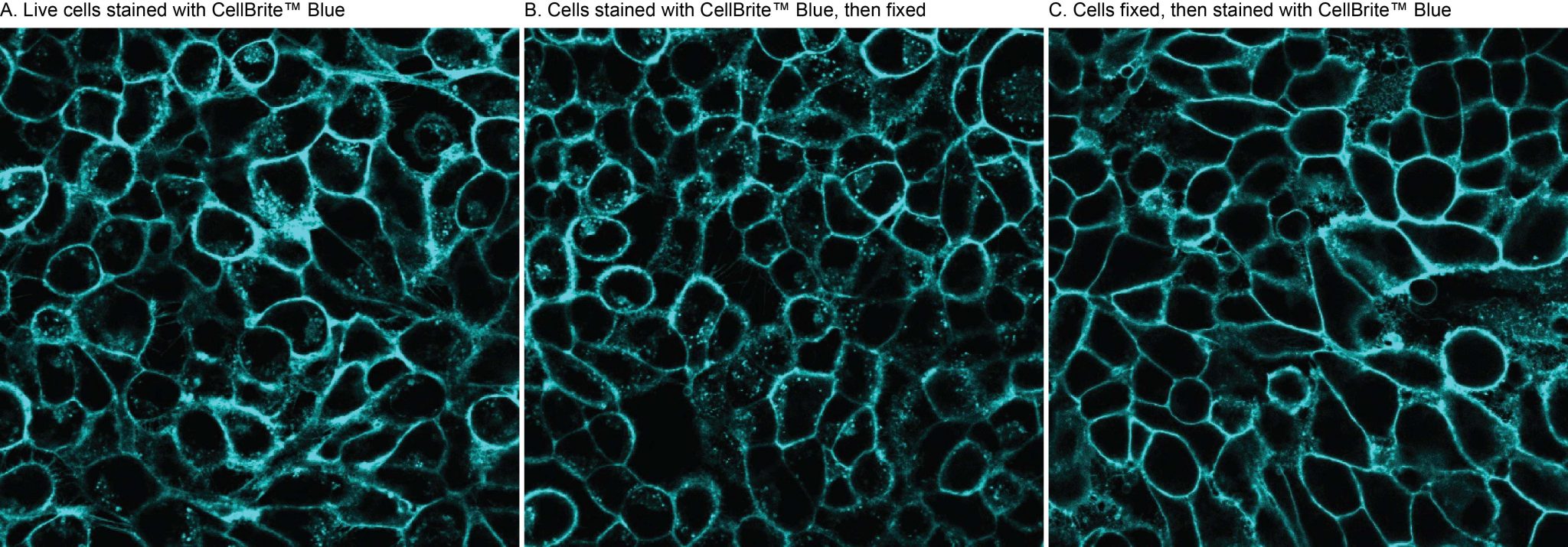
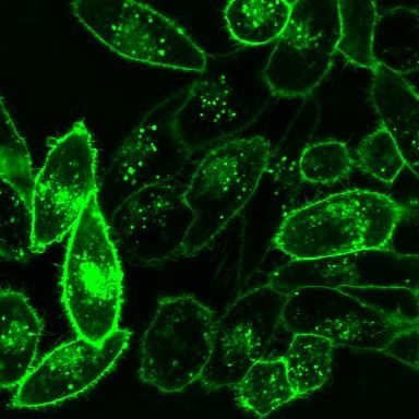
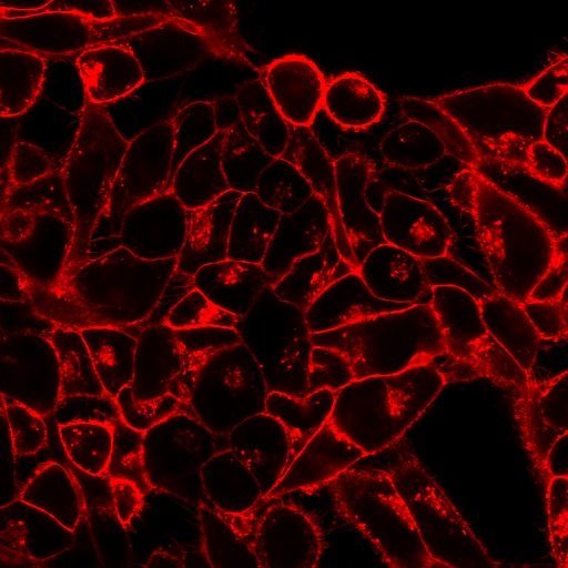
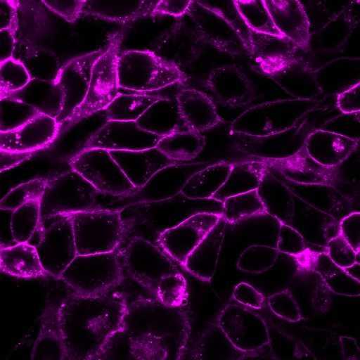
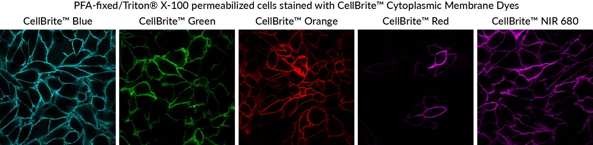
CellBrite® Cytoplasmic Membrane Labeling Kits can be used to label cell cytoplasmic membranes with fluorescent dyes, available in blue, green, orange, red, and near-infrared. The labeling is nontoxic and suitable for long-term tracking of cells. The dyes also can be used to stain membranes of formaldehyde-fixed cells.
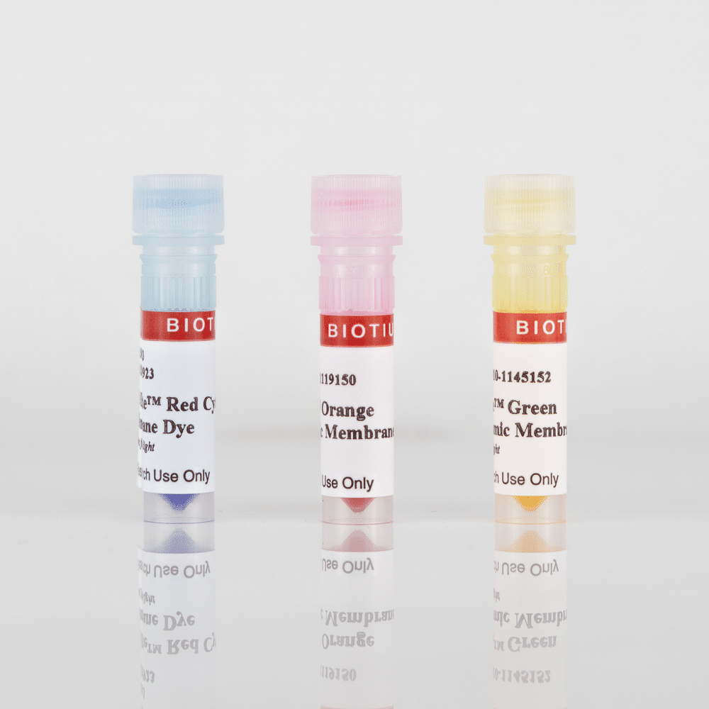
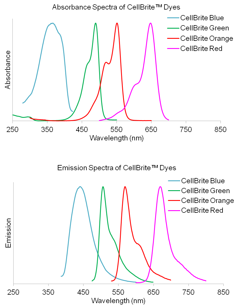
CellBrite® Cytoplasmic Membrane Stains are available as classic lipophilic carbocyanine dyes and novel near-infrared lipophilic dyes. CellBrite® Blue is based on DiB, CellBrite® Green is based on Neuro-DiO, CellBrite® Orange is based on DiI, and CellBrite® Red is based on DiD. These carbocyanine dyes label cytoplasmic membrane and intracellular membrane structures efficiently and permanently. They have been used as tracers in cell fusion, cell adhesion, and cell migration applications due to their properties of low cytotoxicity and high resistance to intercellular transfer. By combining multiple CellBrite® Cytoplasmic Membrane Stains, one can label multiple cell populations with different colors for studies of cell-cell interactions.
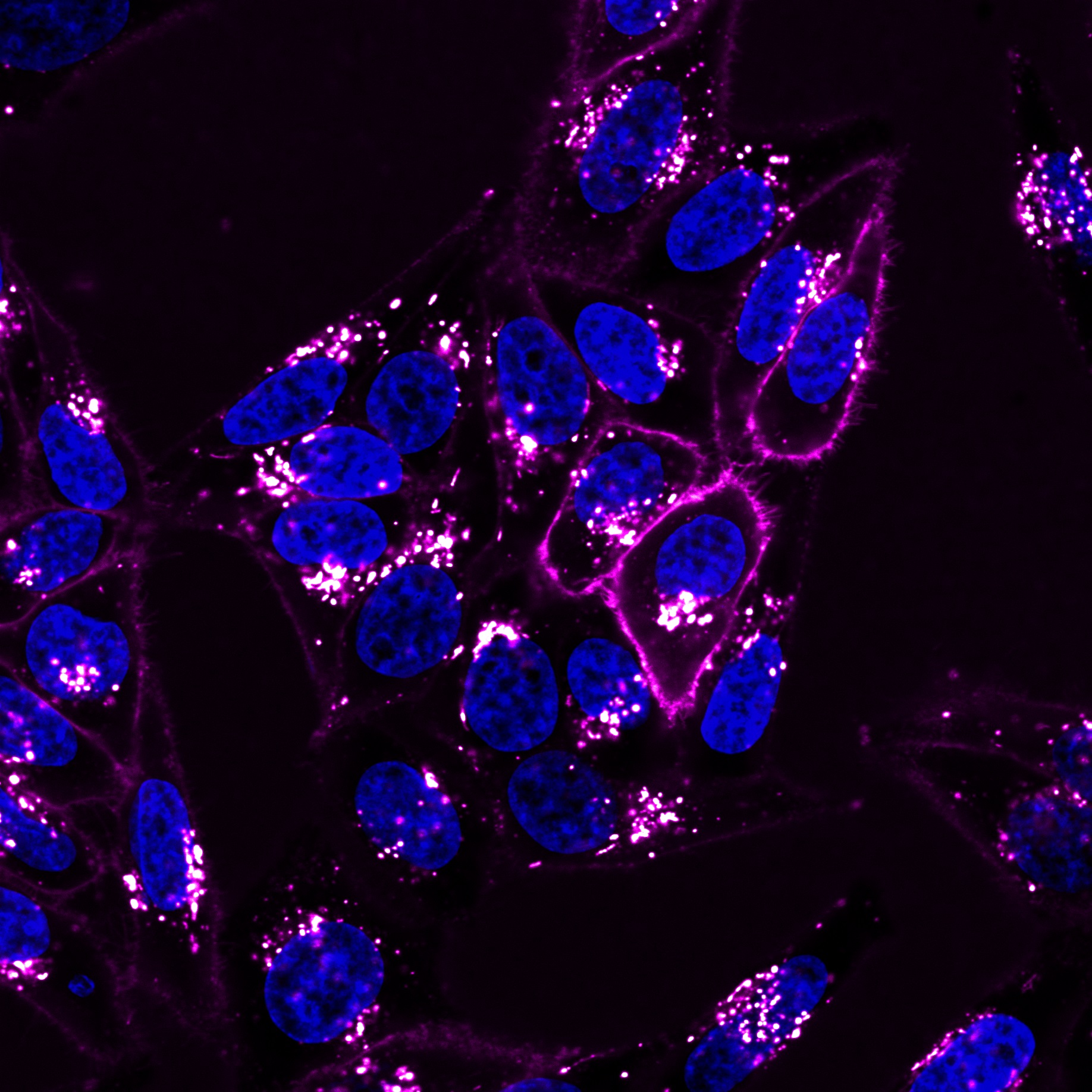
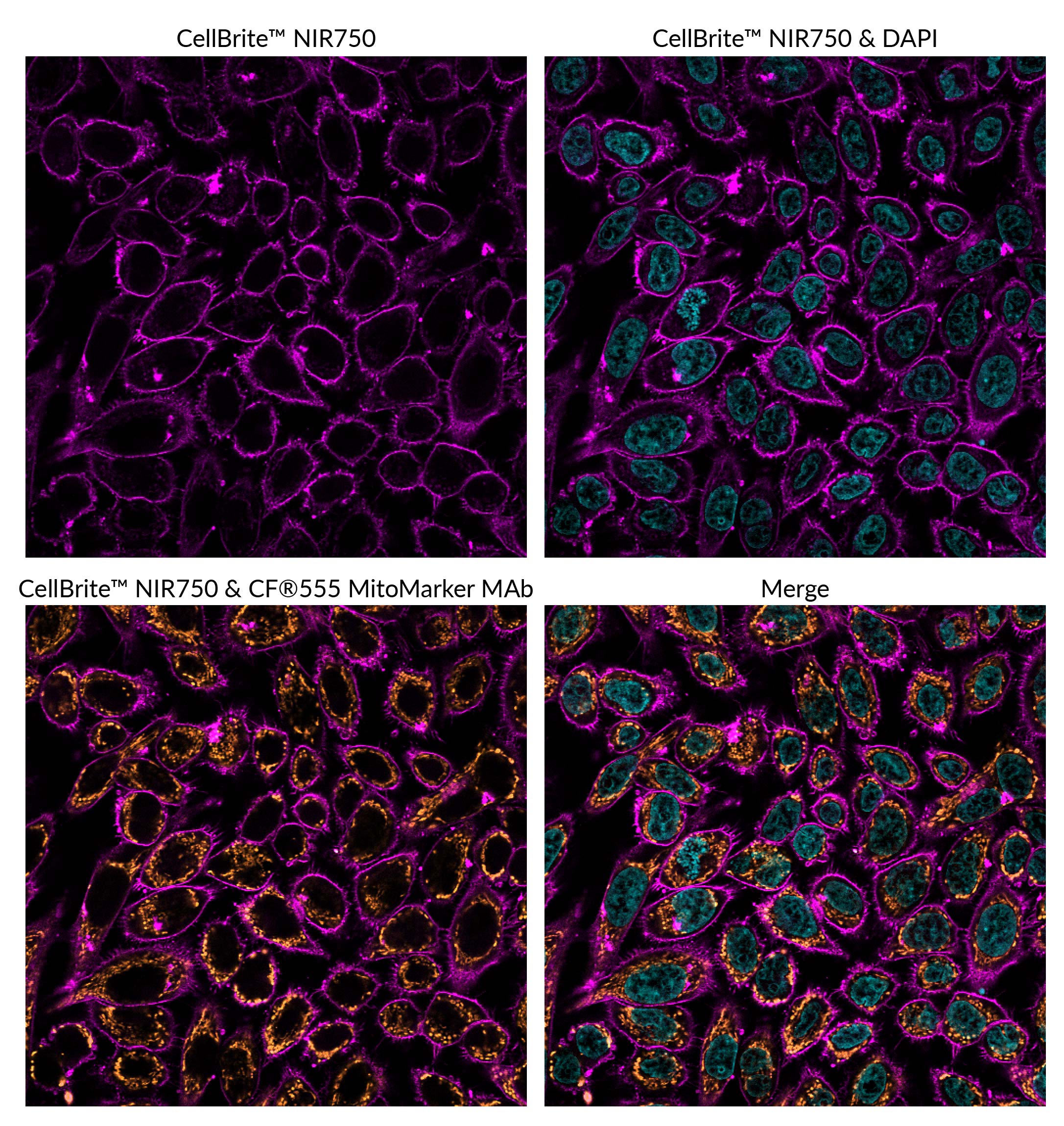
The near-infrared CellBrite® NIR Cytoplasmic Membrane Dyes are unique and due to their long wavelength emission, they can be used to label cells for small animal imaging studies for non-invasive imaging of cell migration and cell homing. CellBrite® NIR Dyes have long 18-carbon hydrophobic tails and an additional water-soluble group. These unique chemical structure elements make the dyes easy to dissolve while providing highly stable cytoplasmic membrane staining, unlike traditional carbocyanine dyes like DiI, DiO, and DiR, which are often difficult to dissolve or prone to precipitation during cell staining.
Biotium's CellBrite® Cytoplasmic Membrane Dyes are ready-to-use and can be added directly to normal culture media to uniformly label suspended or adherent cells in culture. CellBrite® Dyes allow cell populations to be marked in distinctive fluorescent colors for identification after mixing. Double labeling can identify cells that have fused or formed stable clusters.
Note: CellBrite® Dyes primarily localize to cell surface membranes shortly after staining. However, in live cells the stained membranes become internalized by endocytosis, so staining becomes primarily intracellular over the course of a few hours in culture. See "Find the Right Stain for Your Application" below, and our Membrane Staining & Imaging Tech Tip to learn more.
CellBrite® Dyes are non-toxic and stain cells very stably. They can be used to track cells for days to weeks. However, over time the dyes will be internalized by endocytosis, resulting in labeling of intracellular vesicles. A few hours after staining, the dyes will no longer outline the plasma membrane, but will be localized inside the cell. For long-term imaging of cell surfaces in culture we recommend our CellBrite® Steady Membrane Staining Kits which distribute between the cell surface and intracellular compartments, so cells retain cell surface staining over time. For long-term visualization of cell morphology in culture, our stable, non-toxic ViaFluor® SE Cell Proliferation Kits may also be a suitable alternative. ViaFluor® SE dyes covalently label cells throughout the cytoplasm and can be tracked for several days or longer by microscopy or flow cytometry. See our Tech Tip: Using ViaFluor® SE Stains for Cell Tracing and Co-Culture to learn more.
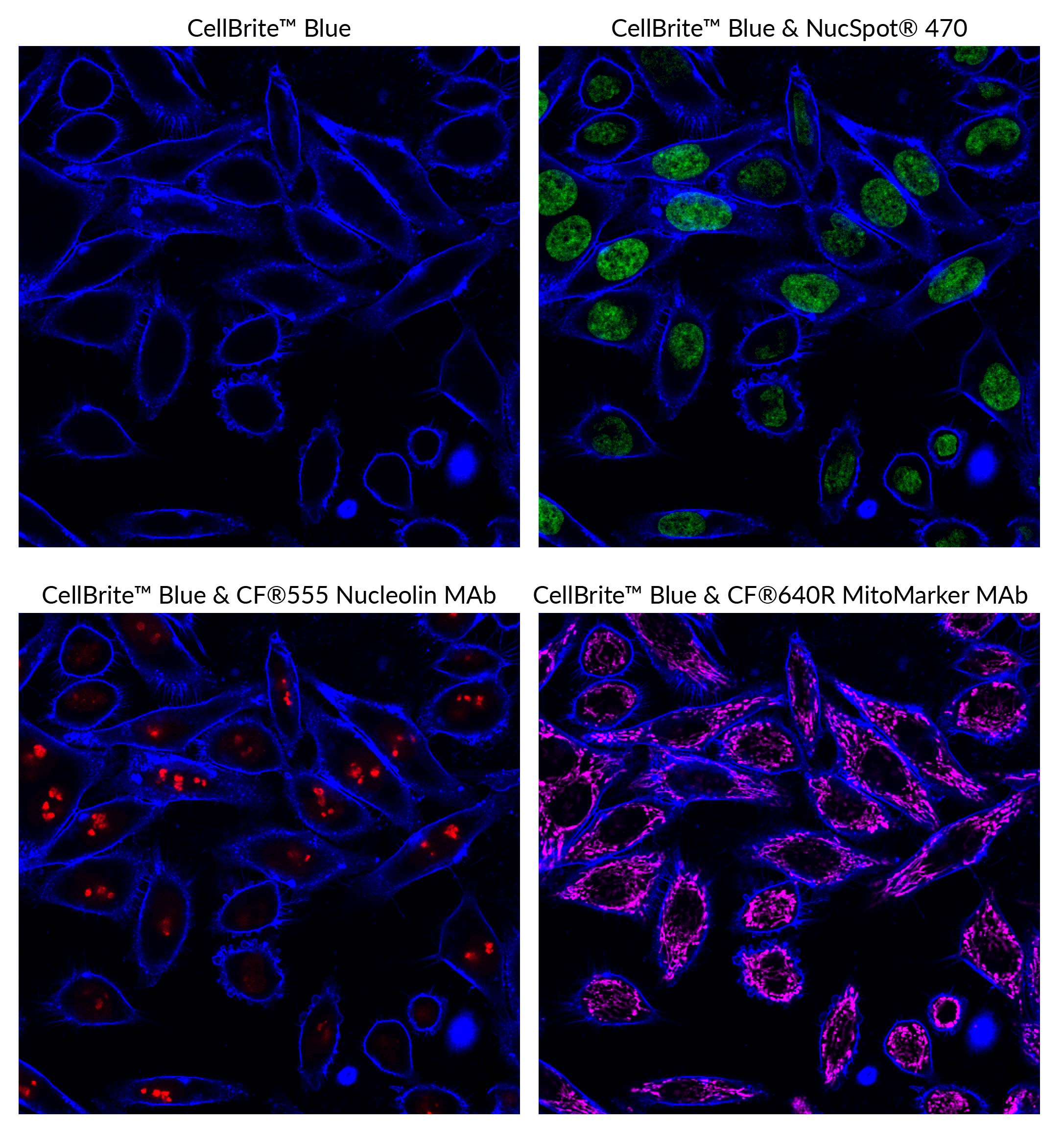
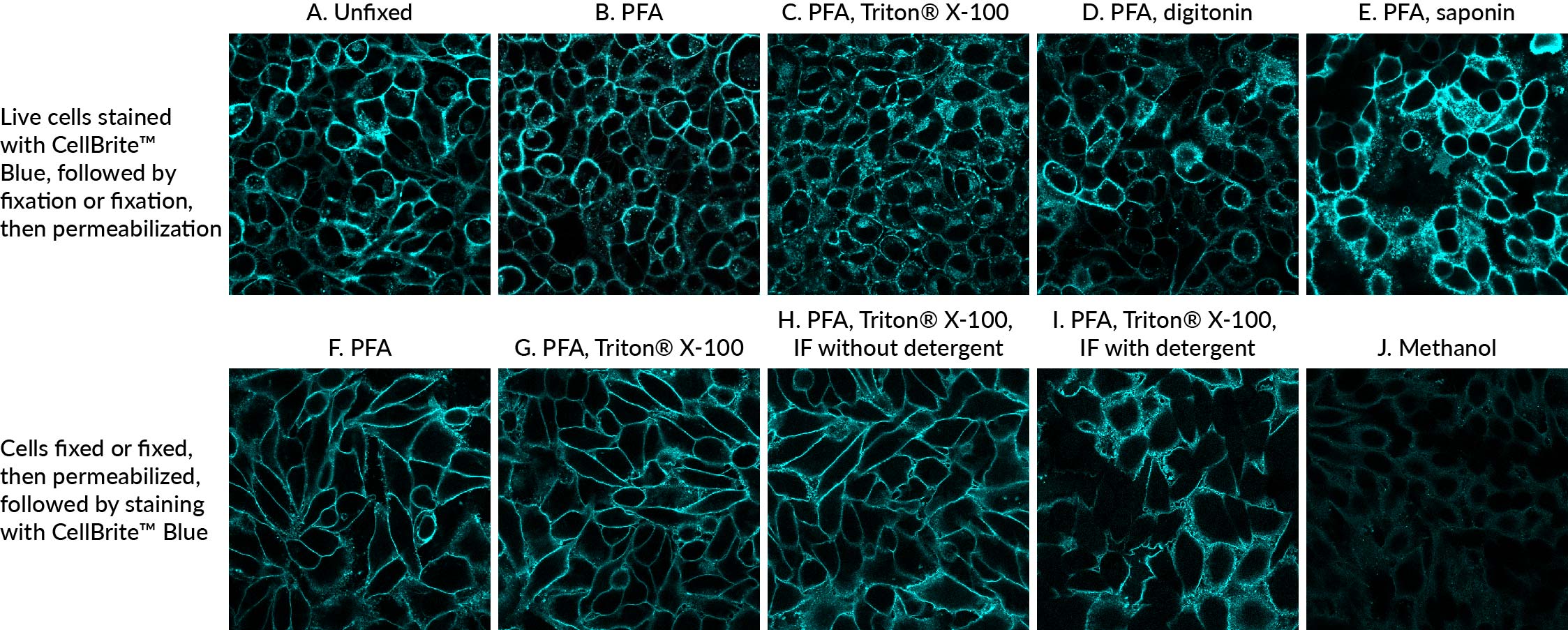
CellBrite® Dyes can be used to stain formaldehyde-fixed cells, but the staining has poor tolerance for methanol fixation or detergent permeabilization. However, permeabilizing fixed cells before staining with CellBrite® Dyes can give good results, see our Tech Tip: Combining Lipophilic Membrane Dyes with Immunofluorescence. For staining formaldehyde-fixed cells we strongly recommend using CytoLiner™ Fixed Cell Membrane Stains which offer more robust and consistent results over the CellBrite® Cytoplasmic Membrane Dyes.
For membrane stains that withstand fixation with either formaldehyde or methanol, as well as detergent permeabilization after staining, see our GlycoLiner™ Cell Surface Glycoprotein Labeling Kits, CellBrite® Fix Membrane Stains and MemBrite® Fix Cell Surface Staining Kits.
CellBrite® Cytoplasmic Membrane Dyes do not stain yeast or bacteria. See our Cellular Stains Table for more information on how our dyes stain various organisms.
Watch our video where Technical Applications Scientist II, Jacqueline Steenhuis PhD answers your top questions about Biotium's various membrane stains for fluorescence microscopy.
For additional support or product recommendations, contact us at [email protected].
| Dye | Ex/Em | Format | Size | Catalog No. |
|---|---|---|---|---|
| CellBrite® Blue | 360/441 nm | 250 uL of Labeling Solution, 200 X in DMSO 250 uL of Cell Loading Buffer, 200X in DMSO | 50 assays | 30024 |
| CellBrite® Green | 489/506 nm | 200X in EtOH | 1 mL | 30021 |
| CellBrite® Orange | 551/569 nm | 200X in EtOH | 1 mL | 30022 |
| CellBrite® Red | 648/670 nm | 200X in EtOH | 1 mL | 30023 |
| CellBrite® NIR680 | 683/724 nm | 2 mM in DMSO* | 100 uL | 30070 |
| CellBrite® NIR750 | 748/781 nm | 2 mM in DMSO* | 100 uL | 30077 |
| CellBrite® NIR770 | 767/806 nm | 2 mM in DMSO* | 100 uL | 30078 |
| CellBrite® NIR790 | 787/820 nm | 2 mM in DMSO* | 100 uL | 30079 |
Download a list of curated CellBrite® and MemBrite® references.
Download a list of curated CellBrite® and MemBrite® references.
Advances in gene therapy increasingly depend on understanding how viral vectors behave within complex, multilayered human tissues. While retinal organoids serve as a powerful model for studying AAV efficacy, their dense, light-scattering architecture has historically limited the ability to visualize and quantify transduction at single-cell resolution. Conventional nuclear stains suffer from rapid photobleaching, cytotoxicity, and shallow imaging depth which hinder repeated live imaging and prevent accurate 3D cell segmentation throughout the organoid. Conventional membrane dyes also pose challenges for staining organoids due to poor penetration, uneven labeling, and rapid internalization by endocytosis.
In a 2025 Small Methods publication, Rogler et. al. developed a longitudinal imaging and deep-learning pipeline to map single-cell AAV transduction dynamics in intact human retinal organoids. This approach required robust and photostable live-cell stains compatible with deep (>100 µm) confocal imaging and repeated imaging over many days. To meet this need, the authors selected Biotium’s far-red NucSpot® Live 650 Nuclear Stain, which provides bright, uniform labeling with minimal phototoxicity and exceptional light penetration compared to blue- or green-excitable DNA dyes. CellBrite® Steady 550, a unique stain for long-term labeling of membranes in live cells, was also used for manual quantification of transduced cells to gauge the performance of their deep-learning method.
Using NucSpot® Live 650, the team captured high-contrast 3D nuclear signals across entire organoids and enabled the use of Cellpose, a deep-learning segmentation algorithm. Paired with GFP-expressing AAV reporters, this allowed precise quantification of transduced cells, as well as quantification of how transduction patterns evolve over time and spatial depth.
The end result revealed heterogeneous AAV penetration profiles, cell-type-specific susceptibility, and spatial gradients of transduction that would have been obscured using conventional methods. Biotium’s NucSpot® Live 650 Nuclear Stain and CellBrite® Steady 550 Membrane Stain enabled high-fidelity, longitudinal imaging in thick living tissues, making quantitative AAV mapping in 3D retinal models possible.
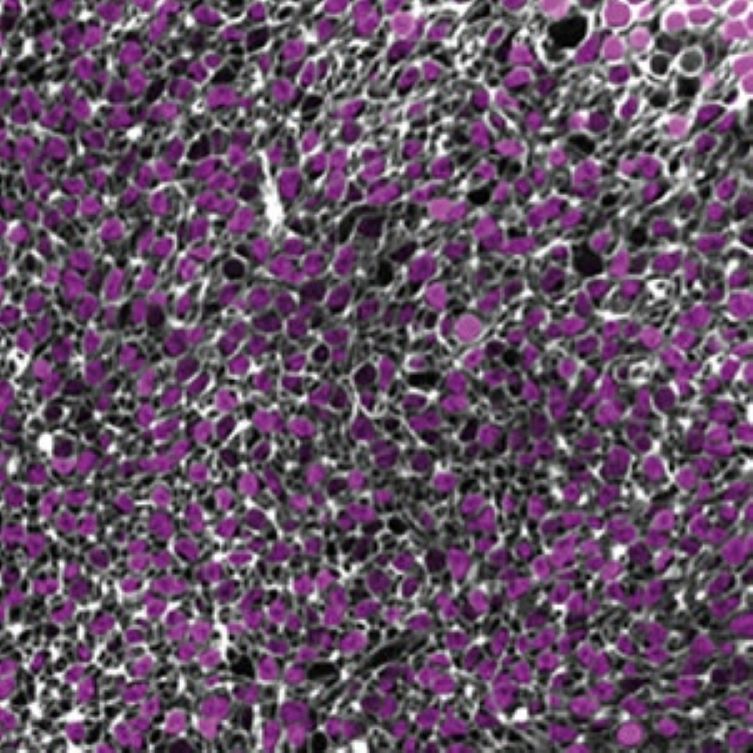
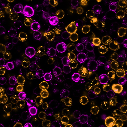
Confocal image of the center plane of the 3D stack of a 264 days old human retinal organoid without virus, stained with NucSpot Live 650 (magenta) and CellBrite Steady 550 (white). Credit: Rogler et al., Small Methods (2025). Reproduced under CC BY 4.0.
Biotium offers an extensive portfolio of bright and specific nuclear and membrane stains, with color options in the near-infrared for deep imaging. View our full selection of cell stains compatible with organoids and other 3D cultures.
Full Citation:
Rogler, T. S., Salbaum, K. A., Brinkop, A. T., Sonntag, S. M., James, R., Shelton, E. R., Thielen, A., Rose, R., Babutzka, S., Klopstock, T., Michalakis, S., & Serwane, F. (2025). 3D quantification of viral transduction efficiency in living human retinal organoids. Small Methods, 2025 Jun 12, e2401050. https://doi.org/10.1002/smtd.202401050
While CellBrite® Cytoplasmic Membrane Dyes can stain formaldehyde-fixed cells, they generally do not give good results in cryosections, possibly because the cell membrane integrity is disrupted, exposing other membrane structures to the dyes. Some customers have reported success using these dyes with vibratome sections.
CellBrite® Cytoplasmic Membrane Dyes are not suitable for membrane staining in FFPE samples as membrane lipids are extracted during the dewaxing and rehydration process. Similarly, acetone or methanol fixation of cryosections will extract lipids, leading to poor staining.
CellBrite® Fix, MemBrite® Fix, and CellBrite® Steady are recommended for use on live cells only. In fixed cells or sections they will label intracellular structures.
In some tissue types, lectins such as CF® Dye WGA Conjugates, CF® Dye Concanavalin A Conjugates, or CF® Dye PNA Conjugates may be useful for staining cell boundaries in FFPE or frozen sections. However, the staining pattern of lectins is highly dependent on cell and tissue type, so we recommend consulting the literature before trying these stains for your tissue of interest.
Alternatively, immunostaining using cell surface-specific antibodies could be done.
So far we have not found a universal plasma membrane stain for tissue sections. This is an application of interest to us and our customers, so we are working to find new solutions.
CellBrite® Cytoplasmic Membrane Dyes are too prone to aggregation to efficiently stain EVs. Some of the CellBrite® Fix, MemBrite® Fix, and CellBrite® Steady dye options have been reported for this application, however we do not recommend them. For optimal staining of exosome membranes we recommend our ExoBrite™ True EV Membrane Stains, which are novel lipophilic membrane dyes specifically designed and optimized for efficient staining of EV membranes with minimal dye aggregation. See our Extracellular Vesicle Research page for more information about our complete line of EV stains and antibodies.
To date, we have not identified a fluorescent cellular stain that will detect bacteria but not mammalian cells with high specificity, or vice versa. While some mammalian cell stains show weak staining of bacteria, they usually do show some signal, and will frequently stain dead bacteria more intensely than live bacteria.
We offer a selection of antibodies for specific bacterial antigens, which potentially have applications for differential staining of bacteria vs. mammalian cells, but we have not validated them in co-culture models.
Also see our Viability PCR Technology Page to learn about how PMA dye can be used for highly specific detection of microbial cell viability in complex samples.
CellBrite® and MemBrite® Stains were originally developed for staining mammalian cells in culture, but some of the stains also have been validated for other organisms and applications. For dyes to stain yeast or bacteria membranes, see Cellular Stains in Different Organisms. For information on staining other organisms or cell types, please see our Tech Tip: Researching Applications for Membrane Dyes.
The CellBrite® Cytoplasmic Membrane Dyes do not stain bacteria. The reactive CellBrite® Fix dyes stain both gram-positive and gram-negative bacteria, while the MemBrite® Fix dyes stain only gram-positive bacteria. However we have not tested these dyes for cell division tracking in bacteria.
There is literature describing the use of CFSE to track bacterial cell division, the ViaFluor® SE cell proliferation dyes are likely to work in a similar manner, but we have not tested this.
See our Cellular Stains Table for a comprehensive list of cellular stains with their ability to stain various cell types.
