Tech Tip: Combining Lipophilic Membrane Dyes with Immunofluorescence
By: Shalini Sharma
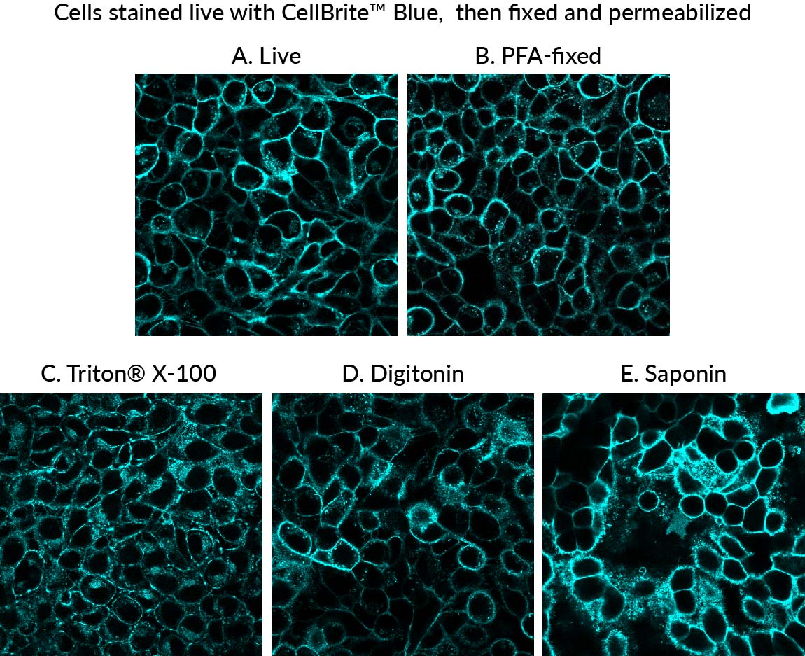
CellBrite® Cytoplasmic Membrane Dyes are membrane dyes that are useful for visualizing cell outlines. However, because they are lipophilic, the dyes tend to redistribute after detergent permeabilization (Fig. 1). For membrane staining of live cells followed by fixation and immunofluorescence, we recommend our newer CellBrite® Fix and MemBrite® Fix fixable cell surface stains. However, these stains cannot be used on cells after they are already fixed. In contrast, our original CellBrite® Cytoplasmic Membrane Dyes can be used to stain cells after formaldehyde fixation. However, their poor tolerance for detergent can make it challenging to incorporate them into an intracellular immunofluorescence protocol.
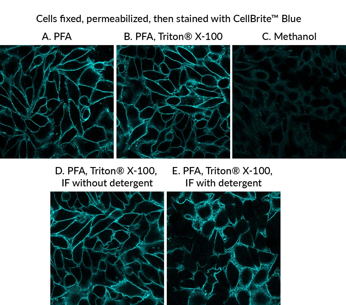
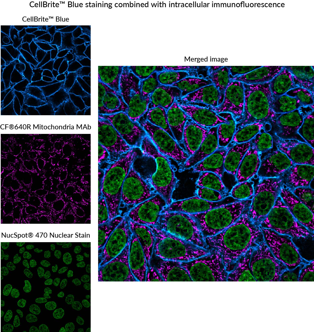
We have developed the following protocol to fix and permeabilize cells before staining with CellBrite® Cytoplasmic Membrane Dyes. Even though the cells are permeabilized before the membrane dye is applied, staining still primarily localizes to the plasma membrane, allowing visualization of cell boundaries (Fig. 2). This protocol works well with a variety of CellBrite® Dyes (Fig. 3), and the staining is well-retained during a typical antibody staining protocol (Figs. 2, 4). However, to avoid dye redistribution, it is important to avoid using detergents after the CellBrite® staining step (see Fig. 2, D-E).
Protocol:
Rinse live cells 3X with HBSS buffer.
Fix cells with 4% paraformaldehyde in PBS, 20 minutes at room temperature.
Note: You may use your preferred formaldehyde fixation protocol, but alcohol or solvent fixatives are not recommended (see Figure 2C).
Rinse cells 3X with PBS.
Permeabilize cells with 0.1% Triton X-100 in PBS for 10 minutes at room temperature.
Note: Permeabilization may be performed at 4°C if preferred.
Add CellBrite® staining solution to the cells and incubate in the dark for 10 minutes at room temperature.
Rinse cells 3X with PBS.
Proceed to blocking, antibody incubation, and washing according to your usual immunofluorescence protocol.
Important: Do not include detergent in any of the buffers used after the CellBrite® staining step.
Note: Protect samples from light.
Image cells in PBS. Glycerol-based mounting medium is not recommended for use with CellBrite® dyes.
Figure 1. Comparison of fixation/permeabilization protocols for CellBrite® Blue staining. Live HeLa cells were stained with CellBrite® Blue, then fixed with paraformaldehyde (PFA), followed by permeabilization with the indicated detergent at 0.1% in PBS for 10 minutes at room temperature. Cell surface staining was well-preserved after fixation (B), but intracellular staining increased after permeabilization with commonly used detergents (C-E). Click image to enlarge. Figure 2. Staining of cell boundaries was observed when HeLa cells were fixed with formaldehyde (PFA), permeabilized with 0.1% Triton® X-100, then stained with CellBrite® Blue (A-B). Poor results were obtained when methanol-fixed cells were stained with CellBrite® Blue (C). Staining was well-preserved after a typical immunofluorescence staining protocol when detergent was omitted from buffers (IF without detergent, D). Using buffer with 0.1% Triton® X-100 for subsequent blocking and antibody incubation steps resulted in dye redistribution (IF with detergent, E). Click image to enlarge. Figure 3. Cells were fixed with formaldehyde (PFA), permeabilized with Triton® X-100, then stained with the indicated CellBrite® Cytoplasmic Membrane Dye according the tech tip protocol. Click image to enlarge. Figure 4. HeLa cells were fixed, permeabilized, and stained with CellBrite® Blue according to the tech tip protocol. Cells were then stained with CF®640R conjugated Mitochondrial Marker Antibody Clone 113-1 (magenta) and NucSpot® 470 nuclear stain (green). No detergent was used during staining after the initial permeabilization step. Click image to enlarge.
 New Products
 Earth-Friendly Products
 Biotium Choice Antibodies
 Special Offers