Kidneys perform several essential functions. They filter out toxins, reabsorb essential electrolytes from the blood, and maintain the acid-base balance in the body. Kidney (or renal) disease is common, affecting around 10% of the adult population, and each year millions die of complications related to Chronic Kidney Diseases (CKD). Renal function is acutely dependent on the renal microstructure which includes the nephron, a long and morphologically segmented tubule where blood is filtered and electrolytes are reabsorbed into surrounding capillaries. There are approximately one million nephrons in an average adult human kidney and about 20,000 in a mouse. Due to the critical importance of the renal microstructure, structural alterations to kidney tubules generally reflect renal damage.
In a recent study published in The FEBS Journal, the authors Kumaran and Hanukoglu have for the first time generated a detailed map of the actin cytoskeletal architecture in sections of a whole mouse kidney by high-resolution confocal microscopy. Actin filaments, an important structural feature of the nephron tubular network, were stained with CF®488A-labeled phalloidin. Various tubular segments of the nephron could be clearly identified and localized to specific regions of the kidney and distinct cytoskeletal profiles could be associated with specific tubule segments. Additional markers including aquaporin, cytokeratin 8-18, CD34 and CF®555-labeled WGA lectin were used to corroborate these observations. The approach used in the study could be incredibly useful for characterizing structural damage in kidney tubules in various renal diseases and animal models of kidney disorders.
Full citation:
Kumaran GK, Hanukoglu I. Identification and classification of epithelial cells in nephron segments by actin cytoskeleton patterns. FEBS J. 2019 Oct 12. doi:10.1111/febs.15088.
Learn more about Biotium’s fluorescent cytoskeletal probes and dyes for staining cellular membranes and organelles for live and fixed cell staining.
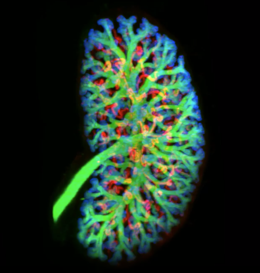
Light sheet imaging of whole labelled kidney at E15.5 (credit: Short, M. Kieran et. al, eLife 2018)