 New Products
 Earth-Friendly Products
 Biotium Choice Antibodies
 Special Offers


Content #1
Content #1
Content #1
With the advent of microchips, computers that once spanned whole rooms now fit into the palm of our hand. Cutting-edge science aims to scale down research experiments in a similar manner using microtechnology. Lab-on-a-chip (LOC) technology was born from this goal in 1979 and has evolved and grown in popularity significantly since then.1
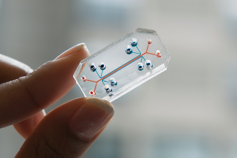
A lung-on-a-chip microdevice. Image courtesy of Wyss Institute/Design Museum.
Three-dimensional (3D) cell culture technologies are still often used for drug development as these paradigms allow for more biologically relevant cell networks and high-throughput screening.4 However, there are many functional aspects of cells and organs that 3D culture models cannot replicate. These limitations include a lack of tissue-to-tissue interfaces, spatiotemporal gradients of chemicals, and mechanically active components. The application of LOC technology breaks through these barriers.5
Even tricky problems, such as mimicking physically separated biological structures, can now be tackled with LOC. Yamamoto et al. showed this in their use of microdevices to create precisely compartmentalized co-cultures of motor neurons and skeletal muscle fibers to form viable neuromuscular junctions. They showed that these cells are functionally identical to their in vivo counterparts in the presence of an excitatory neurotransmitter, inhibitory chemicals, and mechanical force. These cells can be labeled with fluorescent dyes and imaged on-chip with minimal adjustments to standard protocols, as shown by the use of lipophilic carbocyanine dyes, like CellBrite® Green, to stain motor neuron axons in this system.6
As scientists gained the ability to generate increasingly complex systems on chips, they began creating organoids, leading to the birth of the organ-on-a-chip (OOC) subfield.3 With these stem cell-derived, self-organizing miniature organs, researchers seek to replicate the critical structural and functional characteristics of their in vivo counterparts. Functional OOC could provide cost effective, high-throughput disease model systems as an alternative for drug testing in animals or human tissue.7
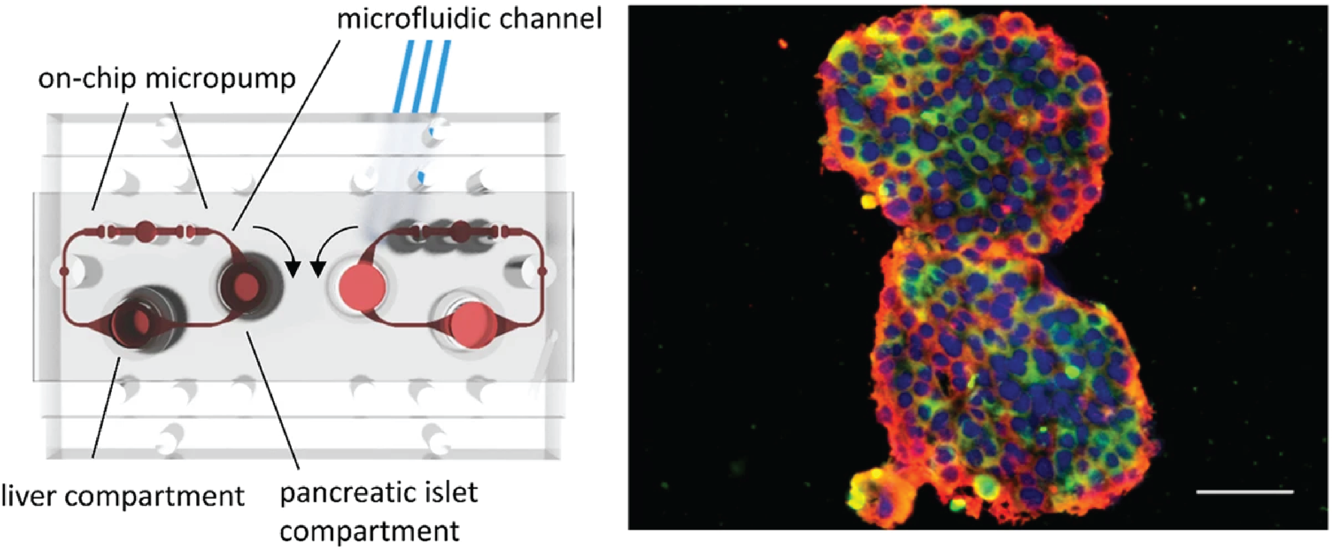
An illustration of the two-organ-chip used by Bauer, et al. with media circuits, respective culture compartments, and micropump valves highlighted in red. The panel on the right shows the pancreatic islet microtissues and liver spheroids used in the experiment immunostained for insulin (pseudocolored red) and glucagon (CF®594, pseudocolored green). Credit: Bauer, et al. https://doi.org/10.1038/s41598-017-14815-w reproduced under the Creative Commons license.
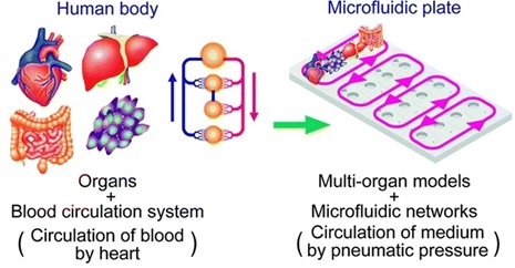
Experts hope that multi-organ chips will eventually lead to the creation of patient-on-a-chip technology. This precise approach would entail creating a complex, individualized multi-organ chip model allowing clinicians to develop treatment plans tailored to each patient’s unique biology. It would also create an attractive alternative to the use of animal models. While many limiting factors still exist, including the expense of the components, the need for universal cell culture media, and the challenge of managing and interpreting data from such a complex system, the potential reward of revolutionizing our approach to personalized medicine and laboratory research keep scientists pushing forward.1
Julianne Davis earned an MSc in Behavioral Neuroscience from the University of Washington, Seattle, where she examined the role of memory in cost-based decision-making. She has also studied sensory integration at the Allen Institute and the neural basis of feeding, thirst, and metabolism at the University of California, San Francisco as a research scientist. Currently, she is a Technical Writer and Support Scientist at Biotium.
