 New Products
 Earth-Friendly Products
 Biotium Choice Antibodies
 Special Offers


Content #1
Content #1
Content #1
Flow cytometry is capable of distinguishing individual cells based on size, shape, and expression levels of dozens of fluorescently-labeled proteins as they are zipped past a light source and various detectors. This technique can measure hundreds of thousands of individual cells in minutes—its power is recognized by scientists and at least one former president.
Cells in suspension are pumped through the flow cytometer, where hydrodynamic focusing presses them into a single-file line for measurement. Conventional flow cytometry uses dichroic mirrors and point detectors (photomultiplier tubes or avalanche photodiodes) to measure specific wavelengths after illumination with a laser. For excitation with multiple lasers, the cells are illuminated by one laser at a time in an assembly line, with each laser accompanied by its own set of dichroic mirrors and detectors.
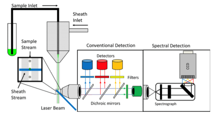
Figure 1. Conventional vs spectral flow cytometry (Nolan & Condello, 2013). Conventional detection uses dichroic mirrors and detectors to measure each color. Spectral flow cytometry uses a spectral detector to measure the whole spectral picture after dispersion with a prism or grating.
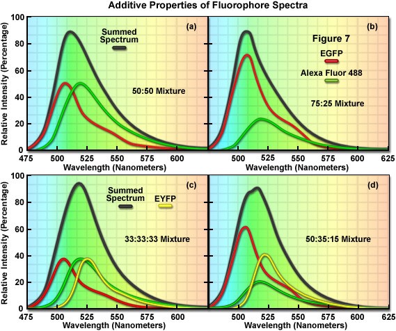
Figure 2. Example of spectral unmixing with two and three fluorophores. The shape of the summed spectrum depends on the fluorophores in the sample and their ratios. Source: Zeiss microscopy online campus.
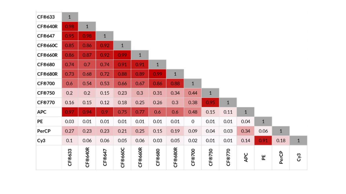
Figure 3. The similarity indices of long-wavelength CF® dyes, with far-red and beyond highlighted. Lower values mean spectra are more different. Source: Biotium
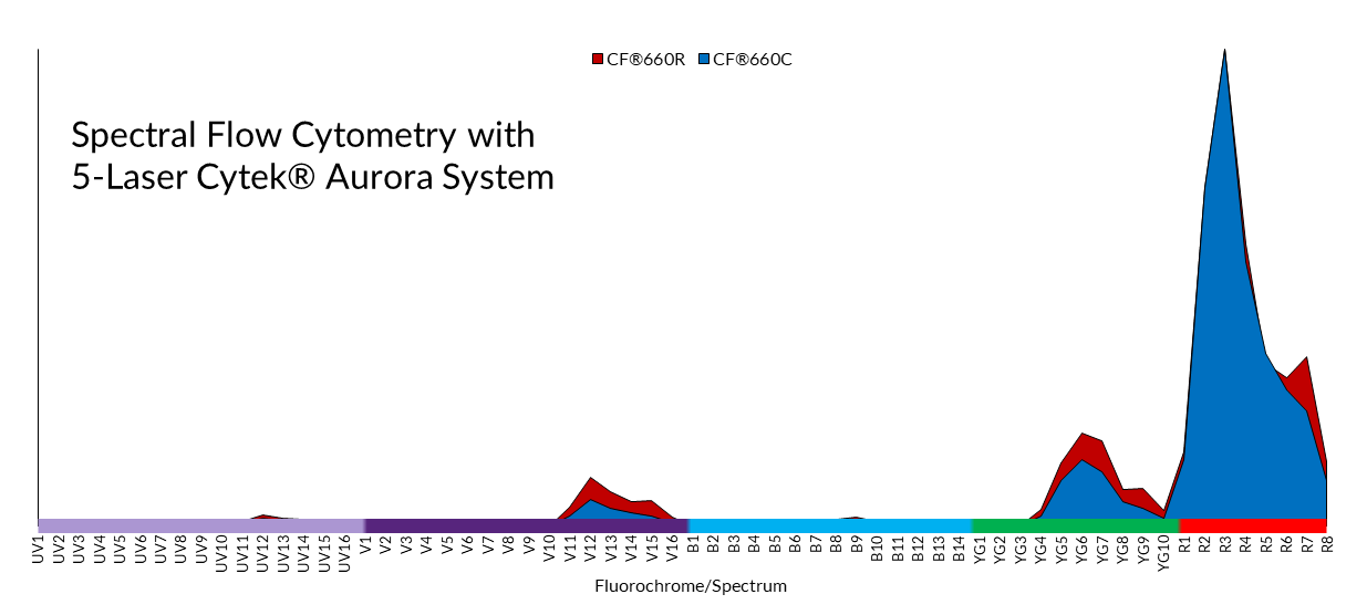
Figure 4. Spectra of CF660R and CF660C on 5-laser Cytek™ Aurora System. Even with a similarity index of 0.99, these two dyes are distinct enough for tandem use in spectral flow cytometry.
Spectral flow cytometry makes it possible to measure many dyes without having the user perform mathematical compensation and subtraction. This “whole picture” of the spectrum can be broken down into its constituent parts to distinguish 40 or more labels in a single experiment, limited only by the dyes available. CF® dyes offer labels across the spectrum that are compatible with spectral flow cytometry and fill in the “spectral gap” in the far-red and longer wavelength region to enhance experimental possibilities!
For further reading, check out Biotium’s spectral flow cytometry page.

Christopher Pratt earned his PhD in cell biology and neuroscience from Carnegie Mellon University in 2016 under the guidance of Marcel Bruchez, PhD. For his thesis, he developed and applied novel fluorescent probes to understand synaptic vesicle and ion channel trafficking in the brain, making him a ardent microscopist. With a passion for communicating science and data, Christopher is a regular contributor to Biotium’s Full Spectrum Blog and is a freelance science and medical writer, analyst, and research consultant based in Chicago, Illinois. Visit his website, twitter, or LinkedIn and get in touch!

Content #1
Content #1
Content #1
Content #2
Content #3