 New Products
 Earth-Friendly Products
 Biotium Choice Antibodies
 Special Offers


Content #1
Content #1
Content #1
Imaging in visible light hits a wall when it comes to imaging in tissue for two main reasons: visible light is absorbed and scattered by tissue and tissue autofluorescence occurs chiefly in the visible spectrum. Both factors limit the usefulness of visible light for looking deep in tissue. Moving into the far red and near-infrared (NIR) region of the spectrum allows light to penetrate deeper into tissue with less absorption, scattering, and autofluorescence, improving signal-to-noise and depth of imaging. NIR spans from 650-2000 nm and contains two spectral “windows” for biologic imaging where water absorption is minimal: NIR-I ranges from 650-900 and NIR-II ranges from 1000-1300, overlapping with short-wave IR (SWIR), which spans 1000-2000 nm. NIR and SWIR imaging are not without drawbacks: spatial resolution decreases with longer wavelengths of light and reduced but present scattering can still degrade image quality. Fortunately, advances in imaging technology including adaptive optics and deep learning can mitigate these limitations.1–5
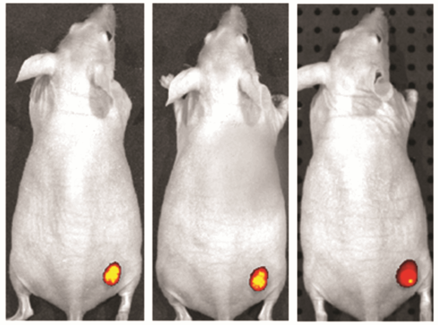
CF®750 for in vivo small animal imaging. Tumors in mice were imaged using an IVIS® imaging system (Perkin Elmer) 24 hours (left), 48 hours (center), and 96 hours (right) after IV injection of CF®750 Avastin® conjugate. Image courtesy of Caliper Life Sciences.
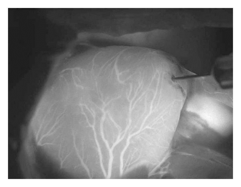
Indocyanine green labeling of coronary arteries in a rat heart. Credit: Alander, et al. https://doi.org/10.1155/2012/940585 reproduced under the Creative Commons license.
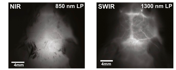
Imaging in NIR-I (left) compared to imaging in SWIR using ICG, showing markedly higher contrast in SWIR despite lower ICG emission. Credit: Carr, et al. https://doi.org/10.1073/pnas.1718917115 reproduced under the Creative Commons license.
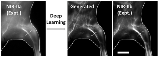
Deep learning models can approximate a NIR-IIb (1500-1700 nm) image, with its improved signal-to-noise, from a NIR-IIa image (900-1300 nm, left). “Generated” shows the AI-created image, while “expt” shows an actual NIR-IIb image for comparison. Scale bar 5 mm. Credit: Ma, et al. https://doi.org/10.1073/pnas.2021446118 reproduced under the Creative Commons license.
Another developing technology that can improve image quality is adaptive optics. Adaptive optics were originally developed for astronomy to counter distortions by the atmosphere, but the principle can be used in biologic imaging to counteract the image produced by scattering in the tissue, even permitting super-resolution imaging at depth.2
For clinical use, several new NIR dyes have been undergoing clinical trials for use in human surgery and photodynamic therapies, largely in oncology. Biocompatible materials will lead the way in NIR dye development, both in and out of the clinic. The advantages of NIR in basic biological research are also gaining traction as volumetric microscopic imaging techniques such as light sheet become more common. Imaging in NIR-II and SWIR is also expected to become more common, as the InGaAs (indium gallium arsenide) sensors required to detect long wavelength photons become more commonplace.3,4,7,10

Christopher Pratt earned his PhD in cell biology and neuroscience from Carnegie Mellon University in 2016 under the guidance of Marcel Bruchez, PhD. For his thesis, he developed and applied novel fluorescent probes to understand synaptic vesicle and ion channel trafficking in the brain, making him a ardent microscopist. With a passion for communicating science and data, Christopher is a regular contributor to Biotium’s Full Spectrum Blog and is a freelance science and medical writer, analyst, and research consultant based in Chicago, Illinois. Visit his website, twitter, or LinkedIn and get in touch!

Content #1
Content #1
Content #1
Content #2
Content #3